Chapter 3: The Eye and the Retina
Larry Hester - was diagnosed with genetic disorder of the eye called retinitis pigmentosa. He first noticed that his vision was rapidly declining.
Bionic eye
an array of electrodes implanted in the back of the eye and through camera that is mounted in eyeglasses it sends signals to the visual system about what is “out there” in the world
it does not completely restore vision but it allows the person to see contrasting lightness versus darkness
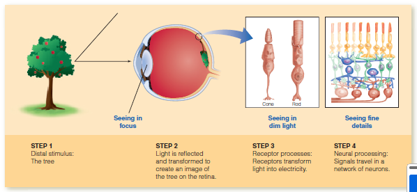
Four steps of visual process
Distal stimulus (the tree);
Light is reflected from the tree and enters the eye to create the proximal stimulus on the visual receptors;
Receptors transform light into electrical signals; and
Electrical signals are “processed” as they travel through a network of neurons.
3.1 Light, the Eye and the Visual Receptors
Light: The Stimulus for Vision
Vision is based in visible light, which is a band of energy within the electromagnetic spectrum.
Electromagnetic spectrum - is a continuum of electromagnetic energy that is produced by electric charges and is radiated as waves
Wavelength - the distance between the peaks of the electromagnetic waves.
The wavelengths in the electromagnetic spectrum
range from extremely short-wavelength gamma rays
(wavelength 5 about 10212 meters, or one ten-billionth of a
meter) to long-wavelength radio waves (wavelength 5 about
104 meters, or 10,000 meters).
Visible light - the energy within the electromagnetic spectrum that humans can perceive. For humans and some other animals, the wavelength of visible light is associated with the different colors of the spectrum, with short wavelengths appearing blue, middle wavelengths
green, and long wavelengths yellow, orange, and red.
The Eye
Eyes - contain the receptors for vision
Light reflected from objects in the environment enters
the eye through the pupil and is focused by the cornea and
lens to form sharp images of the objects on the retina, the network of neurons that covers the back of the eye and that contains the receptors for vision, also known as photoreceptors
Two types of photoreceptors:
Rods
Cones
Outer segments - are part of the receptor that contains light-sensitive chemicals called visual pigments that react to light and trigger electrical signals.
Signals from the receptors flow through the network of neurons that make up the retina and emerge from the back of the eye in the optic nerve, which contains a million optic nerve fibers that conduct signals toward the brain.
Fovea contains only cones. Objects image falls on the fovea when looked at directly.
Peripheral Retina includes all of the retina outside of the fovea, contains both rods and cones
Macular degeneration - destroys the cone-rich fovea and a small area that surrounds it. (mostly common around old people)
Macula is a medical practice that includes the fovea plus a small area surrounding the fovea
Retinitis pigmentosa - attacks the peripheral rod receptors and result in poor vision. In some cases, the foveal cone receptors are also attacked, resulting in complete blindness.
Blind spot refers to the point where the optic nerve passes through the retina, creating a gap due to the absence of photoreceptor cells.
3.2 Focusing Light Onto the Retina
Light reflected from an object into the eye is focused onto the retina by a two-element optical system: the cornea and the lens.
Cornea - the transparent covering of the front of the eye, accounts for about 80 percent of the eye’s focusing power.
Lens - supplies the remaining 20 percent of the eye’s focusing power, can change its shape to adjust the eye’s focus for objects located at different distances
Ciliary muscles - the action which causes the lens to change shape. It increases the focusing power (ability to bend light) of the lens by increasing its curvature.
Accommodation - the change in the lens’s shape that occurs when the ciliary muscles at the front of the eye tighten and increase the curvature of the lens so that it gets thicker
the increased curvature increases the bending of the light rays passing through the lens to create a sharp image on the retina
by looking around at different objects, the eye constantly adjusting its focus by accommodating especially for nearby objects.
accommodation makes it possible to adjust vision for different distances
Refractive Errors - errors that can affect the ability of the cornea and/or lens to focus the visual input onto the retina.
Presbyopia - an age-related loss of the ability to accommodate. As people get older, their ability to accommodate.
Myopia or nearsightedness - an inability to see distant objects clearly. It can be solved by a corrective lens.
occurs when the optical system brings parallel rays of light into focus at a point in front of the retina, so the image that reaches retina is blurred.
Refractive Myopia - the cornea and/or the lens bends the light too much
Axial Myopia - the eyeball is too long.
Hyperopia or farsightedness - can see distant objects clearly but have trouble seeing nearby objects because the focus point of the parallel rays is located behind the retina.
3.3 Photoreceptor Process
Transforming Light Energy Into Electrical Energy
Transduction - the transformation of one form of an energy into another form of energy. Visual transduction occurs in photoreceptors (the rods and cones) and transforms light into electricity.
Visual pigments are light-sensitive molecules found in photoreceptor cells of the retina. They play a crucial role in the process of vision by absorbing light and initiating the photo transduction cascade, which converts light into electrical signals sent to the brain. Two parts of visual pigments: 1.) Opism, a long protein; and 2.) retinal which is the sensitive component
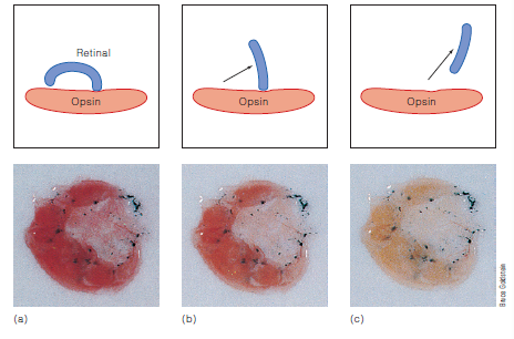
Isomerization is a process where molecules change shape when a light hits the retina and the visual pigment molecule absorbs the light causing the retinal in the molecule change shape from being bent, creating a chemical reaction.
Adapting to the Dark
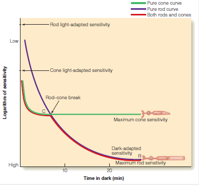
Dark adaptation is the process of increasing sensitivity in the dark. It is measured by determining a dark adaptation curve.
Measuring the Dark Adaptation Curve
Initial Setup: Participant focuses on a fixation point while observing a peripheral flashing test light.
Threshold Measurement: Adjust intensity of the test light until it is barely visible (method of adjustment).
Sensitivity Calculation: Sensitivity = 1/Threshold; high threshold = low sensitivity.
Light-Adapted Sensitivity: Measured with lights on; requires high intensity to see the test light.
Dark Adaptation: After extinguishing lights, participant adjusts intensity to track increasing sensitivity in the dark, resulting in a sensitivity curve.
Measuring Rod Adaptation
Cone Adaptation: The green curve in Figure 3.13 represents cone adaptation, as the test light is focused on the all-cone fovea.
Initial Sensitivity: Cones are more sensitive at the start of dark adaptation, masking rod activity.
Rod Monochromats
Definition: Individuals with a genetic defect resulting in no cones; possess all-rod retinas.
Purpose: Allows study of rod dark adaptation without cone interference.
Dark Adaptation Process
Initial Phase:
Light is turned off; both cones and rods increase sensitivity.
Cones dominate vision initially due to higher sensitivity.
Rods are less sensitive at this stage.
Rod Sensitivity Measurement:
Light-adapted sensitivity measured in rod monochromats reflects rod sensitivity.
Rods are significantly less sensitive than cones when light-adapted.
Adaptation Timeline:
Rod sensitivity increases (purple curve) and reaches maximum in about 25 minutes (Rushton, 1961).
This final sensitivity matches the second part of the two-stage dark adaptation curve.
Key Points:
Rod-Cone Break: The transition point where rods begin to dominate the dark adaptation curve.
Timing:
Cones reach maximum sensitivity in 3-5 minutes.
Rods catch up to cones around 7 minutes.
Rods take 20-30 minutes to reach maximum sensitivity (point R) compared to 3-4 minutes for cones (point C).
Visual Pigment Regeneration
Process: Visual pigment regeneration occurs more rapidly in cones than in rods, explaining the difference in adaptation times.
Dark Adaptation: Initially dominated by cones, transitioning to rods after several minutes.
Importance of Rod Monochromats: Essential for isolating rod adaptation effects without cone influence.
Visual Pigment Regeneration
Visual Pigment Molecule: Composed of retinal (light-sensitive) and opsin (protein).
Light Activation: Light causes retinal to change from a bent shape to a straight shape, leading to separation from opsin.
Visual Pigment Bleaching: The change in shape and separation results in a lighter color of the pigment, rendering it temporarily unresponsive to light.
Process of Bleaching
Initial State: Retinal is bent (Figure 3.10a).
Activation: Upon exposure to light, retinal is isomerized to a straight form (Figure 3.10b).
Separation: Retinal detaches from opsin, leading to bleaching (Figures 3.14a-c).
Color Change: Frog retina shows a transition from red (intact pigment) to lighter shades as bleaching occurs.
Visual Pigment Regeneration
Definition: The process of reattaching retinal to opsin and returning it to its bent shape.
Analogy: Similar to a light switch; once activated (bleached), it cannot signal again until reset (regenerated).
Continuous Process: In normal light, some pigments bleach while others regenerate.
Dark Adaptation
Regeneration in Darkness: In the absence of light, bleached pigments regenerate, increasing sensitivity.
Measurement: William Rushton (1961) demonstrated the correlation between pigment regeneration and dark adaptation.
Cone Pigment: Regenerates in ~6 minutes.
Rod Pigment: Regenerates in >30 minutes.
Sensitivity Relationship:
Light sensitivity depends on visual pigment concentration.
Rate of sensitivity increase in darkness correlates with pigment regeneration speed.
Detached Retina
Condition: Occurs when the retina detaches from the pigment epithelium, hindering regeneration.
Causes: Can result from traumatic injuries (e.g., sports accidents).
Consequences: Permanent blindness in the affected visual field unless reattached via laser surgery.
Spectral Sensitivity
Overview
Rods and Cones: Key photoreceptors in the eye that adapt vision to darkness.
Spectral Sensitivity: The eye's sensitivity to light as a function of wavelength.
Spectral Sensitivity Curves
Definition: Graphical representation of the relationship between light wavelength and sensitivity.
Measurement Method:
Present one wavelength at a time (monochromatic light).
Use filters or a spectrometer to create specific wavelengths.
Determine the participant's threshold for seeing each wavelength using psychophysical methods.
Measurement Process
Threshold Measurement:
Measure sensitivity at regular intervals (e.g., 400 nm, 410 nm).
Higher thresholds at short and long wavelengths; lower in the middle spectrum.
Conversion to Sensitivity:
Sensitivity is calculated as ( \text{sensitivity} = \frac{1}{\text{threshold}} ).
Results in a spectral sensitivity curve showing sensitivity versus wavelength.
Rod vs. Cone Sensitivity
Cone Sensitivity:
Measured by having participants look directly at a test light (stimulating cones in the fovea).
Rod Sensitivity:
Measured after dark adaptation (rods are more sensitive).
Test flashes presented in the peripheral retina (off to the side of fixation).
Key Findings
Sensitivity is highest in the middle of the visible spectrum.
Less light is needed to perceive wavelengths in the middle compared to short or long wavelengths.
Figures
Figure 3.15a: Threshold versus wavelength curve.
Figure 3.15b: Spectral sensitivity curve derived from threshold data.
Conclusion
Understanding spectral sensitivity is crucial for comprehending how we perceive light and color, influenced by the distinct roles of rods and cones in our visual system.
Rod and Cone Spectral Sensitivity
Key Points
Sensitivity Curves:
Rods: Most sensitive to light at 500 nm (blue-green region).
Cones: Most sensitive to light at 560 nm (green region).
Vision Adaptation:
Transition from cone to rod vision occurs during dark adaptation.
This shift increases sensitivity to short-wavelength light (blue and green).
Example: Green foliage appears more vibrant at dusk.
Purkinje Shift:
Enhanced perception of short wavelengths during dark adaptation.
Named after Johann Purkinje, who described this effect in 1825.
Experiment: Close one eye for 5-10 minutes in the dark, then compare color sensitivity between eyes (e.g., blue flower vs. red flower).
Rod and Cone Pigment Absorption Spectra
Absorption Spectrum:
A graph showing light absorption vs. wavelength.
Rod Pigment:
Absorbs best at 500 nm.
Cone Pigments:
Three types, each with distinct absorption peaks:
Short-wavelength (S): Best at 419 nm.
Medium-wavelength (M): Best at 531 nm.
Long-wavelength (L): Best at 558 nm.
Summary
The differences in spectral sensitivity between rods and cones are linked to their respective pigment absorption spectra.
Rods are more sensitive to shorter wavelengths, enhancing night vision and color perception during low light conditions.
Understanding these differences is crucial for comprehending human color vision and adaptation processes.