Genetics Section 2
DATE: 9/15/25
Exams will be handed back at end of class
—worm lab tomorrow. only lesson two — read background from lesson one, BC that’s what quiz will be on
Chapter 6
low 20000s genes in the human genome
little over 1000 genes per chromosomes
Linkage
Two points
-1) two or more genes located on the same chromosome
physically connected to each other (synteny)
each chromosome made up of continuous linear DNA molecule — called linkage groups
Bateson and Punnet:
conducted cross b/t pea differing in flower color and pollen shape
what ratio would you expect for the dihybrid cross?
What ratio did they observe?
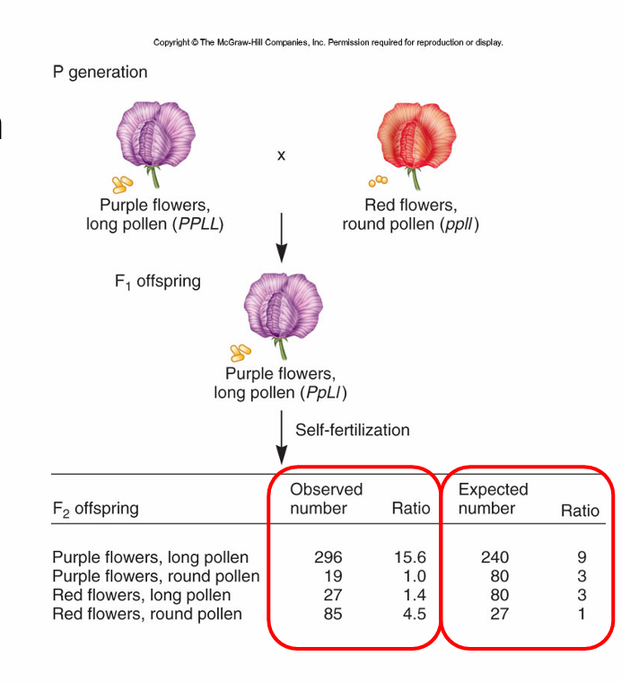
anytime you see gene linkage you will see those things stick together more often
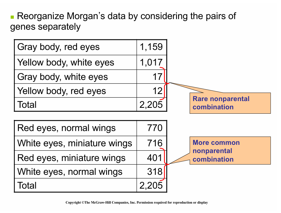
Parental =nonrecombinant
Nonparental = recombinant
Linkage can be altered during meiosis
prophase I of meiosis I
Crossing over
Genetic recombination —nuevo combo of alleles; nonparental
—different combo of traits than parents
parentals - aka nonrecombinants
—same combos of alleles as parents
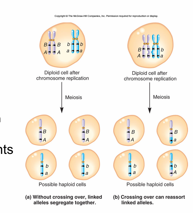
if genes are linked they are going to violate mendals 2nt law unless they cross over
Morgan — evidence that on the same chromosome
studied X-linked traits
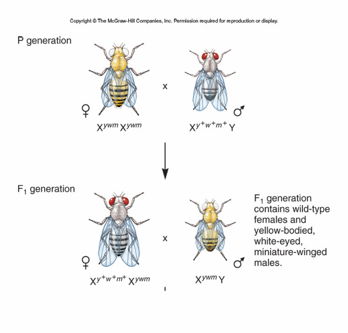
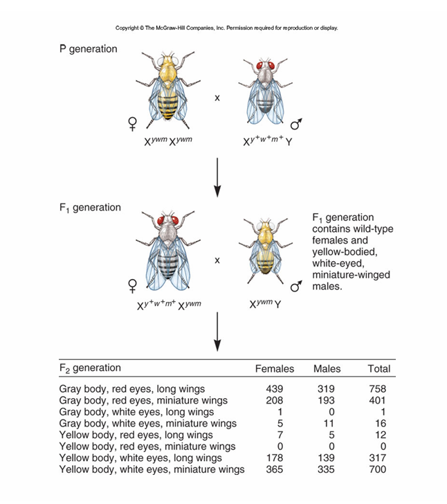
crossed wild type males to females that had yellow bodies, white eyes and miniture wings
what would be the F1 genotype/phenotype
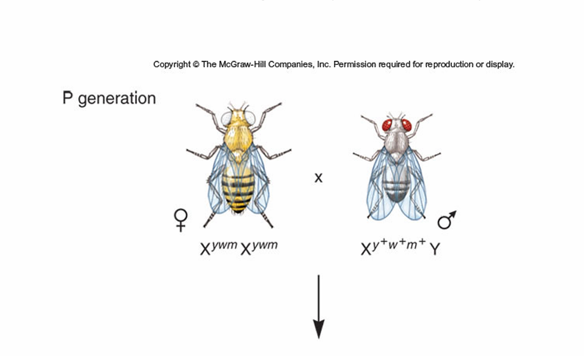
crosses the heterozygous F1 to hemizygous recessive males
what ratios of F2 would you expect
Morgan’s explanation
Janssens — proposed that crossing over involves physical exchanges b/t homologous chromosomes
Realized that crossing over b/t homologous X chromosomes in consistent w/ data
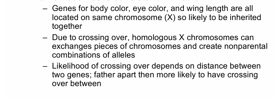
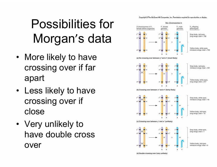
Crossing over becomes more likely the futher abart they are on the chromosome. easier to spilt a log b/t two nails w/ a hatchet the further apart they are
single crossovers can create single recombinants
the least common would be due to the middle being the only different, being the least likely to go down.
in linked genes these will stay linked forever and alwayse unless crossing over occures.
Linked or independent assortment
-can test for this using chi square analysis (use white eye and yellow body dihybrid cross as an example
-step 2 propose a hypothisis
—2 genes are not linked
—propose even if you think they are linked; allows to calculate expected values
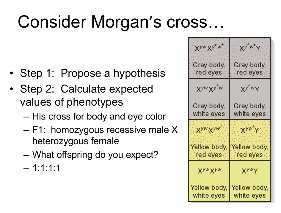
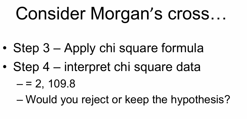
if you noticed the parental genes are over represented then you gotta make the hypothesis that the genes are linked.
Genetic Mapping:
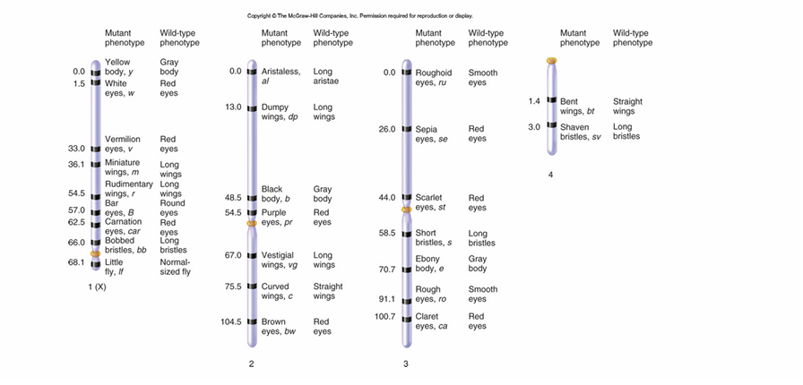
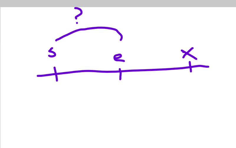
Goal - determine linear order and distance of separation among genes linked on same chromosome
Locus - site where gene is found on chromosome
DATE:09/17/2025
quiz moved to Monday BC no class on Friday
if you have multiple genes that are all linked together it is more likely for genes to cross over and get you a recombinate gene the further apsrt
the least likely is for the two genes on the end to both cross and the middle bing the only diff. one
Mapping Genes:
—goal - determines linear order and distances of separation among genes linked on same chromosome
—locus - site where gene is found on chromosome
You have to know how to do this by monday
Traditional gene mapping:
-distance estemated based on likelyhood that crossover will occie b/t particular genes
—close - crossover unlikely
—Far apart – crossover more likely
Conducting mapping experiment:
to interpret a mapping experiment. the reasercher MUST know if the traits are due to a cross over
—conduct the test cross
Crossover only matters in the heterozygote
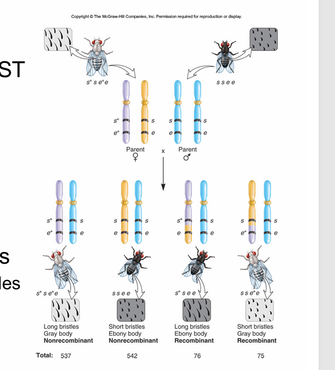
To get the heterozygote, must start w/ true breeding parents
—tells which chromosomes el alleles are on
— tells which are recombinants y nonrecombinats
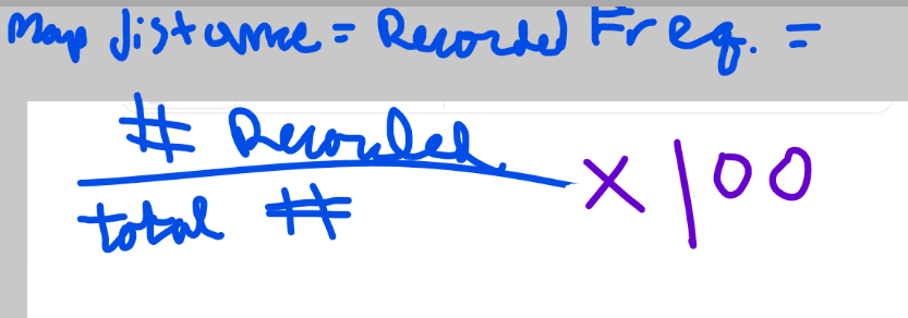
map distance = # of recombinant offspring divided by tot. # of offspring times 100
for this cross, what would that be
units = map units of cM (centiMorgans)
one cM = ~ 1million base pairs of DNA stuff

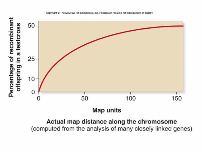
conducted many crosses
assumed that distance would be more acurate b/t closly linked genes
—this is BC when #s get large possible for multiple crossover number of recombinants to underestimate distance
the best data is looking at two genes closer together be cause it can get FrEaKy with the long stuff. I think.
Exam question like this:
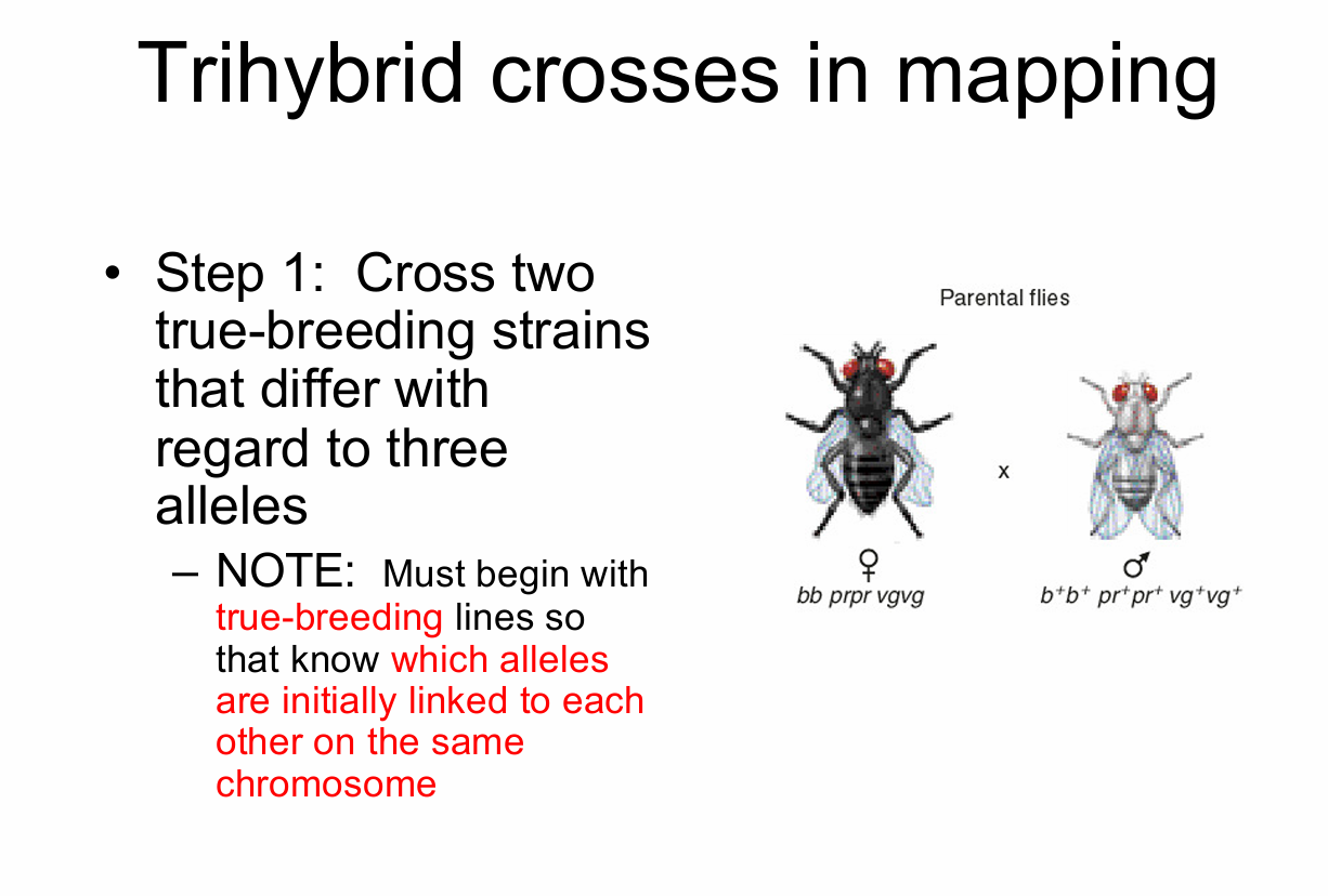
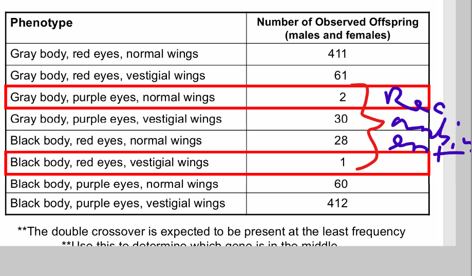
bb= black, prpr=purple, vgvg= vestigial
P gen: Black, Purple, Vestigial x gray, red, normal
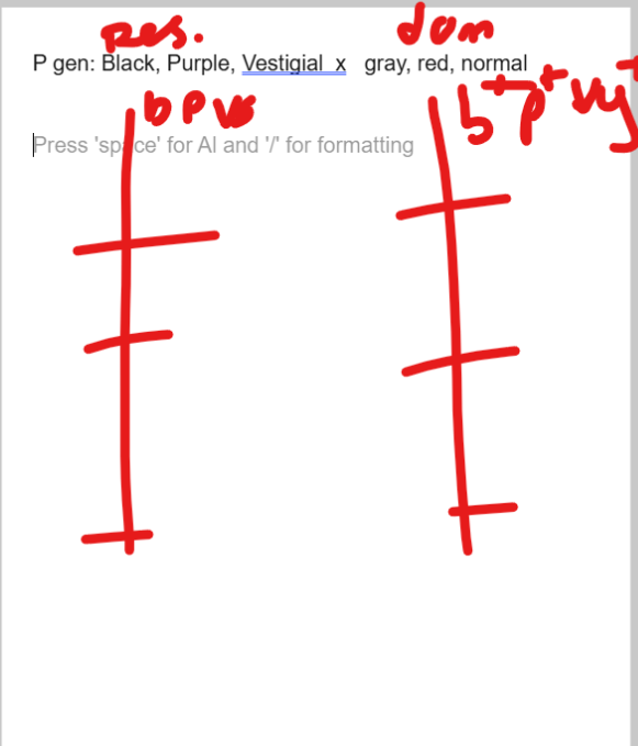
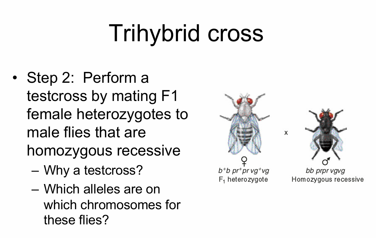
to figure out the one in the middle, find the one switched out the least: those would be the eye colors, you now just put the others on the out side — que esta:
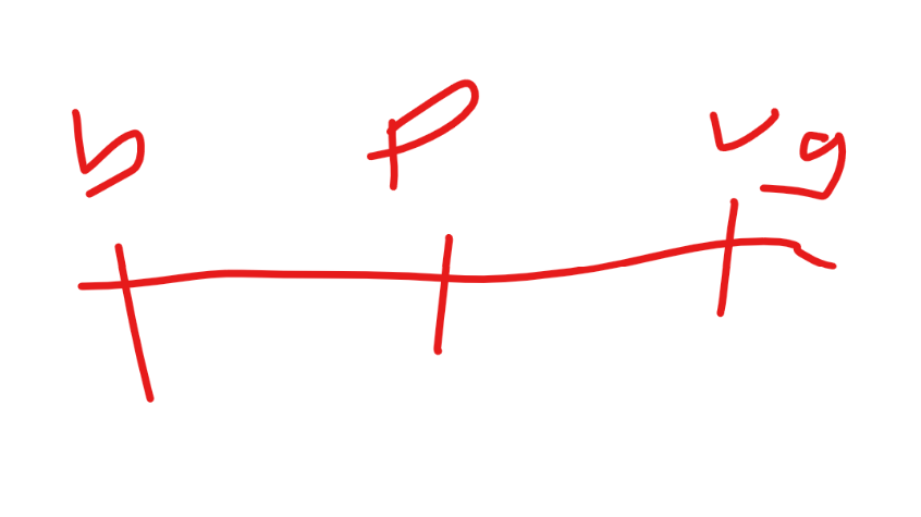
to figure out distance:
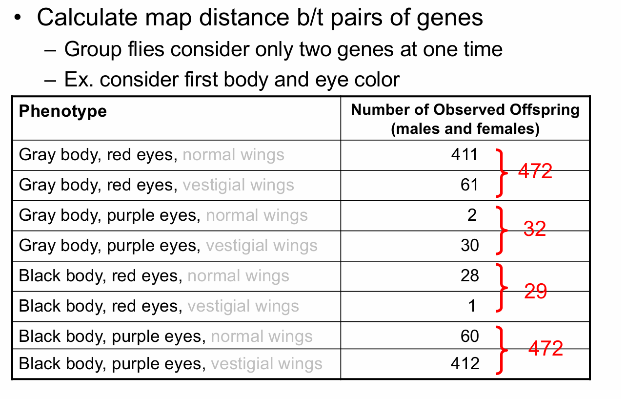

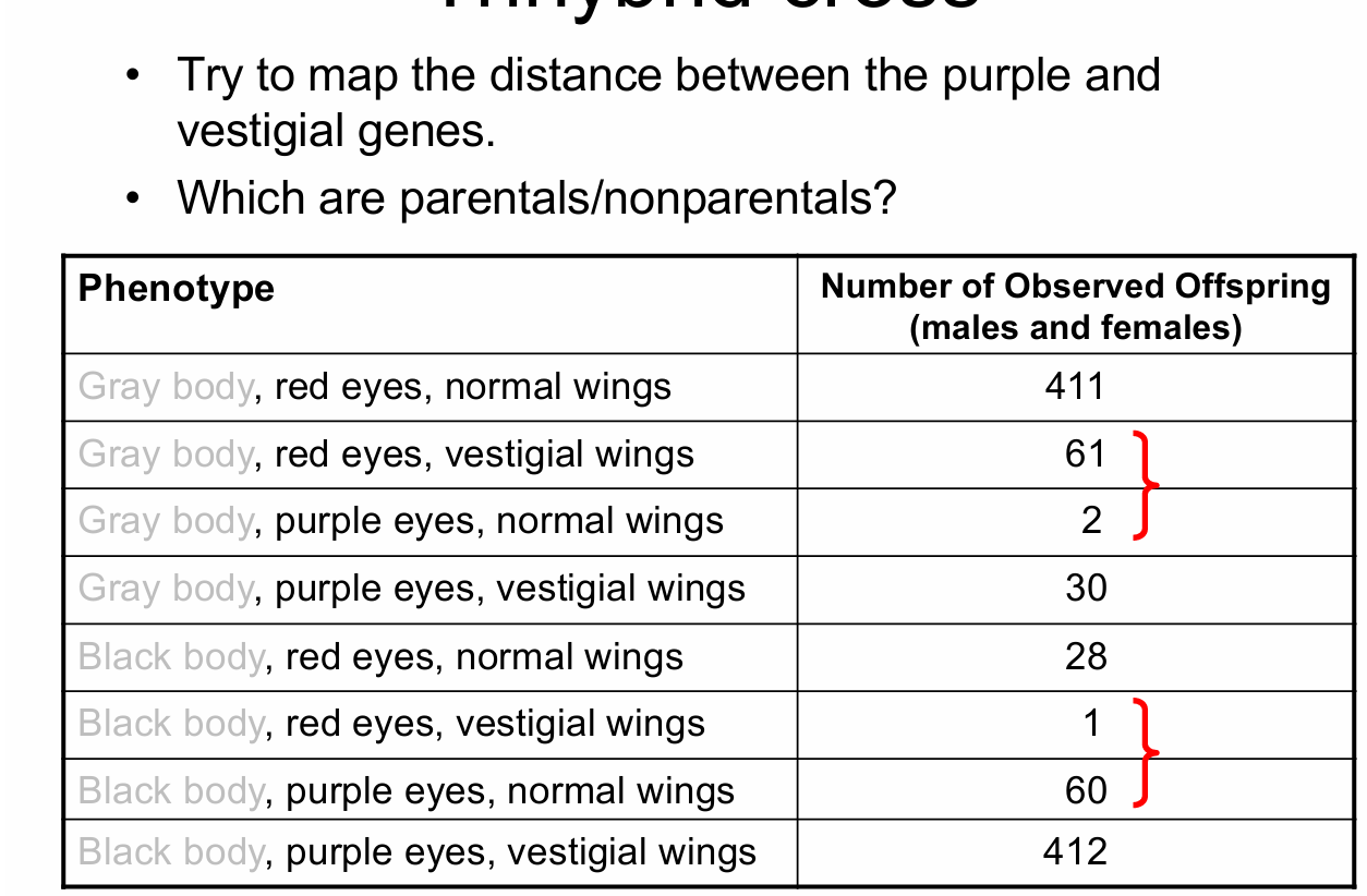

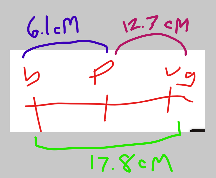
all calculated, the further apart the genes the less acurate, so always start in the midle and work your way out — necisito practicar esta mas
crossing over happens in prophase of mieosis 1 and…
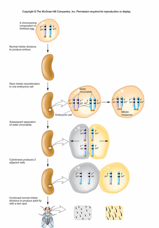
Mitotic recombination:
This is rare, but possible
stern — working w/ strains carrying mutations & bristle morphology
heterozygous females expected to be gray and long bristles
saw patches — twin spot
Explanation — miotic recombination
we aint do the yeast in the book
La fetcha: 9/22/25
(Novembre, venti-dos, en la ano venti-venti-cinco)
Chapter 7:
Genetic Transfer / Mapping in Bacteria
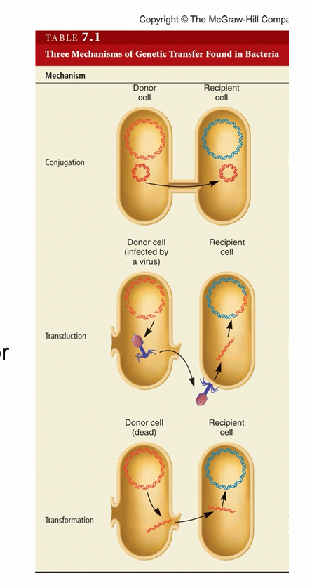
genetic transfer — process where bacteria transfer genetic material
—Three mechanisms:
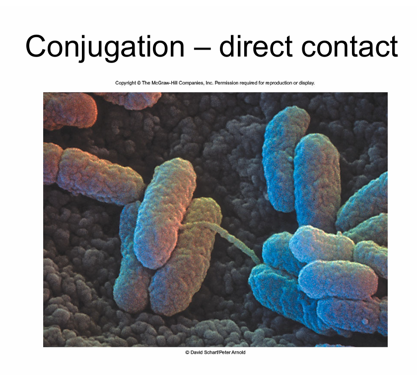
Conjugation – direct contact for exchange
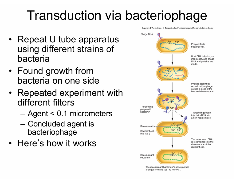
Transduction – via virus
Transformation – from environment — we’ll do this in lab
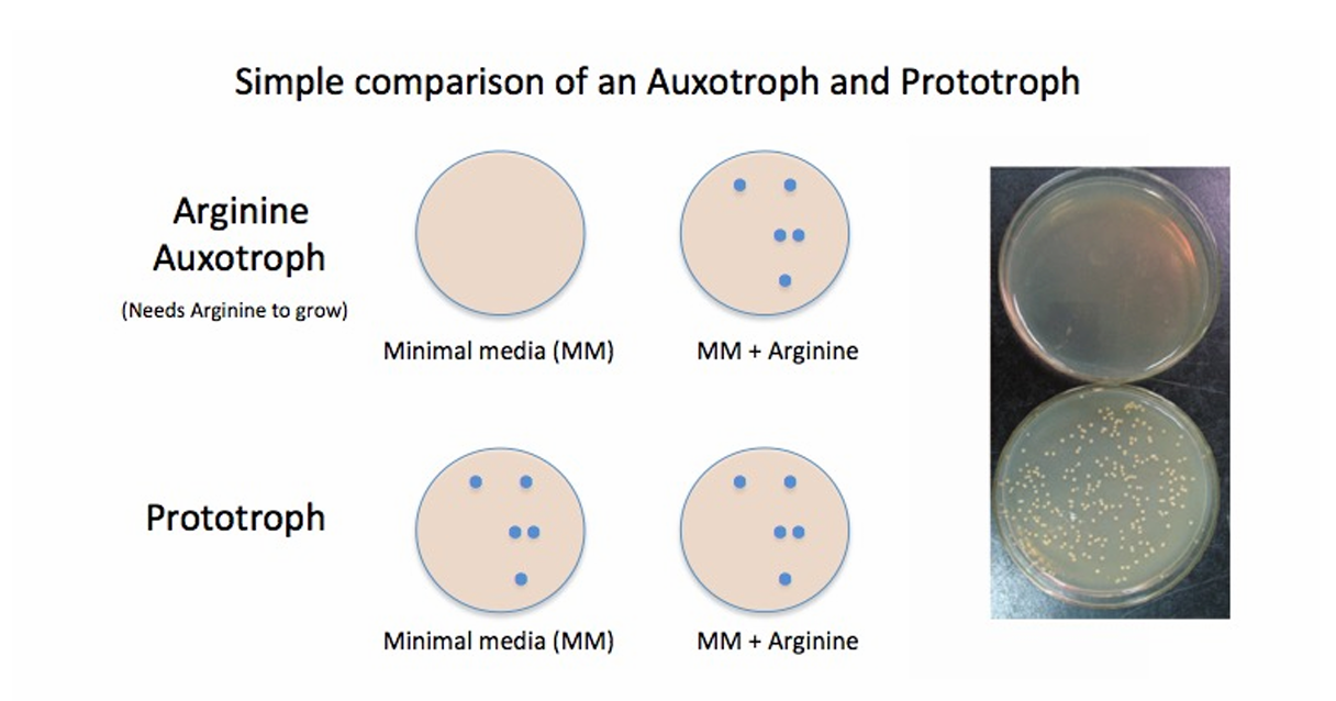
Auxotroph — strain can’t grow on minimal media porque inability to synthesize some essential organic compound requires for its growth (e.f. amino acid, vitamin)
Prototroph — strain can grow on minimal media
Conjugation - EX:
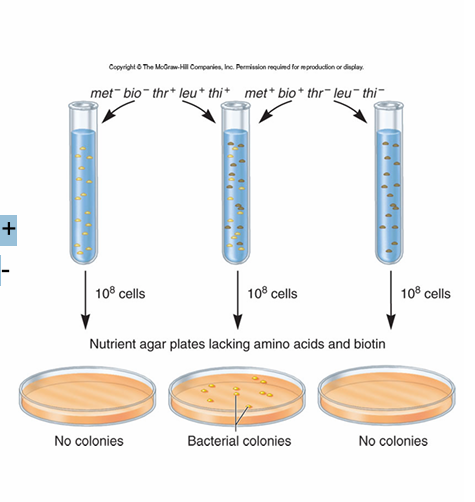
Lederberg and Tatum
—working with auxotrophs
need nutrient supplemented media
One strain: met- bio- thr+ leu+ thi+
One strain: met+ bio+ thr- leu- thi
Mix together à grows on media lacking all supplements
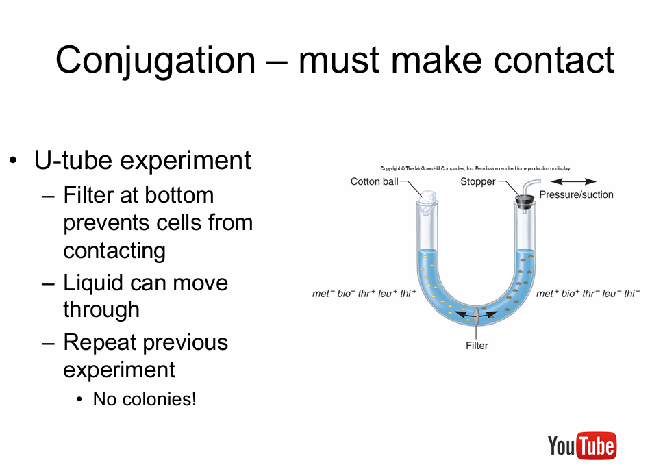
conjugation requires the bacteria actually touch
—not all bacteria can conjugate — only some
must have fertility factor (F) to donate DNA
(F+) have plasmid; (F-) lack plasmid
F factor - genes required for conjugation
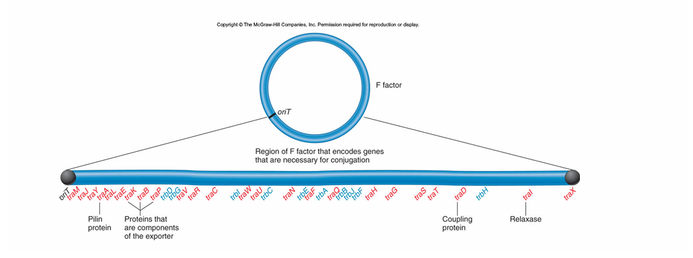
bacteria that are F+ have the thing for the conjugation
it stabs the other with a pilous and gives it the thing to smts. with a F-
if you mix a bunch of F+ and F- and let them be for long enough they all will be F+
F plasmid is the DNA thing in F+
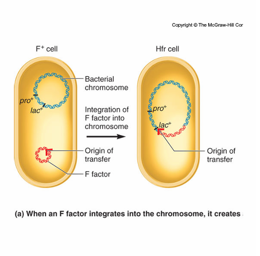
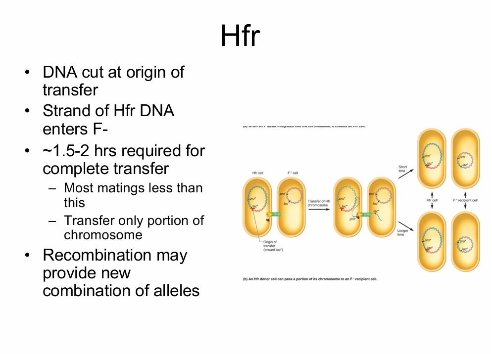
If the F plasmid integrates into the host cell chromosome then it’ll still be F+ (Hfr - high frequency strain), but when it conjugates it will also pass along the other’s chromosomal DNA
takes 2hrs to transfer the whole chromosome
Hfr strains
Very efficient in chromosomal genes into F- strains
• F factor integrated into bacterial chromosome
• Origin of transfer – determines starting point and direction of transfer
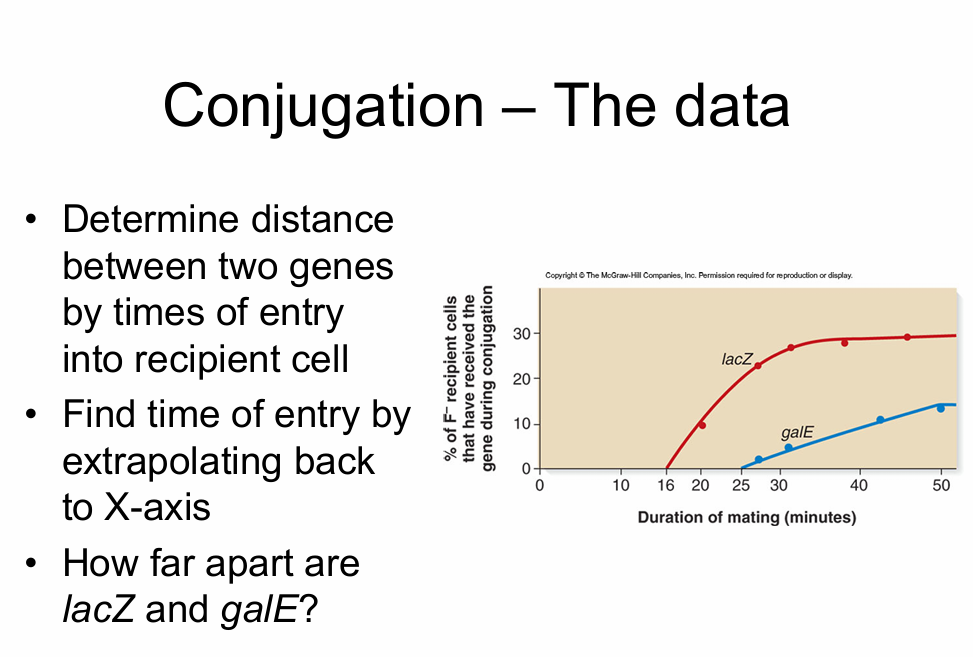
we measure distance on bacteria by minutes, not centimorgans
—just asking when you see it
1st at 16min. second at 25min. so, that means there are 9mins. apart.
they will go in a complete cercle if left alone long enough — this experiment is how they learned E. coli had circular chromosomes.
DATE: 09/24/2025
Chapter 8 - Chromosomal Abnormalities
Nat. variation:
Need to know what normal chromosomes look like:
—Examine chromosomes from several individuals
– Look at actively dividing cells
– From which stage of the cell cycle were these chromosomes obtained?
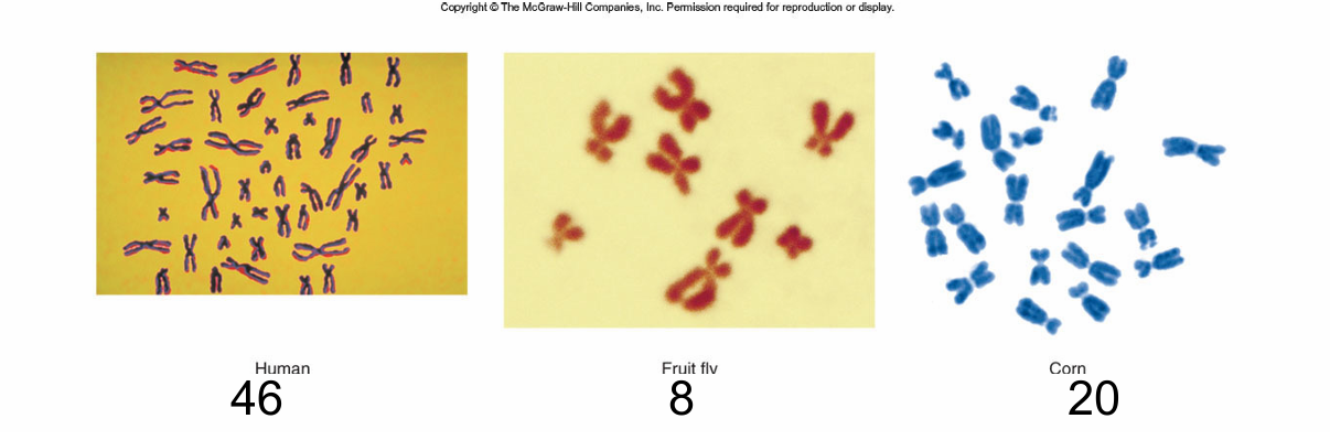
ways to clasify chromosomes:
1) lcation of centromere
2) size
3) banding pattern
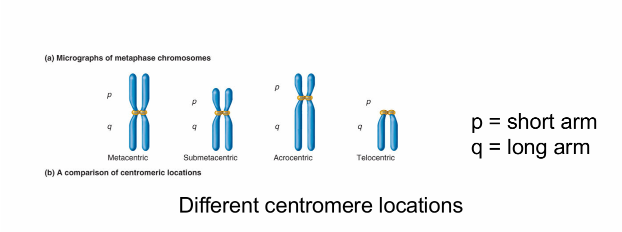
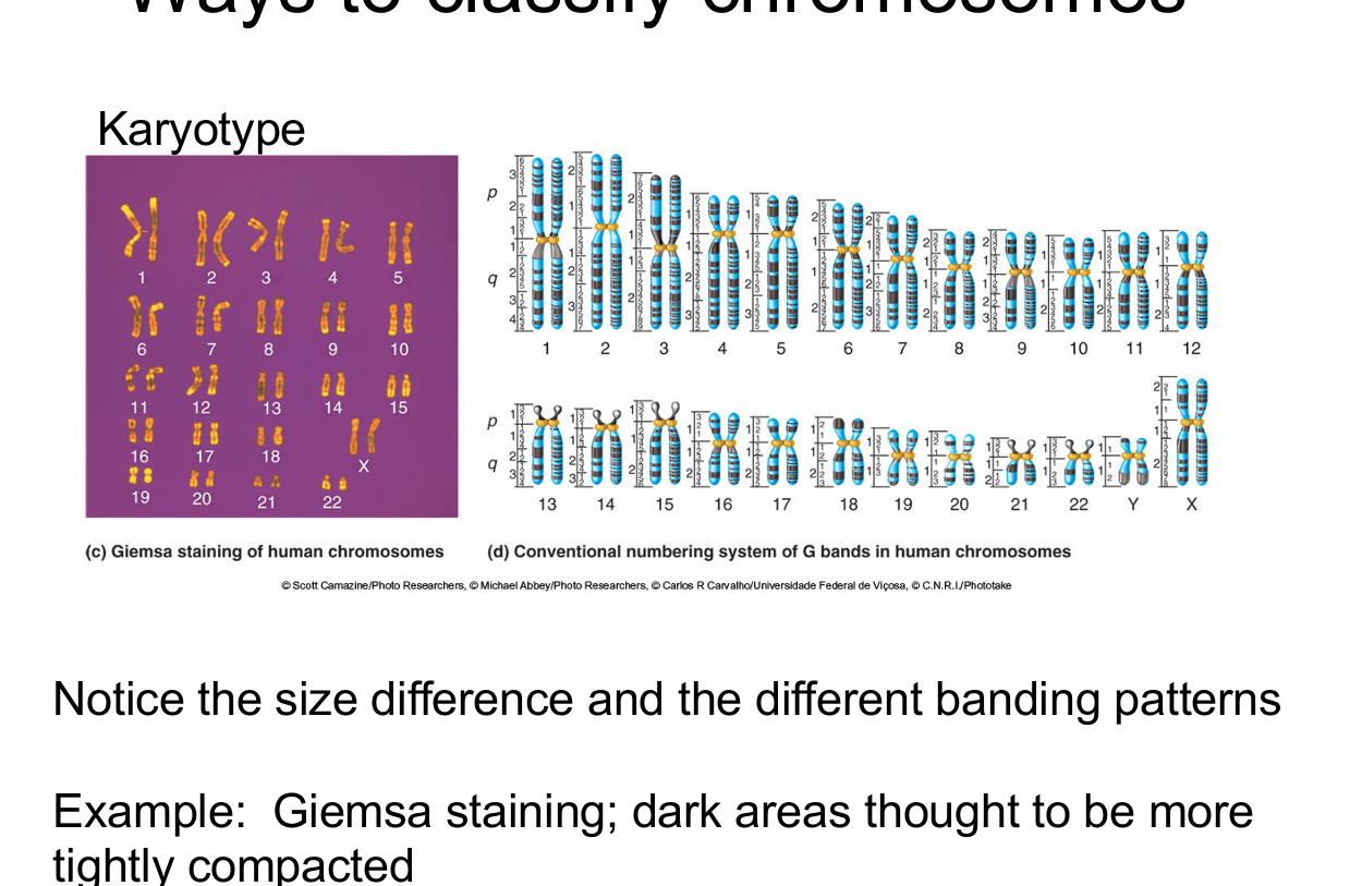
certain parts will stain lighter or darker, and theyre all different, and that’ll let you ID the specific one. Chromosomes are also counted from 1 = largest and 22 = smallest
Why look at the banding patterns?
Helps distinguish chromosomes of similar size and centromere location
Helps detect changes in chromosome structure
Helps address relatedness between specie
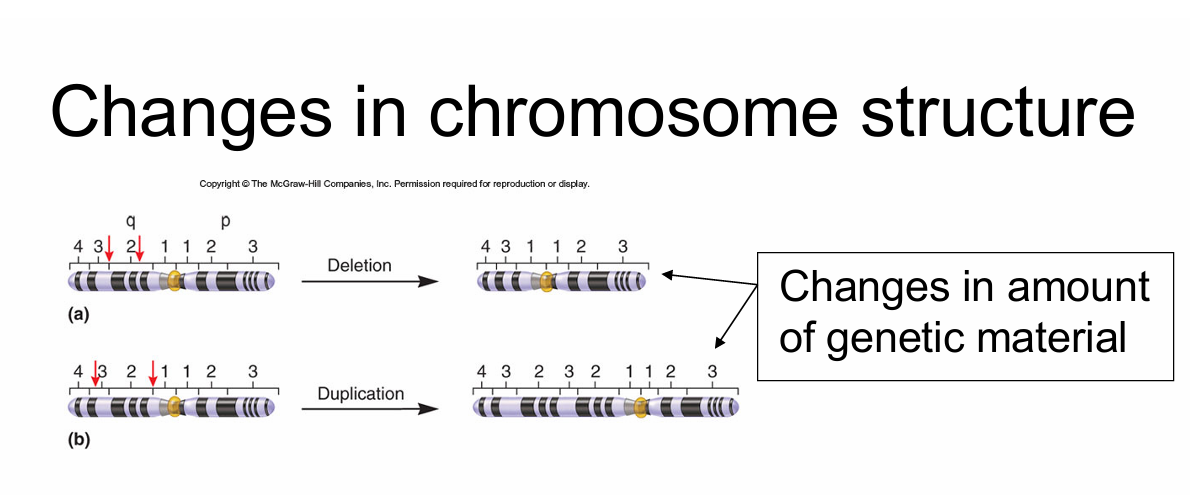
if you can see the difference in genes under a microscope its bad BC it’ll take out many genes, and likely be a phenotype now
DELETIONS:
chromosome breaks; fragments lost
lost piece degraded
dependinging on location and size of deletion there can be phenotypic effects
EX: Cri-du-Chat
—deletion in chr 5
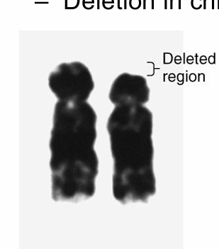
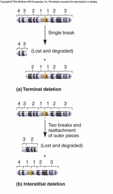
deletions typically cause a phenotype if done eongugh
DUPLICATIONS:
Extra genetic material
usually from abnormal recombination
phenotypic effect
depends on size
usually less harmful that deletions of similar size
many small duplications have no effects
—Lead to formation of gene families
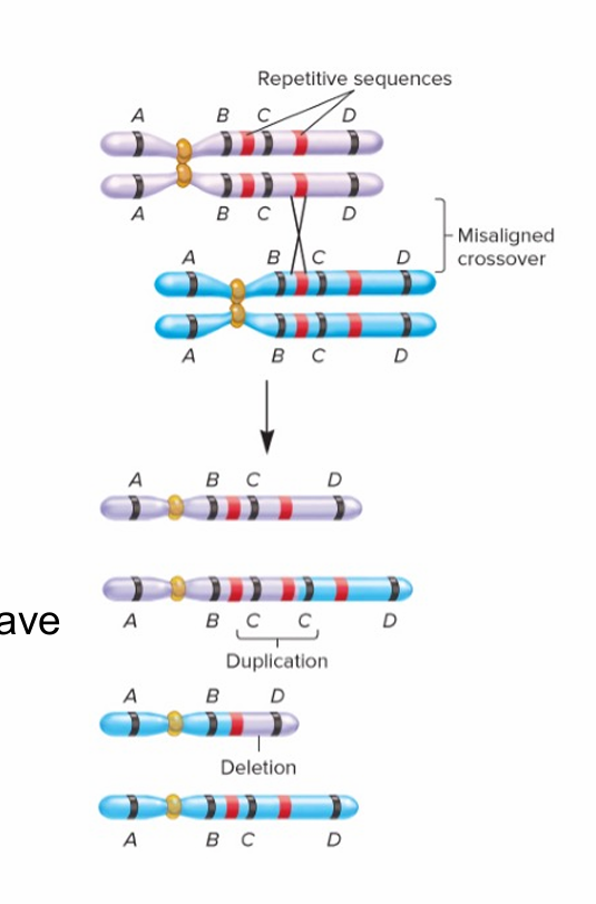
if only a few genes are duplicated probally no phenotypr, but you can get one if enough do. EX: Down’s sendrome
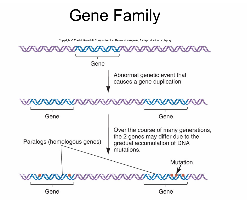
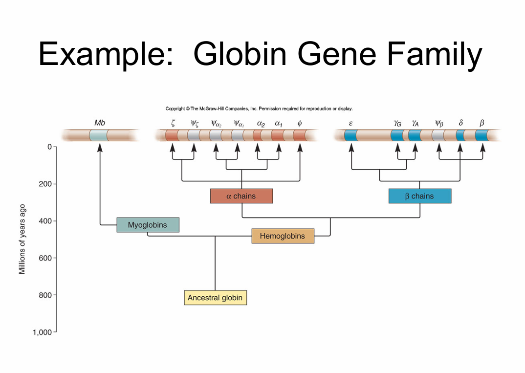
in Biochem you’ll get to study hemaglobin for a loooooooooooong time : )
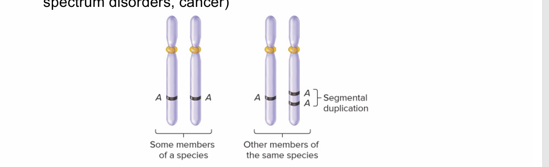
Copy # Variation (CNV)
a segment of DNA that varies in copy # amoung members of the same species
May be missing a particular gene or a duplicate (INDEL = Insertions/DELetions)
Human rate is approximately 0.4%
Associated w/ some complex diseases (e.g. schizophrenia, ASD, cancer)
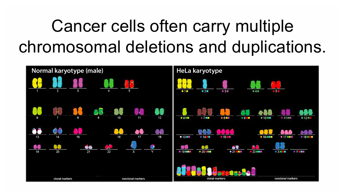
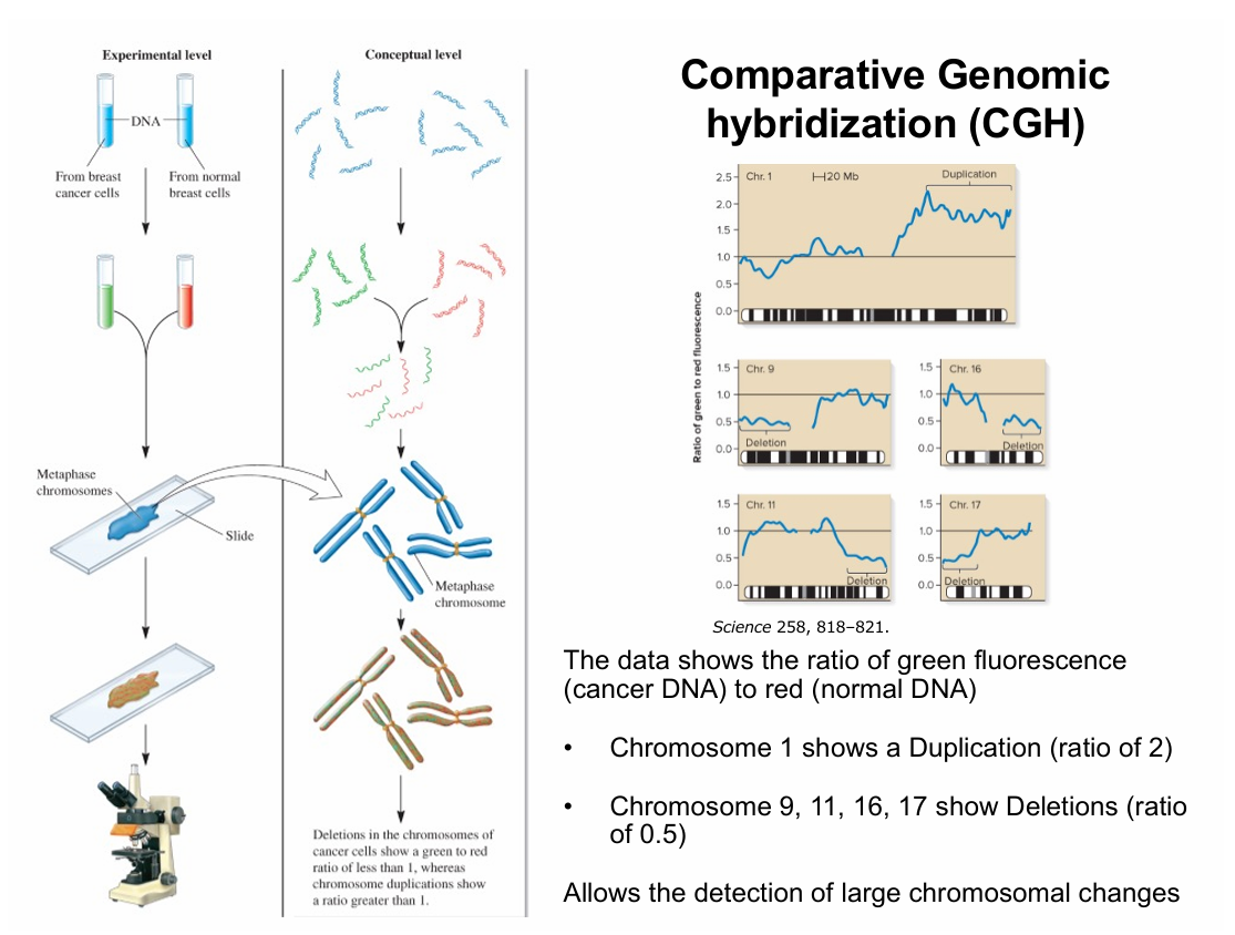
Smaller INDELs may be difficult to detect with karyotype analysis
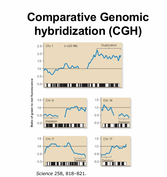
Comparative genomic hybridization can be used
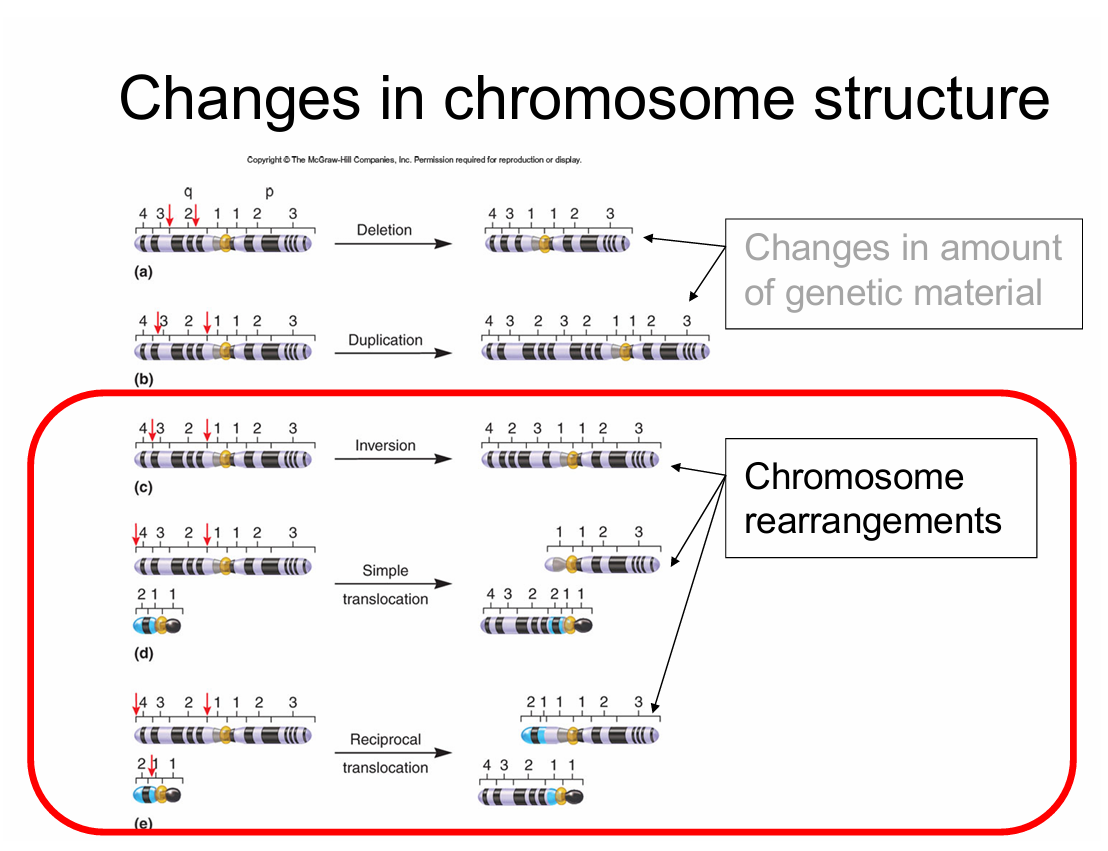
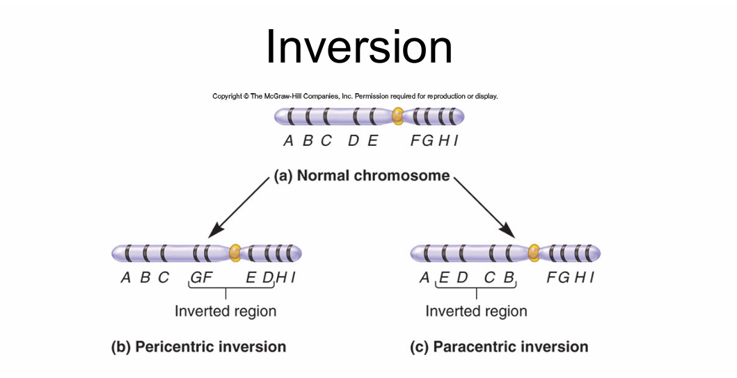
Total amt. of DNA remains the same
Many have no phenotypic consequences (~1 in 50 in human population)
depends on boundaries of inverted segment
Depends on where the break occurs
If you have a chromosomal inversion you’ll probally be fine BC you’ll be heterozygous, but it can show up in your offspring and mess them up — typically causes fatality
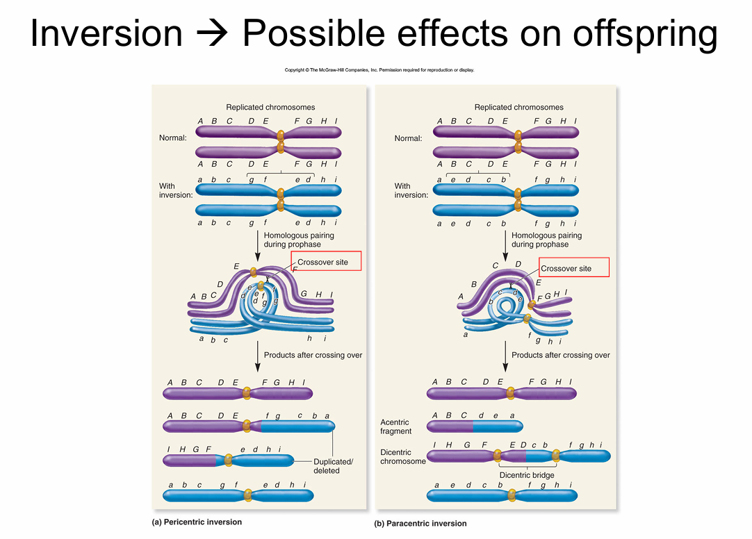
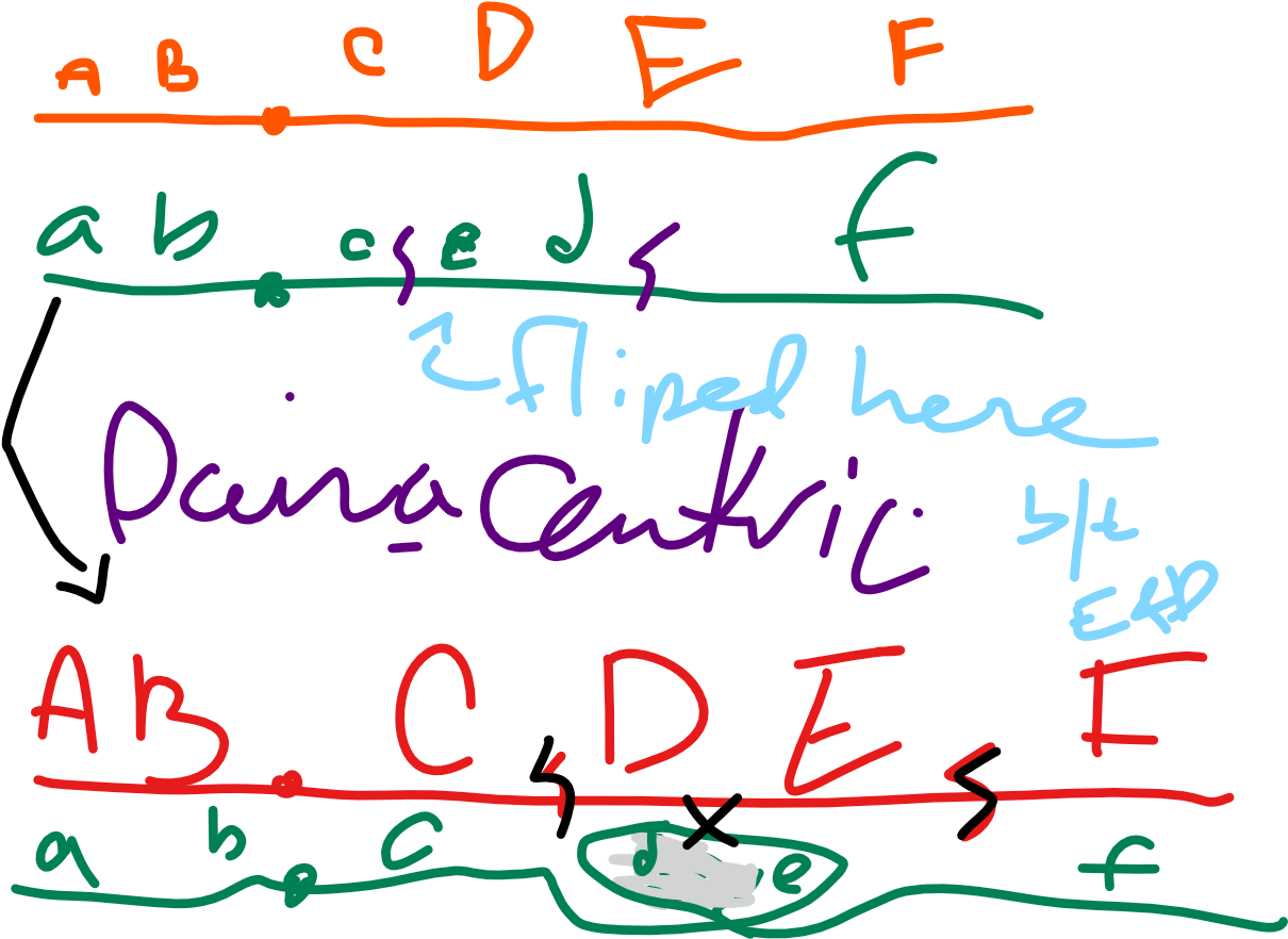
1) A B * C D e c * b a ← f is missing!!! AHHHH
f d E F ← this is missing so much!!!!! AHHHHH
stuff like this is almost alwase fatal to offspring — often results in miscarrage in utero (ass opposed to outero?)
just like w/ the other crossing over, the bigger the gap the more likely it is to get FuNkY
We will have a quiz on friday T-T
DATE:09/26/2025
SIKE! NO QUIZ!!!!
—Do HW. Packet, however.
Crossing on the inversion loops will be Critical on the exam
Translocation:
reciprical translocated are the most common
most of the time no phenotype as the genes are still ther, but in diff. orders.
Telomeres ofter prevent translocations (caps off the end and makes it not wanna touch it)
if end of chromosome is broken, lakes telomere and is reactive
reciprocal translocations also produced by crossing over b/t non.- homologs
—Balances; total amount of DNA not alterd
—Usually no phenotypic effects
may have position effect
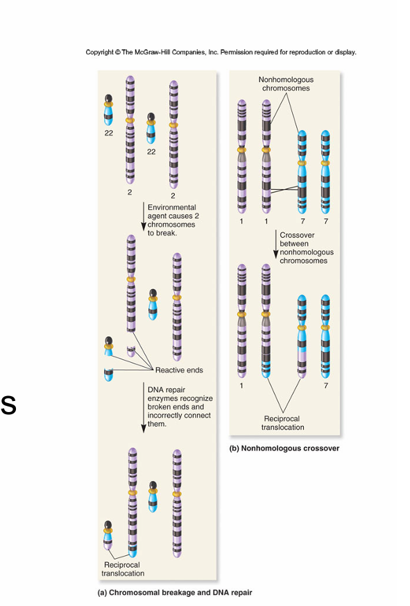
the reason this causes problems is that this causes weird things in miosis
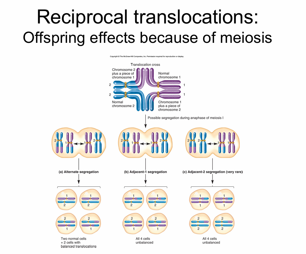
What are the products of miosis for someone who carries a translocation?
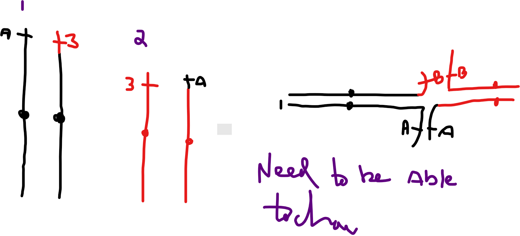
Know the different b/t alternate, adjacent-1, and adjacent-2 segregation
Alternate: 2 normal cells + 2 cells with balanced translocations
Adjacent-1: all 4 cells unbalanced (missing region, and double region. One black and one red in each)
Adjacent-2: All 4 cells unbalanced (rarely occurs) would be if both black ones goes into a thing together. All one thing and hardly any of the other
Alternate can have viable offspring
Adjacent types typically causes non viable offspring
when stuff pairs up like goes with like
5-10% of infertility issues comes from these types of chromosomal abnormalities
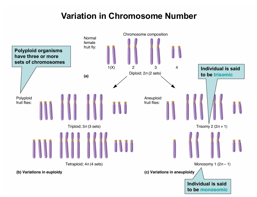
Chromosome numer = ploidy
n - haploid
2n - diploid
3n - triploid
etc, you can have any number
most plants and animals are diploid
polypoids do not survive in humans
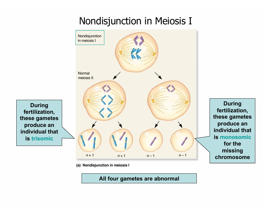
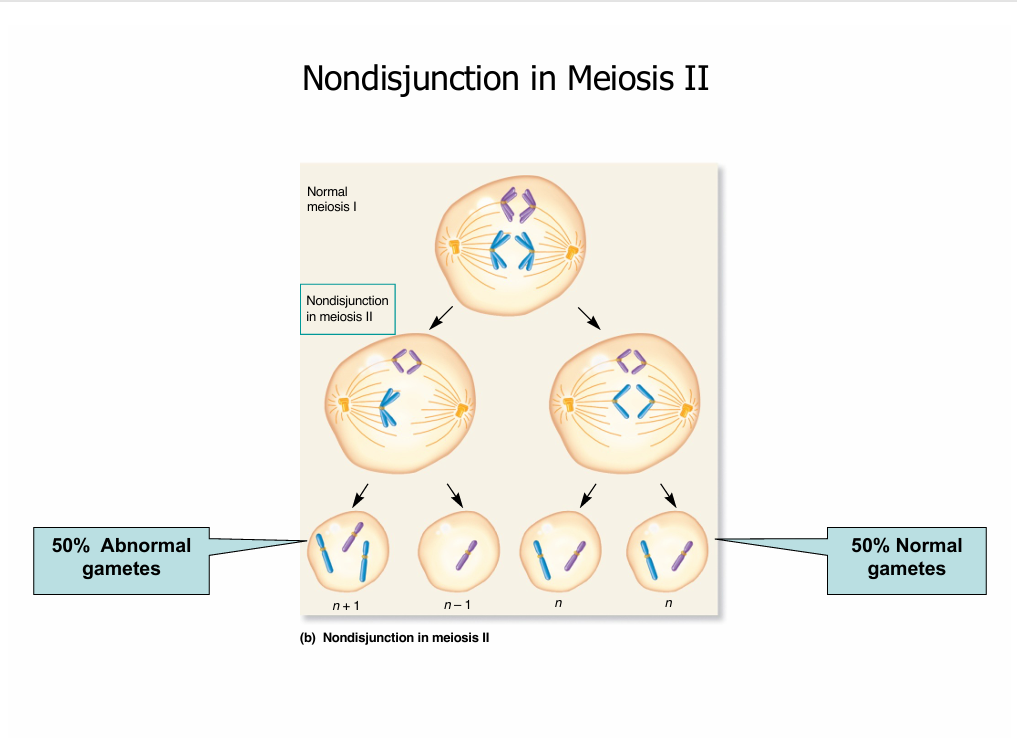
aneuploidy is possible in humans, such as in downs sendrome
Trisomy - (2n + 1) one chromosome is doubled
Monomy - (2n-1) one chromosome is halfed (one is gone)
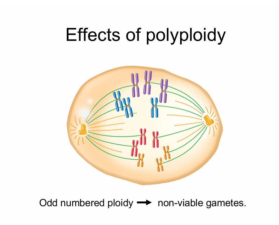
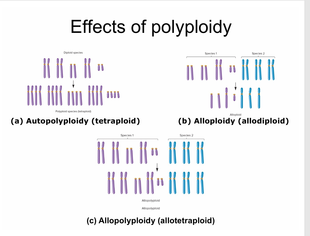
Allopolyploidy = thing that has two different species as parents (liger/mule) - bigger fruits
Aneuploidy = most common cause of misscarage
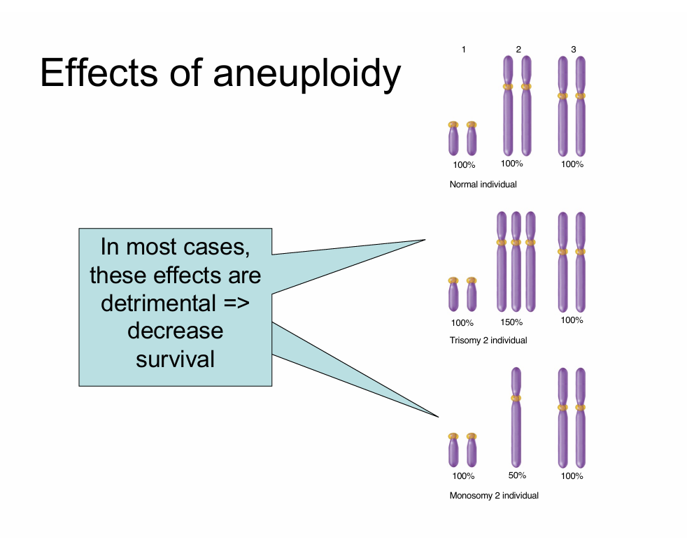
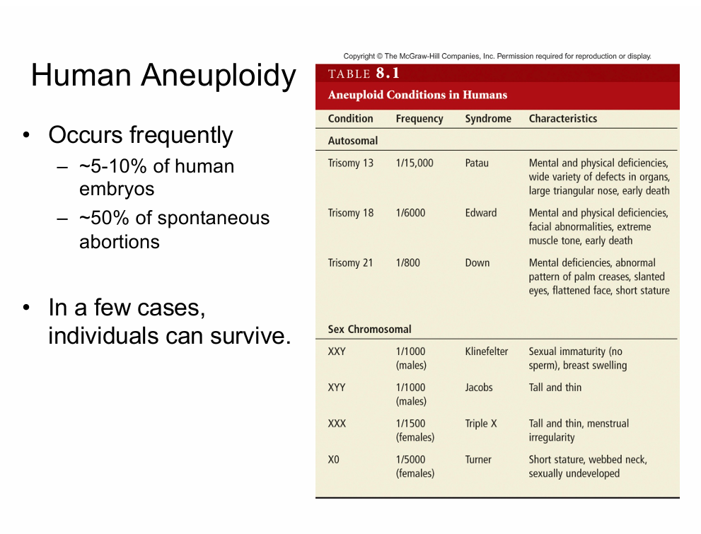
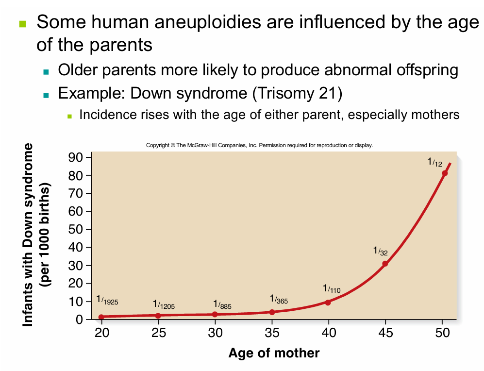
aneuploidies happen with more frequency the older the parents (especilly mothers) are
—this is due to how long the eggs have been present
—theyve been in miosis 1 since utero and spindle fibers can break down and increase chances of chromosomes splitting wrong
DATE: 09/29/2025
Chapter 9:
Molecular genetics
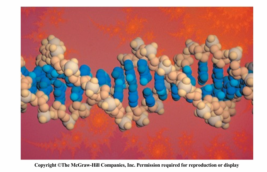
The study of DNS structure and function at the molecular level
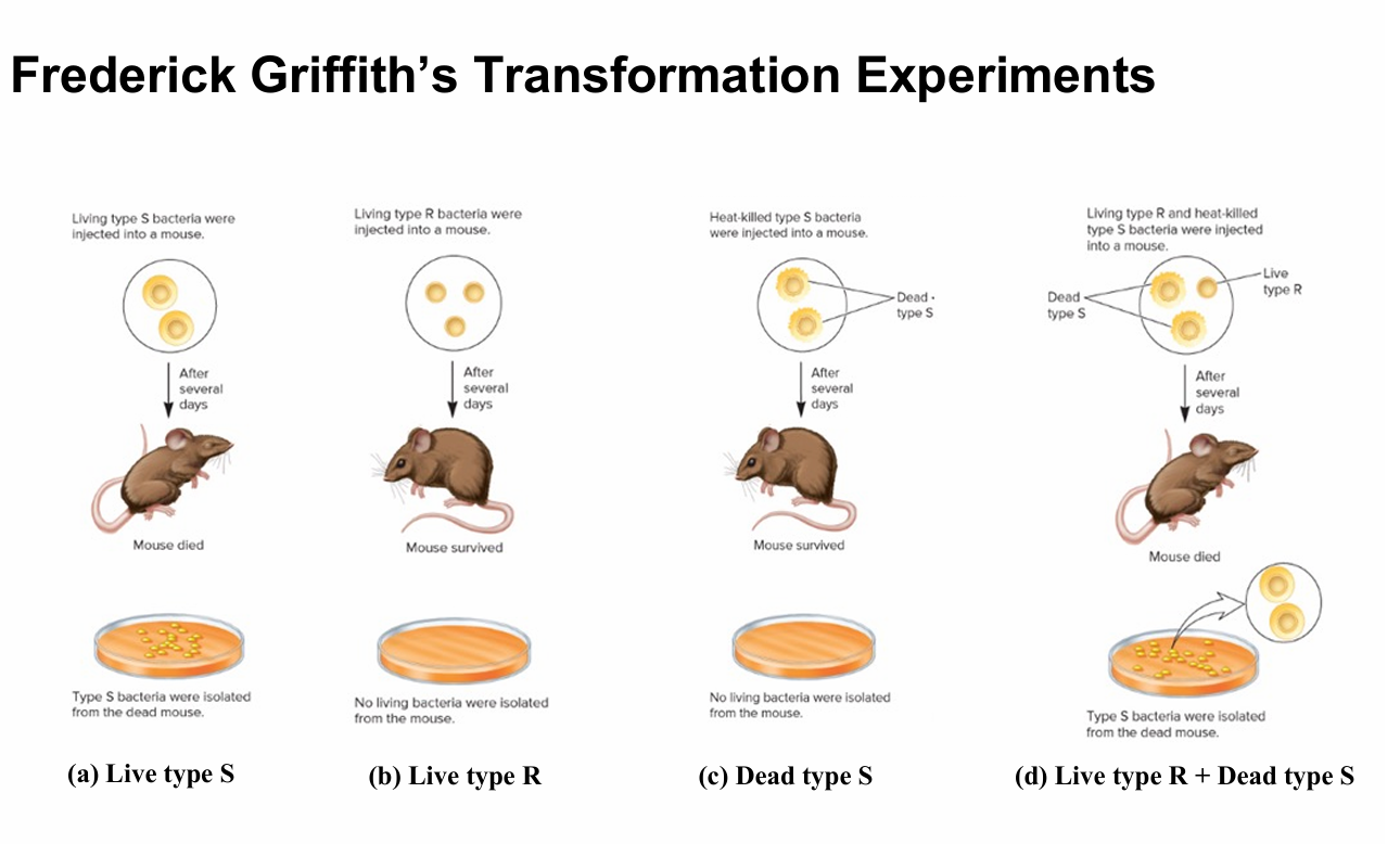
the experiment that proved DNA is the hereditary material as opposed to protein
the term transformation comes from this experiment due to part d where the live bacteria took the DNA from the dead bacteria making it more deadly
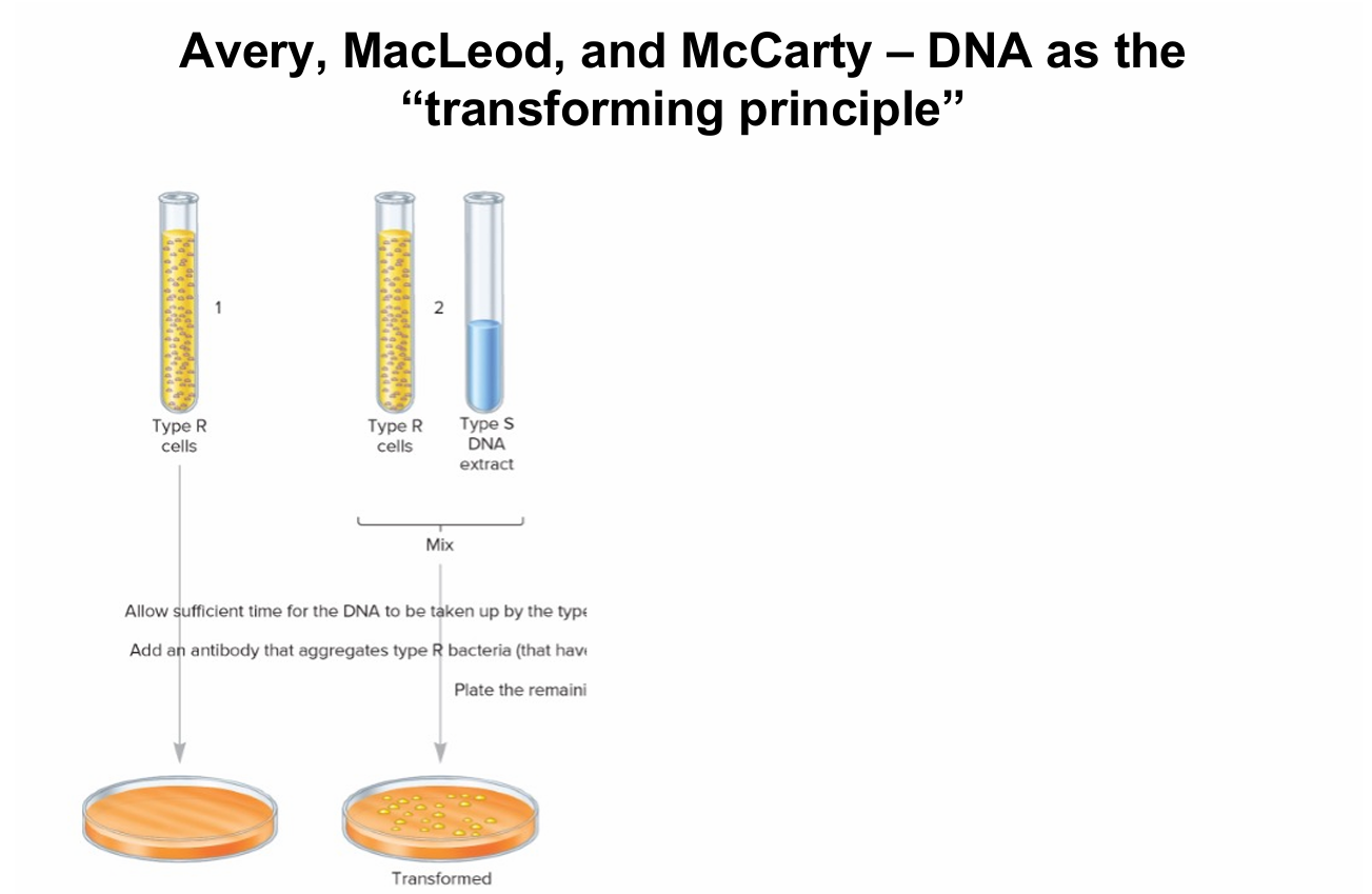
Classic biochemical experiment — Will be on the MCAT
they knew stuff had to be encoded by either DNA, RNA, or Protien
proved that DNA was heretitary thing
degraded the protiens to only see the DNA
Hershey and Chase — DNA as the hereditary material
used bacteriophages
Hershey and Chase provided evidence that DNA is the genetic material of T2 phage
• Used radioisotopes to distinguish DNA from proteins
• 32P labels DNA specifically
• 35S labels protein specifically
• Radioactively-labeled phages were used to infect non radioactive Escherichia coli cells
• After allowing sufficient time for infection to proceed, the residual phage particles were sheared off the cells
• Radioactivity was monitored using a Geiger counter
• Most of the 32P had entered the bacterial cells (DNA)
• Most of the 35S remained outside the cells (protein)
why did they use radioative phosporus/sulfate?
—BC of the phospate groups in the DNA and no protiens have that
—they used radioative Sulfer for protien BC there is not any of that in DNA
You need to know all these experiments and their names
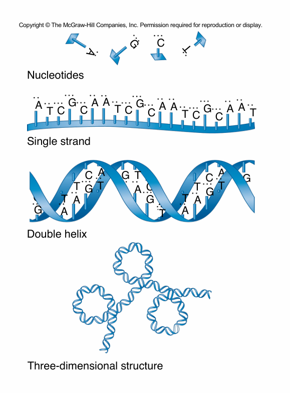
Nucleic Acid Structure
DNA and RNA are large macromolecules w/ several levels of complexity
purines: Adinene and Guanene (Ag - silver is pure)
Pyrimidine : cytosine, uracil, thymine (CUT - cut of py(pie))
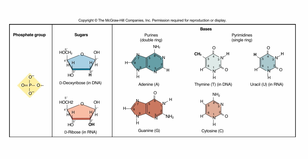
why we use 1’ and not just numers: BC there are also numbers on the nitrogonous bases, meaning that plain numbers are those
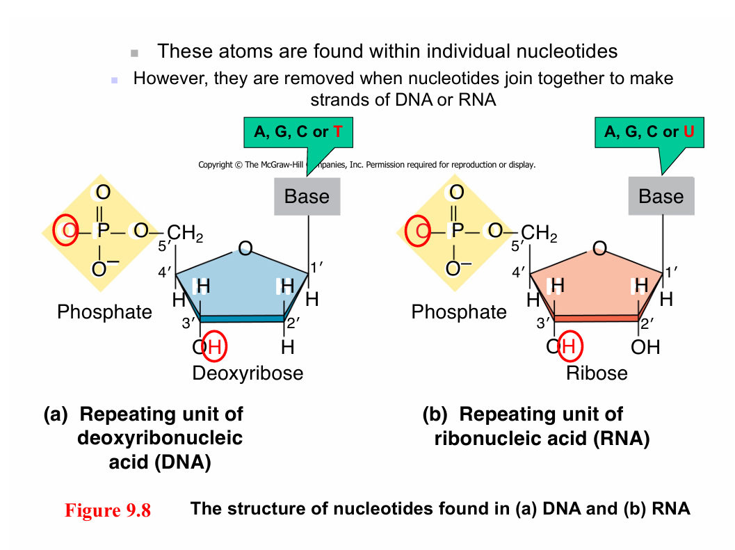
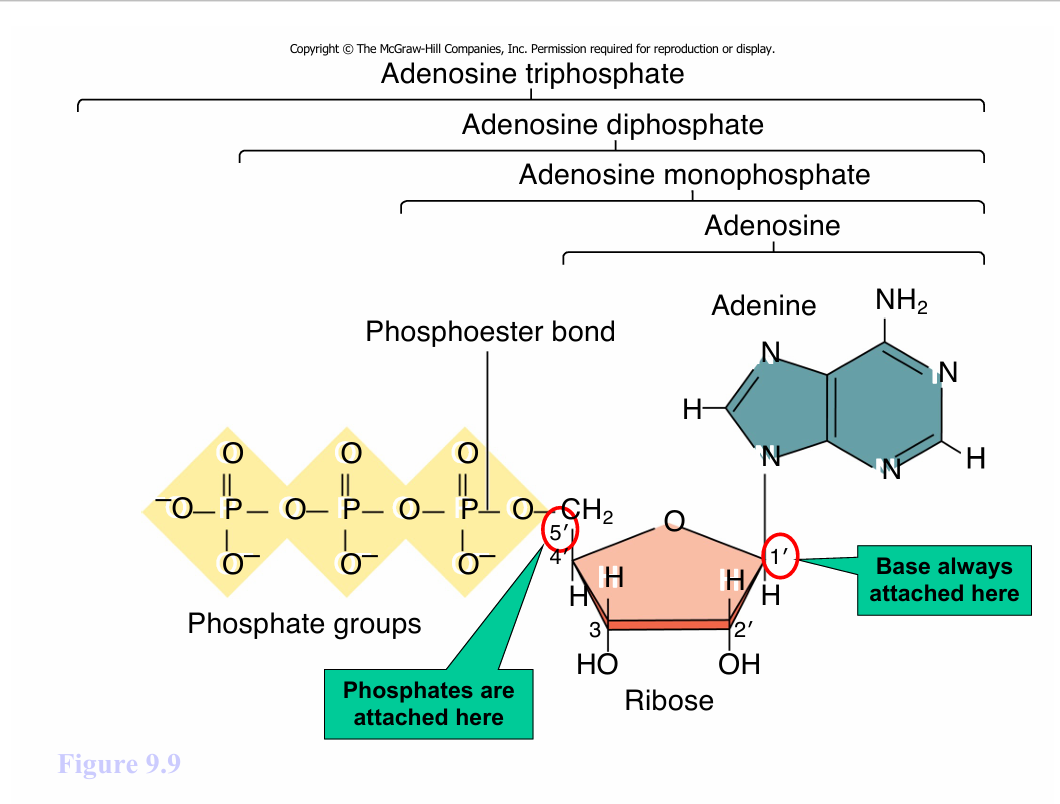
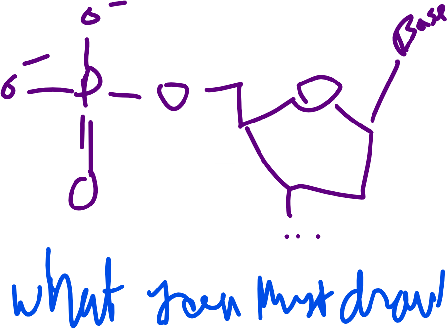
5’ carbon always has a carbon then phospate group
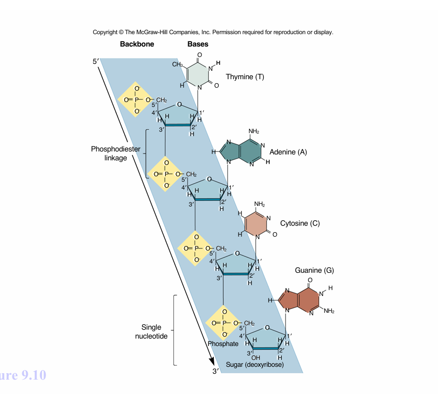
if given no other information you can assume that you start on the 5’ end and end on the 3’ end
you can tell DNA from RNA if theres a T or U for the drawings
IF you know one strain then you also know the second one, EX:
5’ -TACG -3’
3’-ARGC -5’
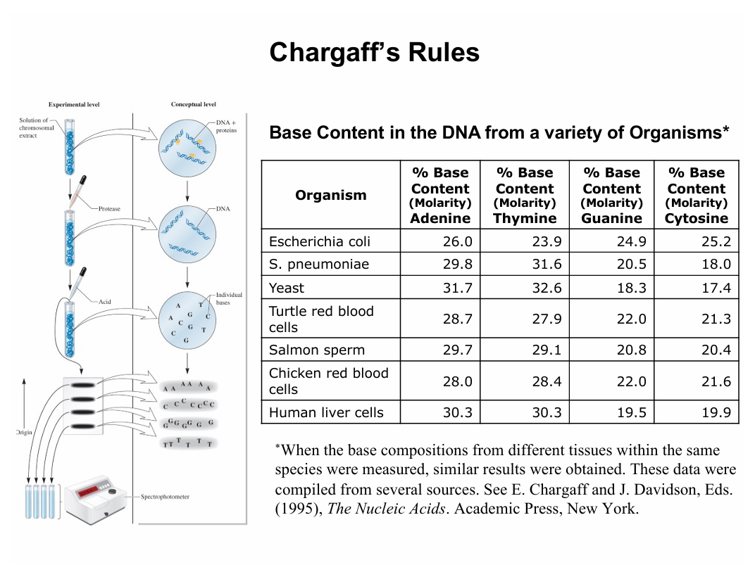
the A and T as well as the G and C are roughly aproximatly BC one typically will have the other
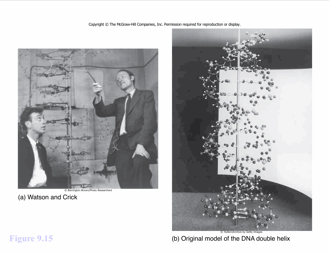
These guys were trained as physical chemists, not this stuff, but uh, life finds a way…
X ray defractions that give X patterns have heliacal structures (double helix baby)
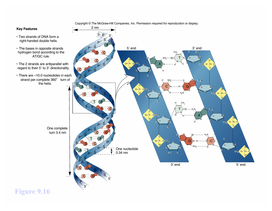
just the DNA w/out protien structures attached means a simple right going double helix
they all have the same phospate back bone
the bases are all tucked on the inside to keep the molecule stable and keep down the mutations
if they’re inside they have less reactivity
the DNA doesn’t have the OH on 2’, making it more stable BC no O
RNA is less stable BC of the OH on EVERY 2’
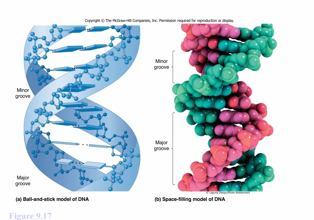
any protien will probally bind to the major grove where it has a better access to the bases
if not there in the neg. chared backbone in the minor grove
only thing holding the two strands togater are H bonding
T-A has 2H bonds
C-G has 3H bonds
A-T rich sequences are where the bonds are more apt to break due to the less amount of the bonds
B - DNA is all you need to know for this class
RNA STRUCTURE:
The primary structure of and RNA strand is much like that of DN
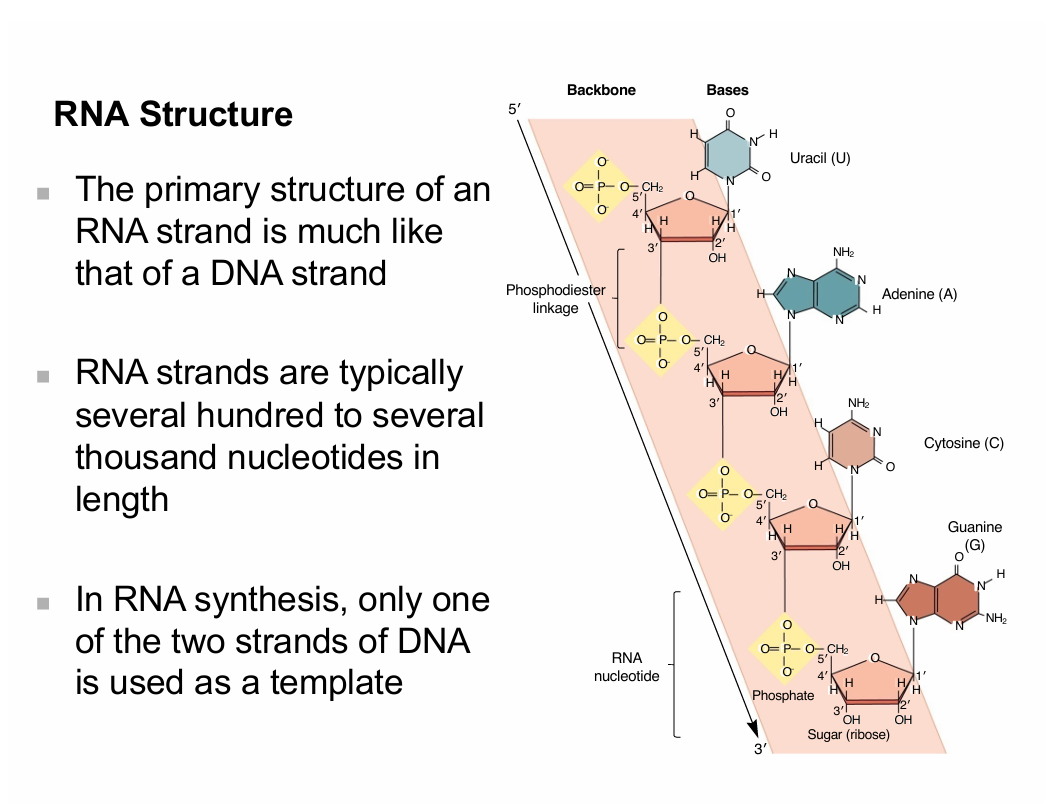
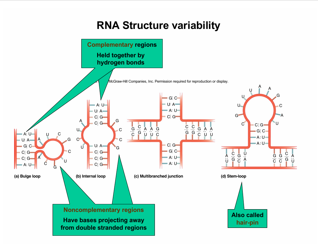
Hair pin in tRNA
DATE: 10/01/2025
Chapter 10:
One week from today = exam 2 (El proximo mircoles)
All these bad bois ties togeter w/ chromosomes
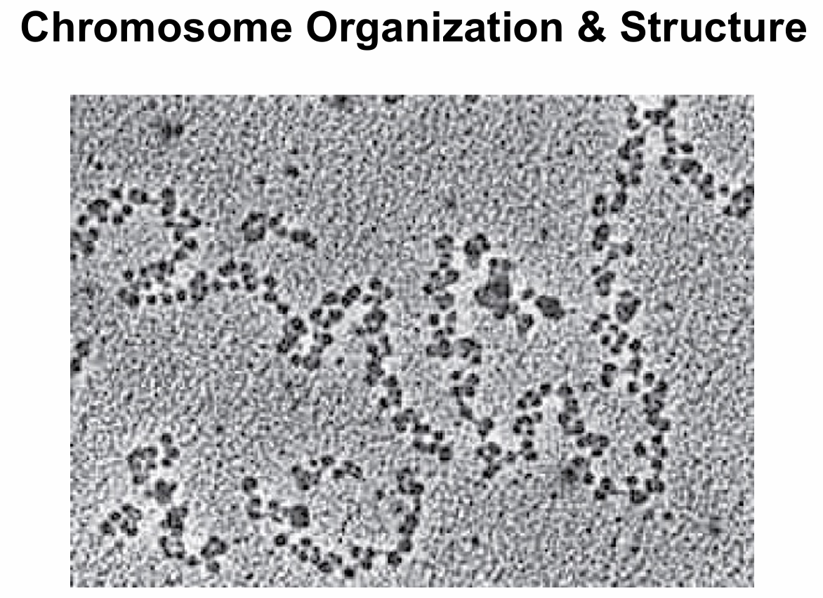
How chromosomes are organized/packaged to fit into our cells: — getting into molecular biology
Genomes:
chromosomes - structures that contain genetic material
genome - all of the genetic material organism has
—bacteria - usually single circular chromosom
—Eukaryote - one complete set of chromosomes w/in the nucleus
mitochondrial genome
chloroplast genome
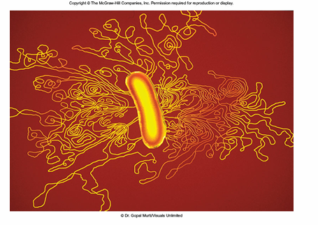
the lines are the one chromosome
—they are far bigger than the cell
like 1 milll. x longer
if humans was done like this would be like 6ft
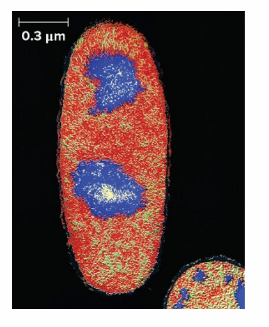
Bacterial chromosomes
located in nucleoid
may have 1-4 copies of chromosome in cell
—growth conditions
—phase of cell cycle
—bacterial species
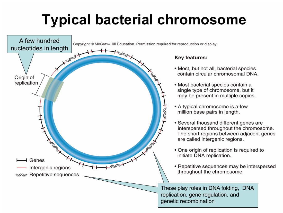
origen of replication = every bacteria chromosome has one of these
repetitive sequences are immportant for packaging
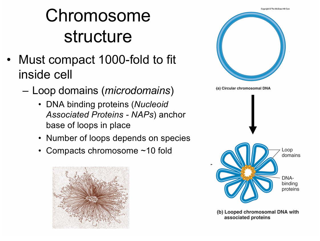
micro/loop domanes must 1st be done
a scaffolding is made to keep it from getting tangles.
it is binded to scaffled protiens at those repetitive sequenced
the sketchy one is E. coli
these are folded 10 fold and scrunched, but its still to big to fit
we miust now do a thing called super coills
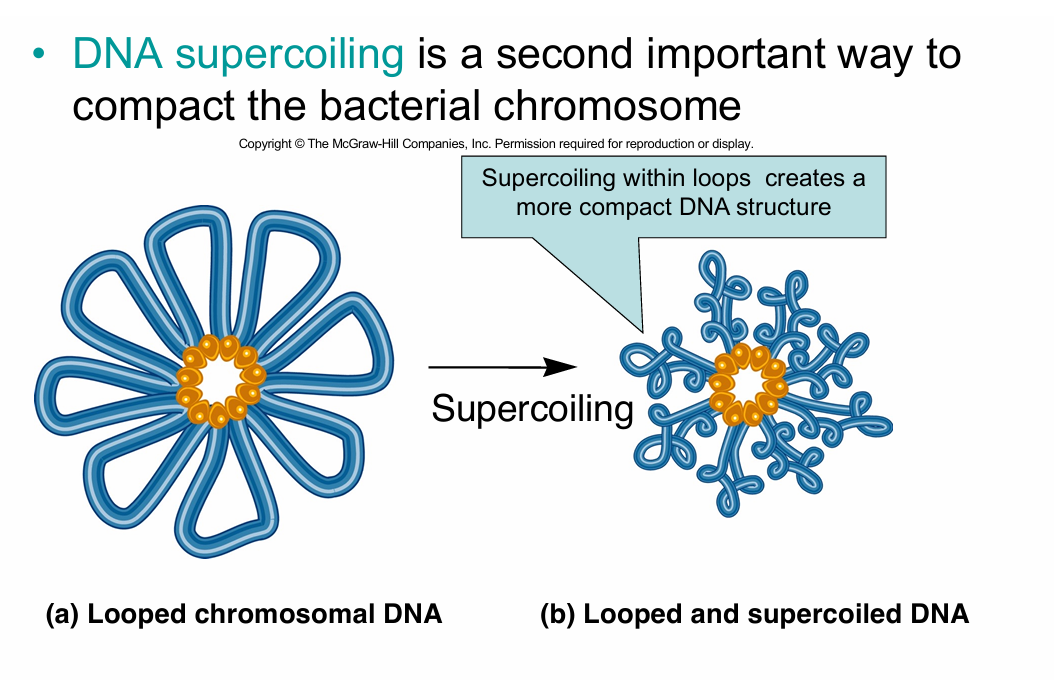
adding more twistes to the already twists of DNA
does not happen by itself, enzymes must assist with it
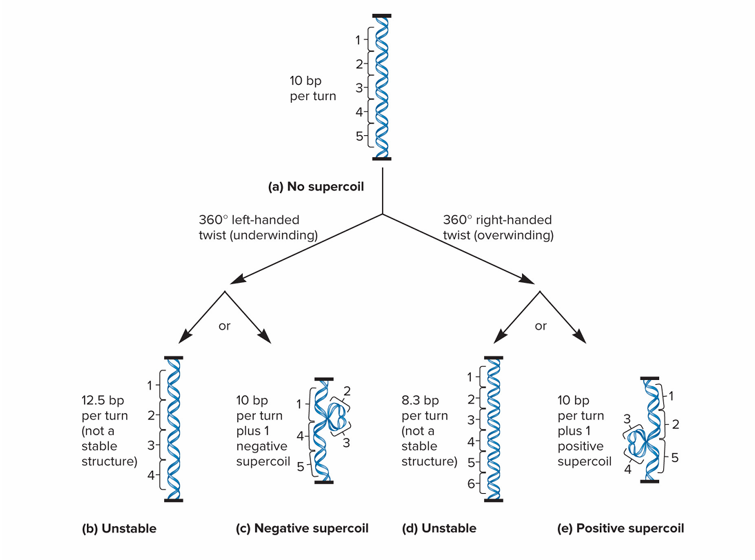
for a complete turn of a helix is 10 base pairs
if you unwind then it causes strain it supercoils and is a neg.
if you wind tighter it becomes positive
these are topoisomers
they are not stable and will go back
if too much pressure then you get double bonded braecks'.
enzyme = ase
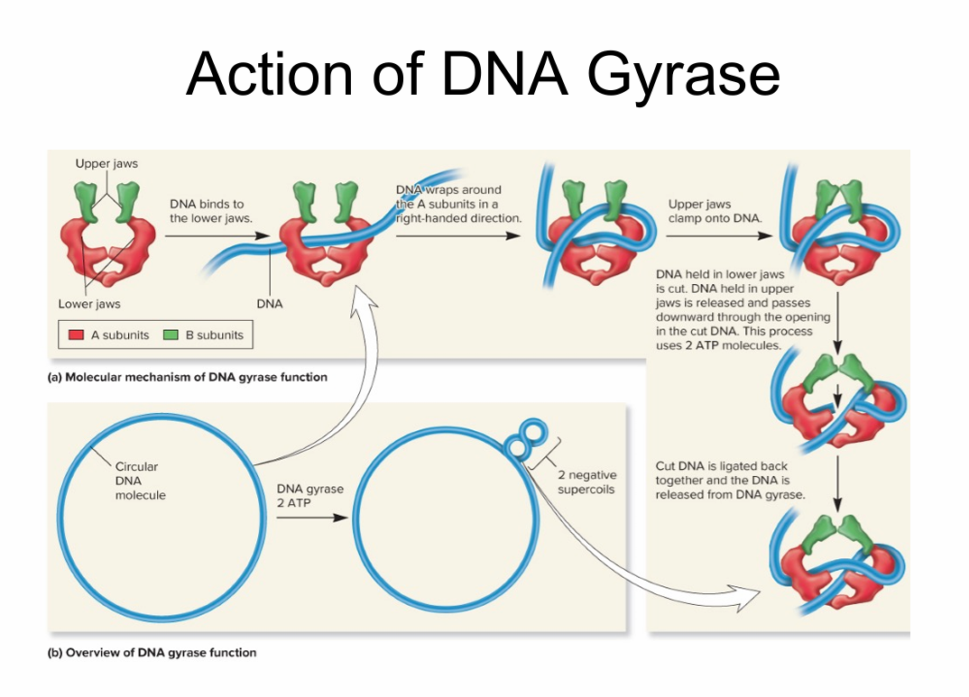
In bacteria topoisomerase (TOPO) is the enzyme that does all this super coiling
there is a topoisomerase-1 and a topoisomerase-2
—gotta know the difference
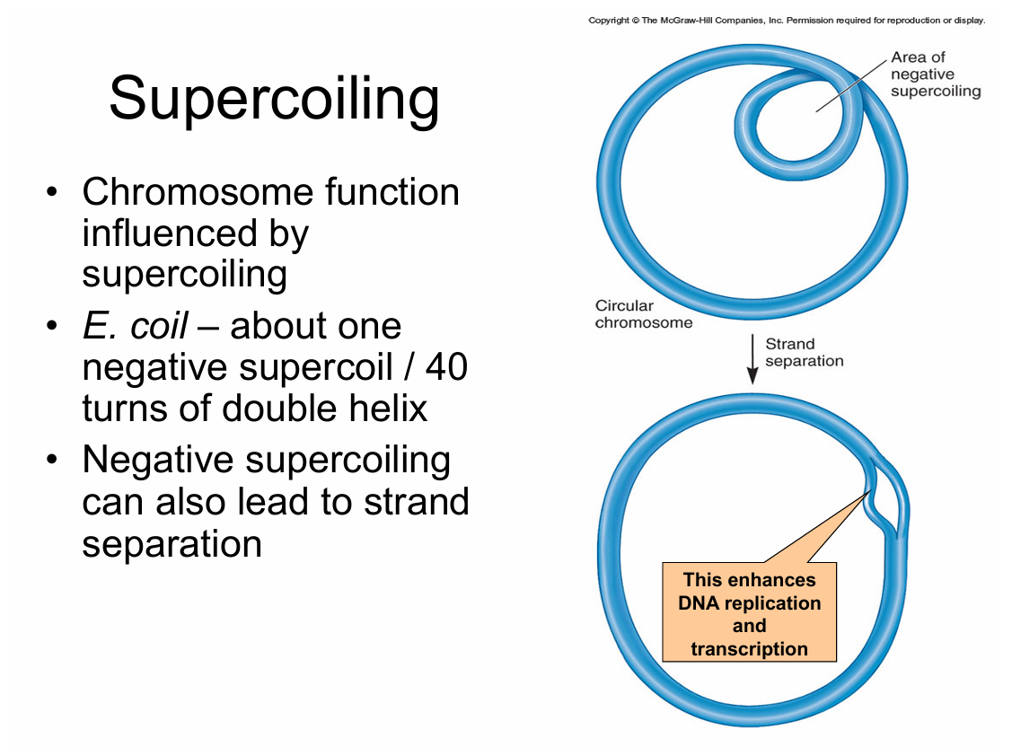
Supercoiling:
Achieved/controled by 2 enzymes
—DNA gyrase (TOPO II)
intos neg. supercoils
relaxes pos. supercoils
—Topoisomerase I
relaxes neg. supercoils
—Coumarins and quinolones – antibiotics that inhibit bacterial topoisomerases (but not eukaryotic)
Ex. cipro
If they can’t supercoil they die, and that’s how some antibiotics work.
Eukaryotic chromosomes:
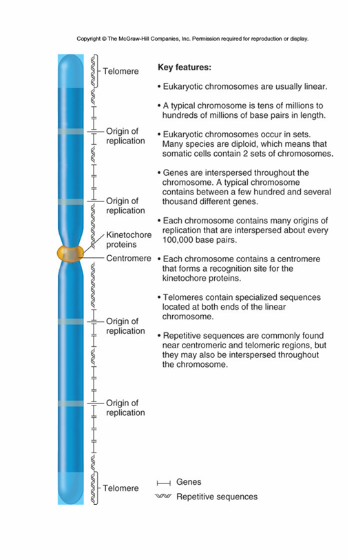
Long liner DNA molecule
three regions for replication and segregation
origin of replication
centromeres
telomeres
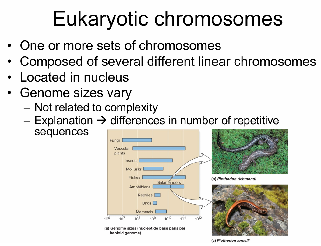
eukaryotic chromosomes are far bigger that prokaryotes, but a salamander has a larger genome than us
humans have smaller genomes than many plants and animals because … tiploid i think?
sequence complexity
number if times base sequence present w/in genome
—uniqe
once or few times
structural genes
—Moderately repetitive
• Few hundred to several thousand times
• Genes coding for rRNA/histones
—Highly repetitive
• Tens of thousands to millions of times
• Relatively short
• Ex. Alu – present in ~1 million copies in human genome– Proposed to have arisen 65 million yrs ago from ancestral gene à 7SLRNA
• Tandem repeats– Short sequence repeated over in ro
50% of human genome is virus genome - more virus then human
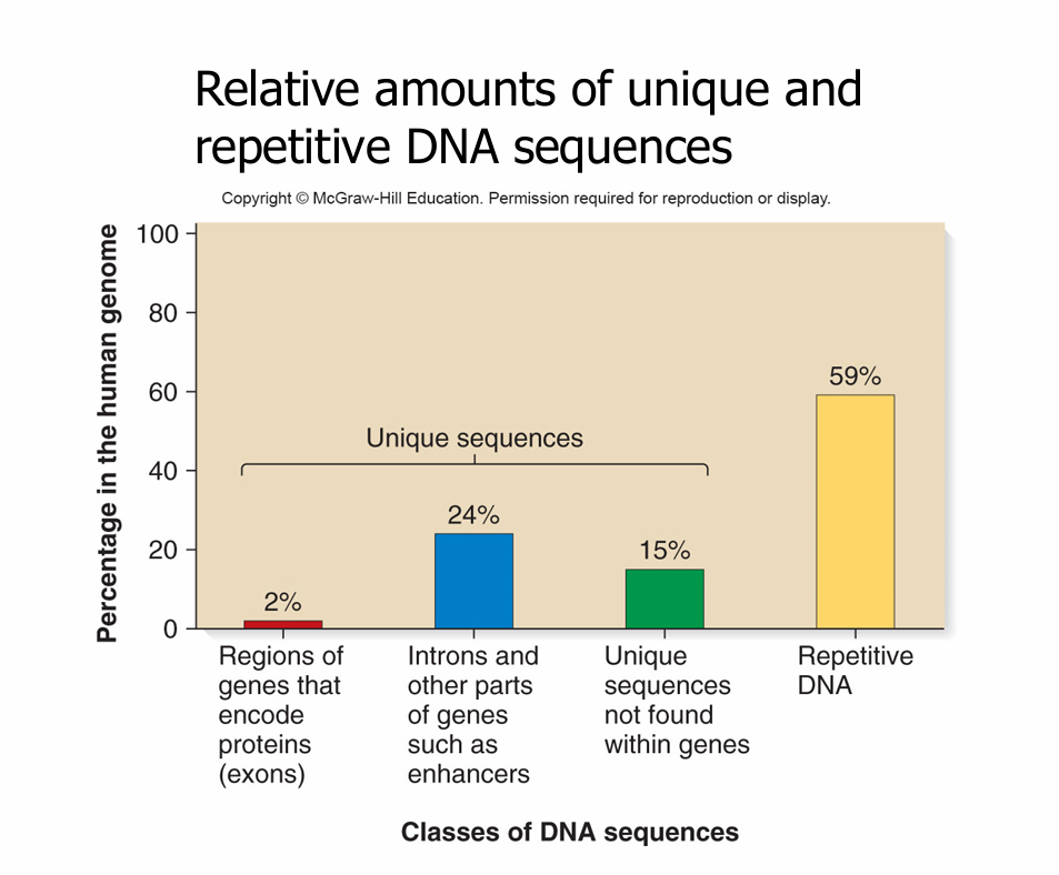
protiens do a lot (most) of stuff that you learn about, but it’s only like2% of genome
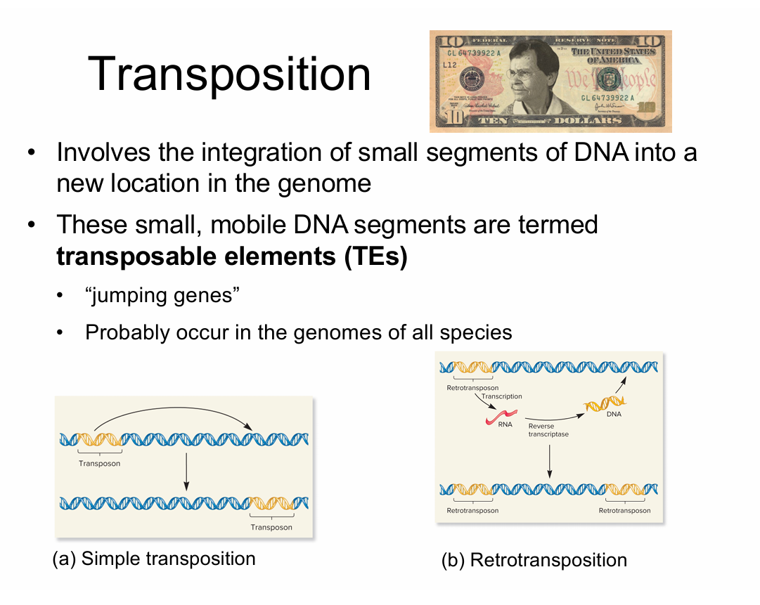
Simple transposons - (cut and paste) — jumps
Retrotransposons (copy paste)- ends w/ 2 copies- seem like the importaint ones for…
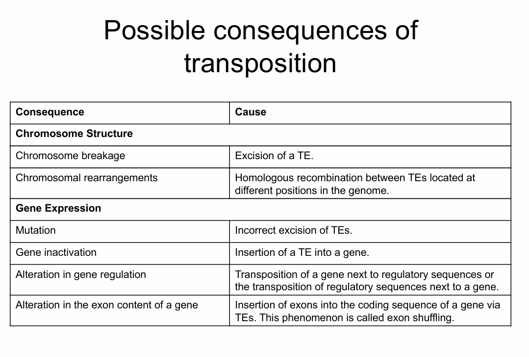
transposons can cause structual abnormalities
—it can acidently cause breakage
—it can cause missalinement depending on how it does
transposons not origonally human, likely from virus, but are still really helpful
really rare in simple organisms
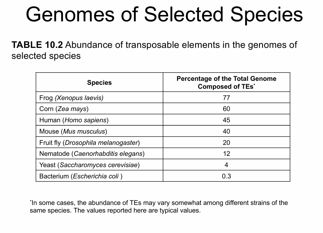
most common sequence you will find in the human genome is transposons
Some repetitive sequences in eukaryotic genomes are due to the proliferation of TEs
in mammles for example:
LINES (Long interspersed elements)
—usually 100 -10000 bp long
—Occur in 20,000 to 1,000,000 copies per genome
— Almost 17% of the human genome
SINEs (Short interspersed elements)
Less than 500 bp in length lements)
Example: Alu sequence present in about 1,000,000 copies in human genome (10% of the genome
Human Chromosomes end to end
—1 meter
Size of nucleus
—2-4 micrometers
• Must be compacted
– involves interactions between DNA and several different proteins
– The DNA-Protein complex is called chromatin
• Alternate between tight and loose compaction states
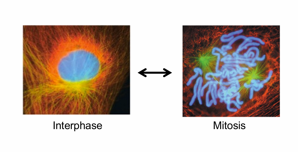
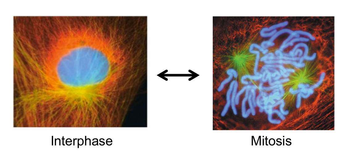
interphase = compacter
mitosis = not compacted
must be able to do it fast and good so no mess ups
Quiz on friday over ch 8 and 9
DATE:10/3/25
NEXT WEDNESDAY TEST OVER CHS> 6-10
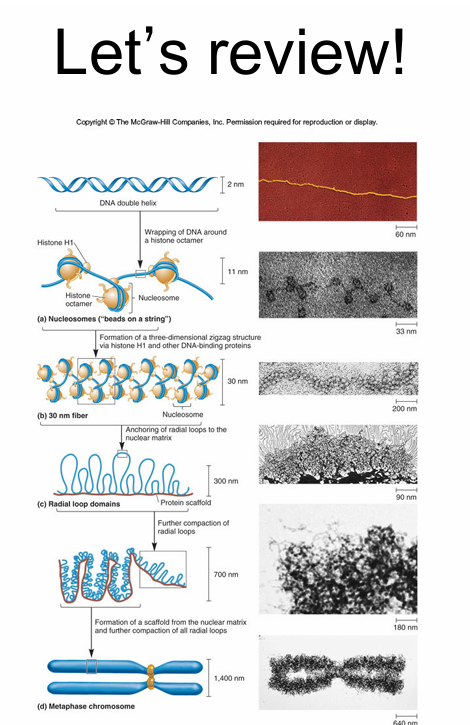
How to get from double helix to a singular chromosome:
Nucleosome
repeating unit in chromatin
DNA wrapped around histone octamer
—”beads on a string”
—Compacts DNA 7 fold
—two copies of each of these:
H2A
H2B
H3
H4
Connected by linker DNA
—20 to 100 bp
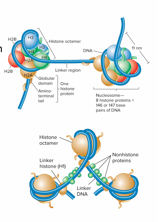
DNA gets wrapped aroung the nucleosome of core histone protienes
it’s getting more compact, but thicker
ends up lookinh like a pearl necklase under microscope
human genome length: 3.2 billion bp
5th histone protiend: Histone H1 it binds in the inbetween of the octomeres
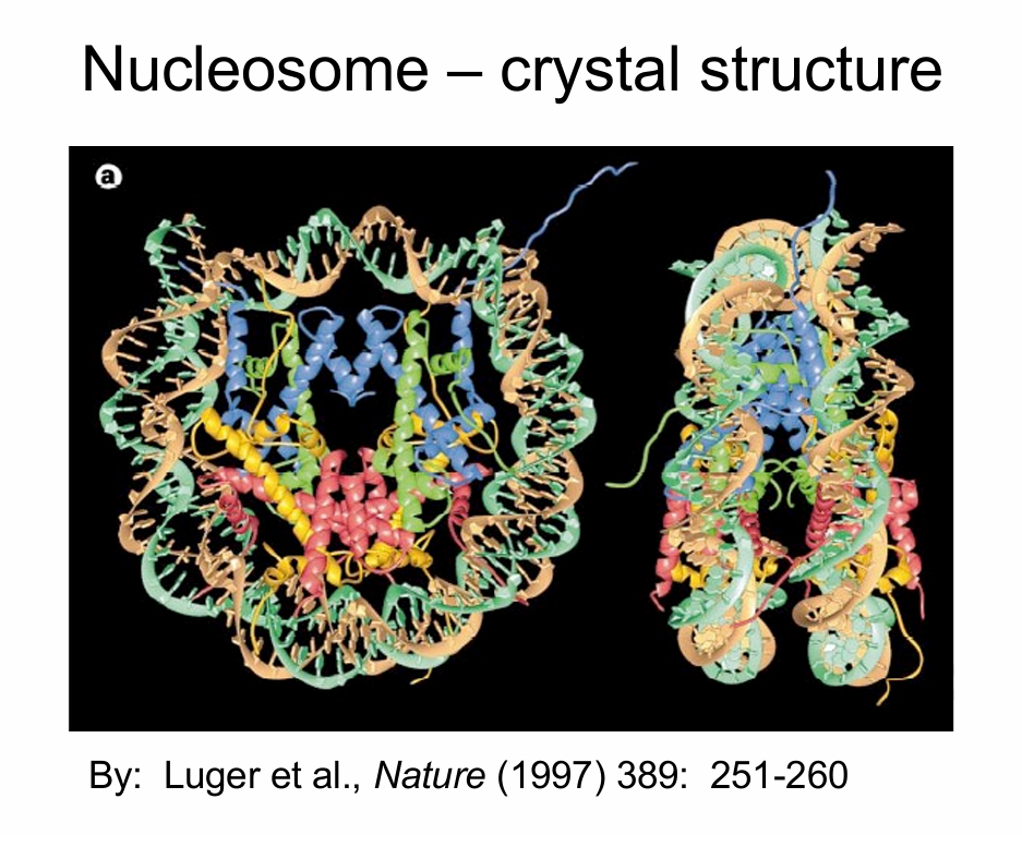
parts of the histone protiens stick out and hold on to the DNA (as you can see the little things hanging out)
Histones:
Globular domain
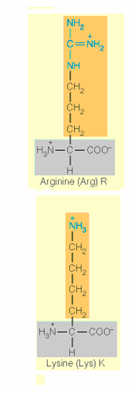
flexible, charged amino terminus (amino terminal tail)
Basic → contain lots of arginine and lysine residues • Arginines à electrostatic and hydrogen bonding interactions with DNA backbone • Core histones: H2A, H2B, H3, H4 • Histone H1– Linker histone– Binds to DNA in linker– May help compact adjacent nuclosomes– Bound less tightly than cor
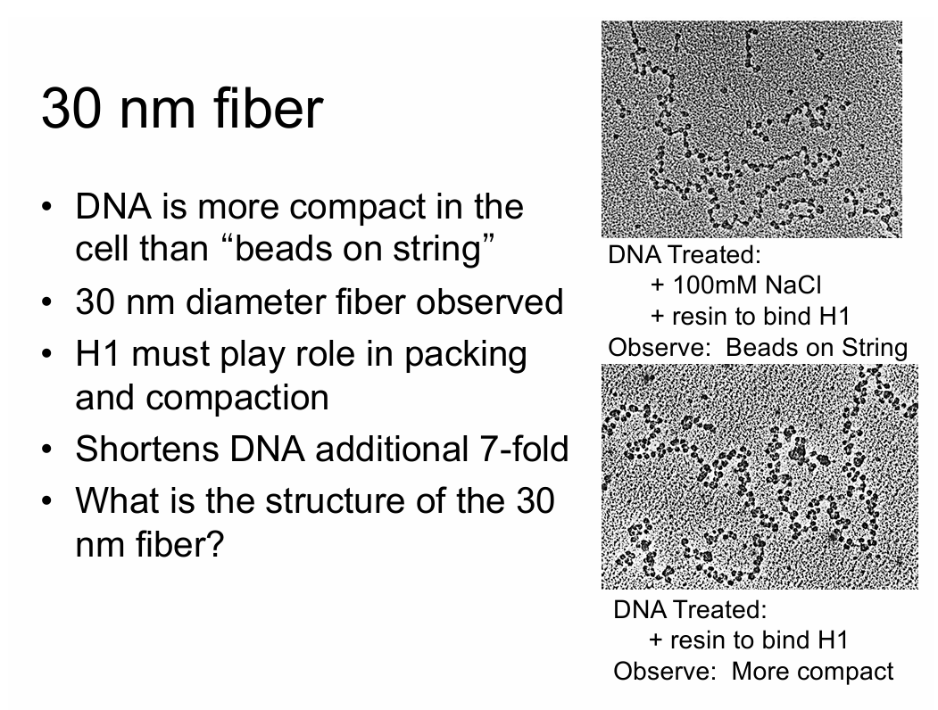
Chromatines aren’t actually smt. we can see. it’s just our best guess
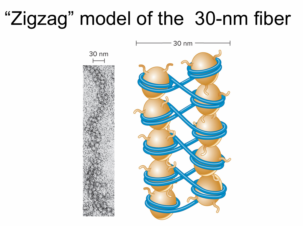
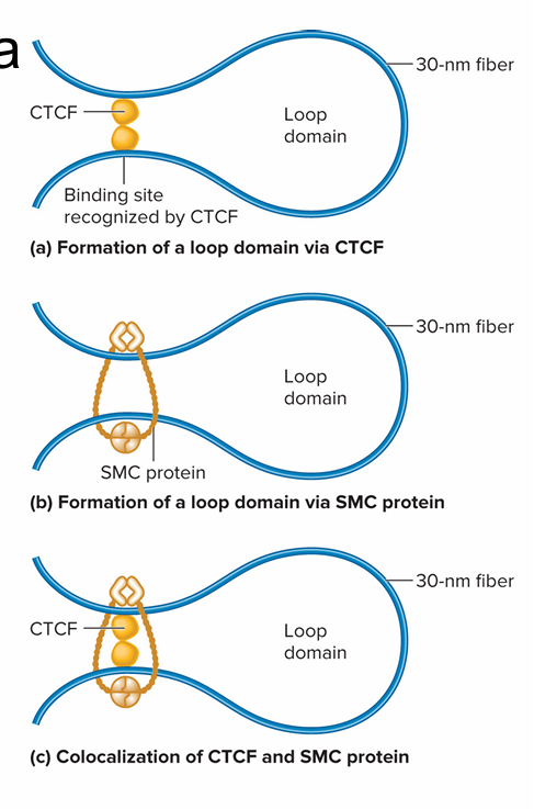
Further compaction via loop domains
CCCTC binding factor
(CTCF) - binds to repeats of the sequence CCCTC
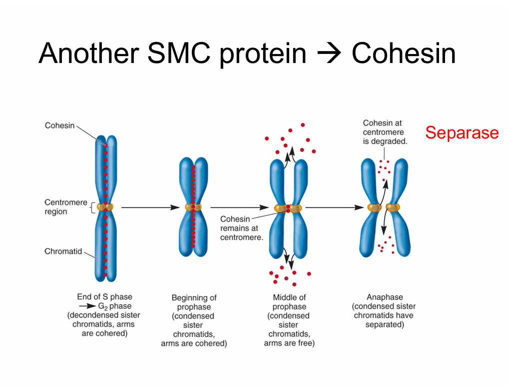
SMC protiens
- structural maintenance of chromosomes
- use ATP to catalyze changes in chromosome structure
- form a dimer that can wrap itself around 2 DNA segments and form a loop
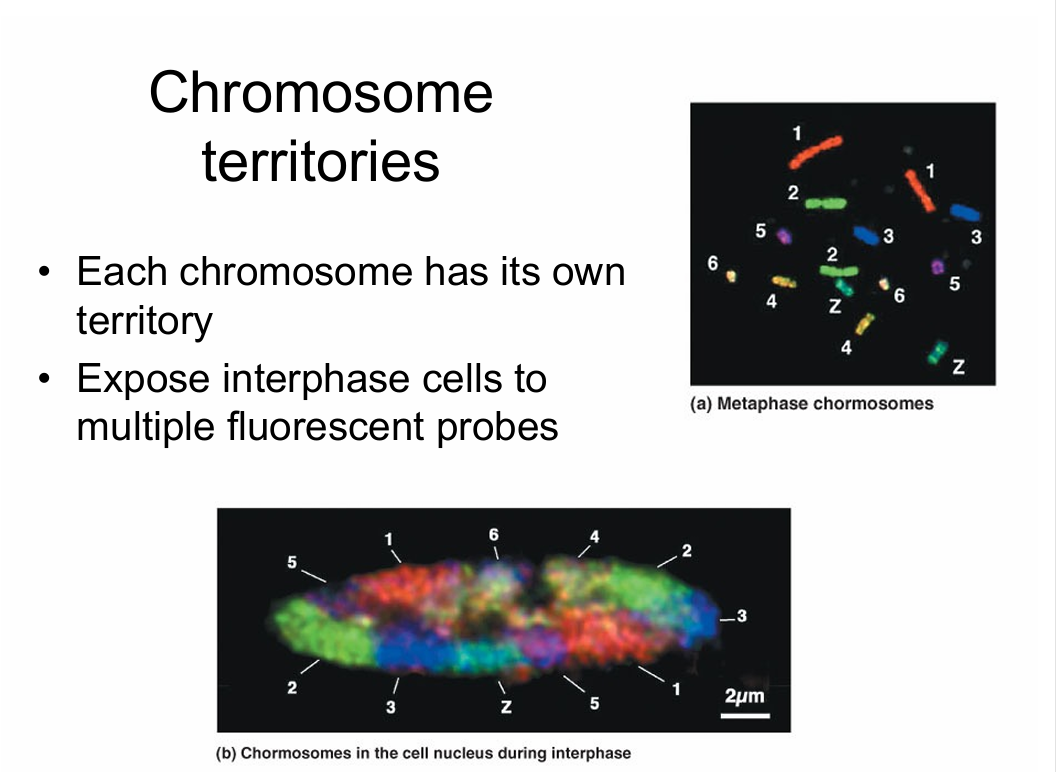
Interphase chromosomes:
Vsr
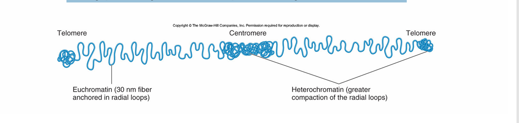
iable levels of compaction
Euchromatin
— radial loops formed by 30 nm fiber
Hererochromitine
—more tightly compacted
—compacted even furthed
– Typically transcriptionally inactive
Heterochromatin
Constitutive heterochromatin
– Always heterochromatic
– Always transcriptionally inactive
– Usually contain highly repetitive sequences
• Facultative heterochromatin
– Occasionally interconvert between euchromatin and heterochromatin
– X inactivation
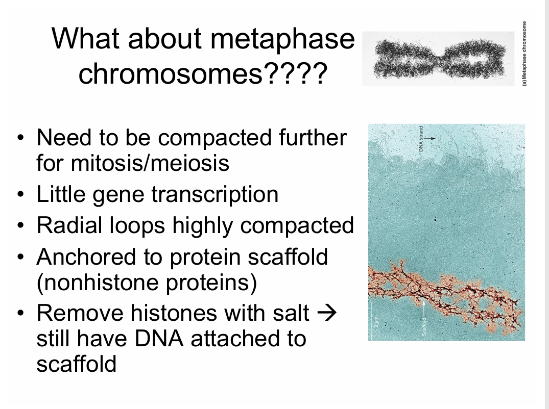
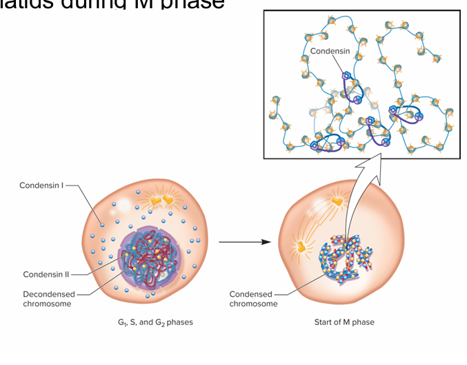
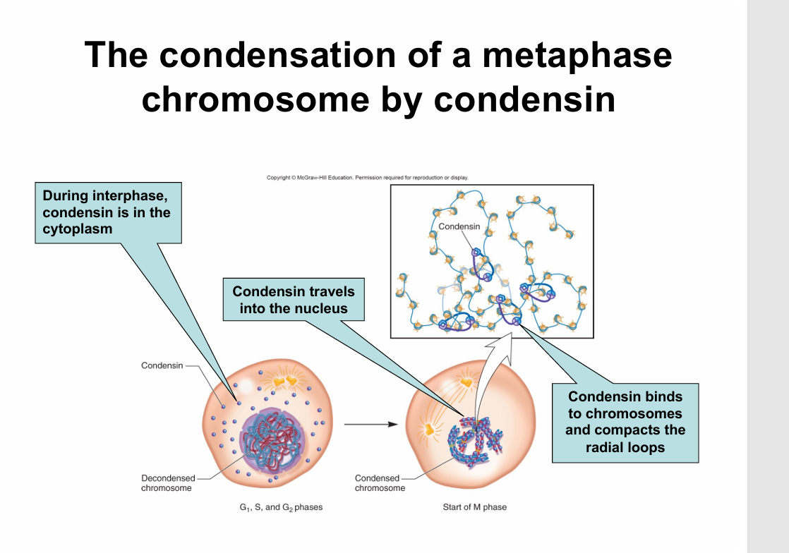
Condensin:
Classified as SMC protein
involved in compaction durring M phase
coates chromatids durring M phase
DATE: 10/06/2025
REVIEW SESH BEFORE EXAM THIS WEDNESDAY AHHHHHHHHHHHHHH
Review activity
AHHHHHHHHHHHHHHHHHHH