5.3 Heart Anatomy and Physiology
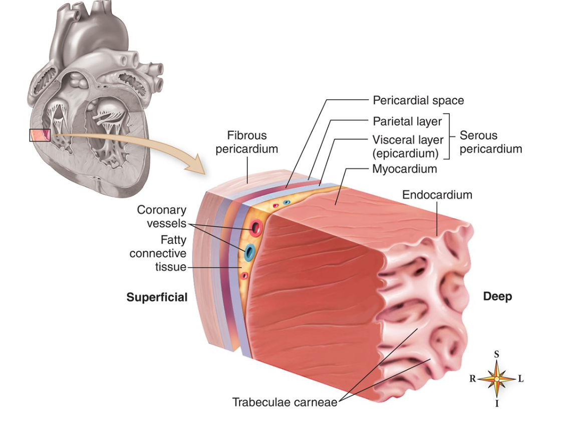
Pericardium and Layers of the Heart
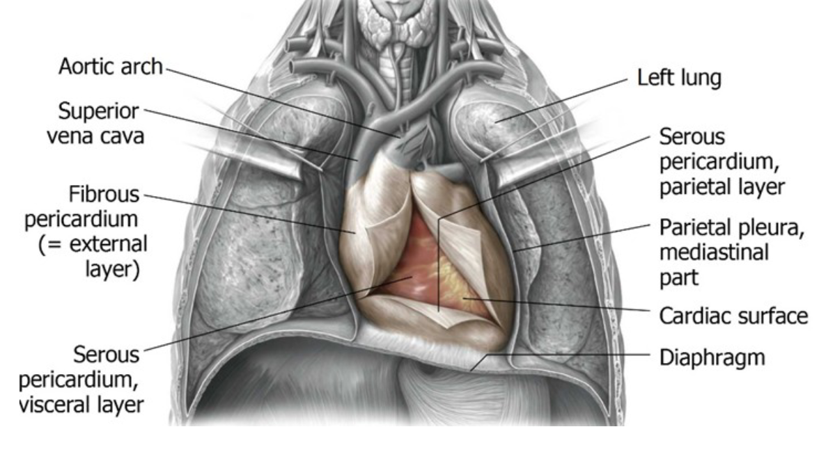
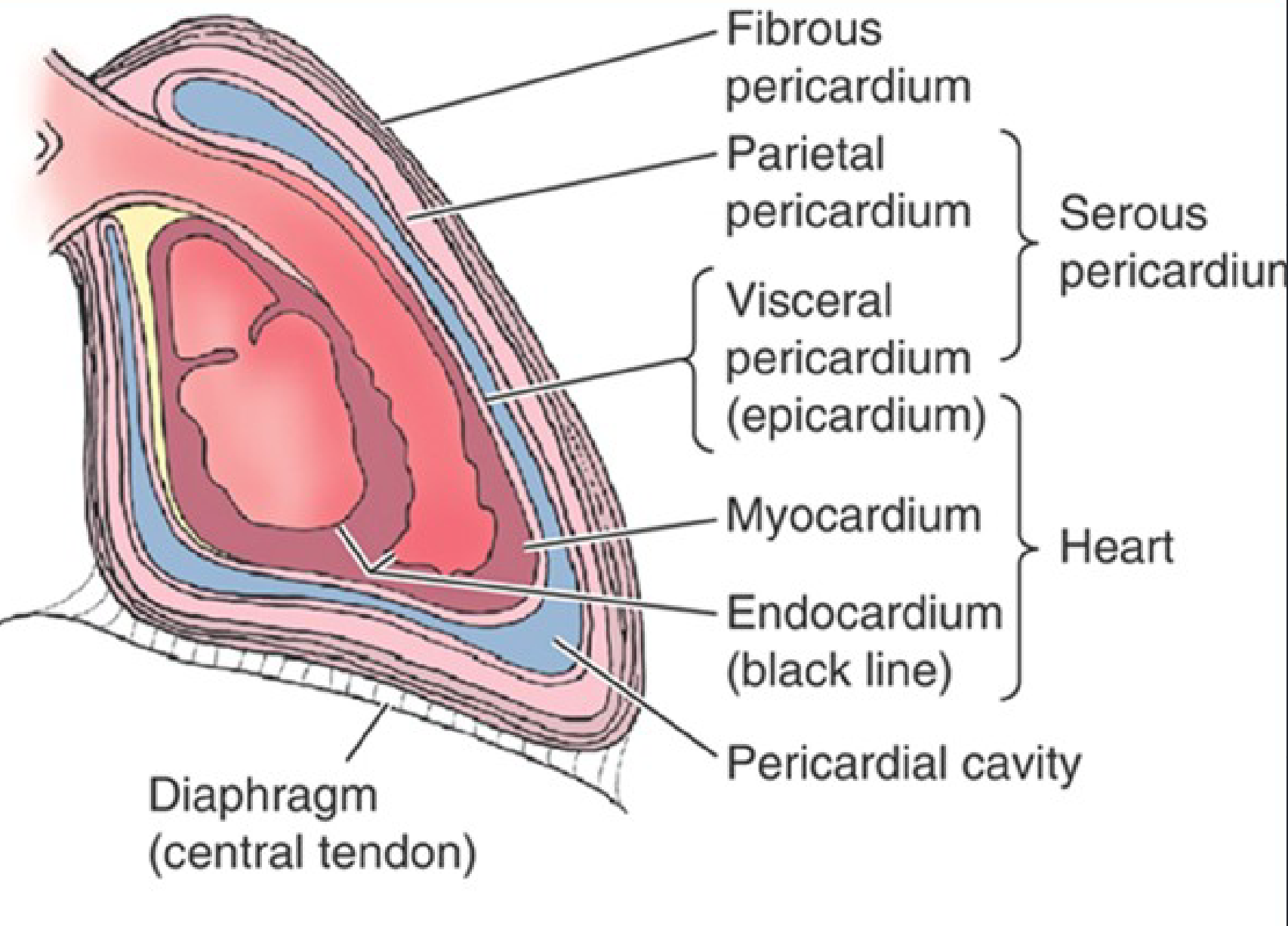
Pericardium: Protective sac surrounding heart.
Fibrous Pericardium: Prevents overfilling, influenced by movements of sternum and diaphragm.
Serous Pericardium: Divided into two layers:
Parietal Layer: Lines the fibrous pericardium.
Visceral Layer (Epicardium): Covers the heart surface.
Pericardial Cavity: Contains serous fluid, reducing friction during heart movement.
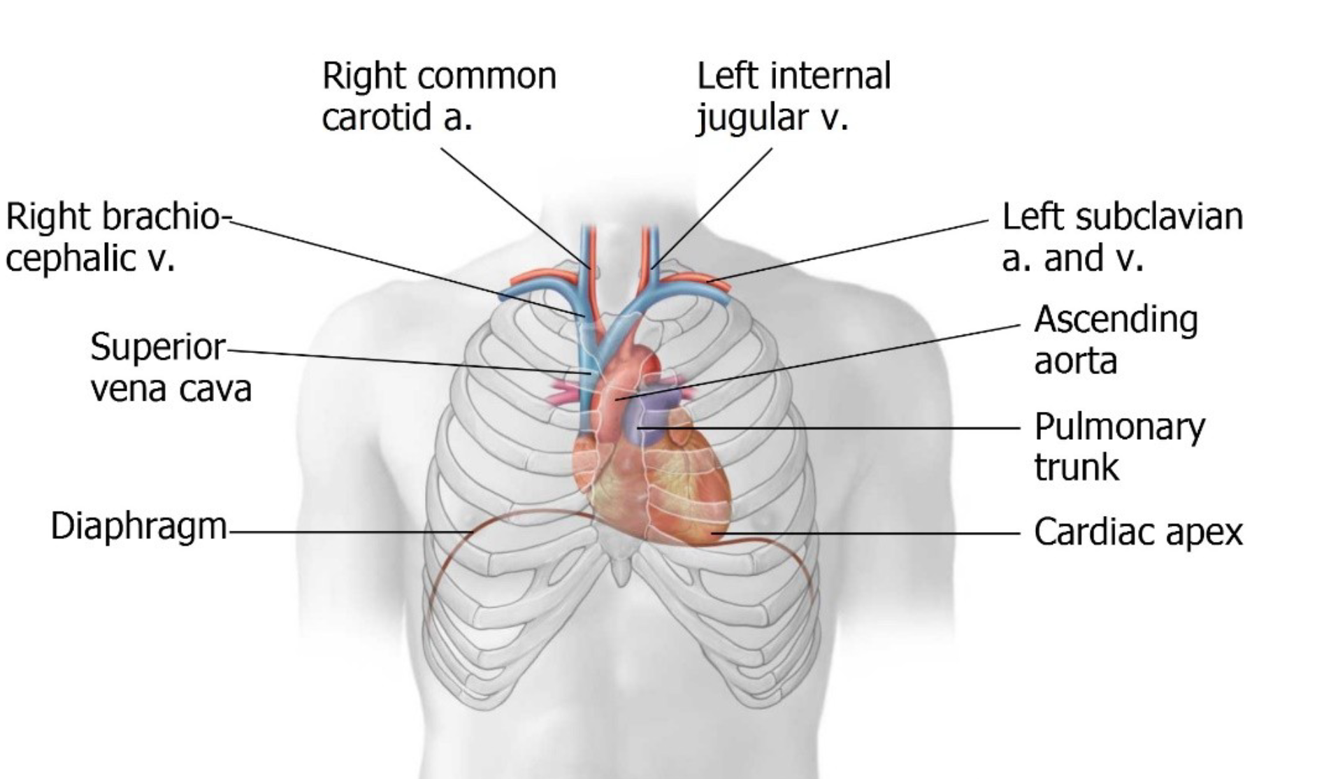
Heart parts/chambers
Mediastinum (central part)
Pericardium (membrain)
Heart
Base (top)
Apex (bottom)
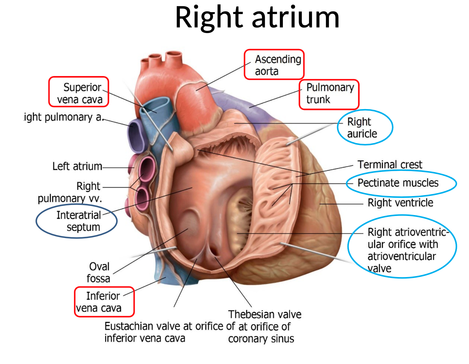
Right atrium
heart's upper right chamber, receiving deoxygenated blood from the body via the superior and inferior vena cavae and the coronary sinus, and then pumps it through the tricuspid valve into the right ventricle
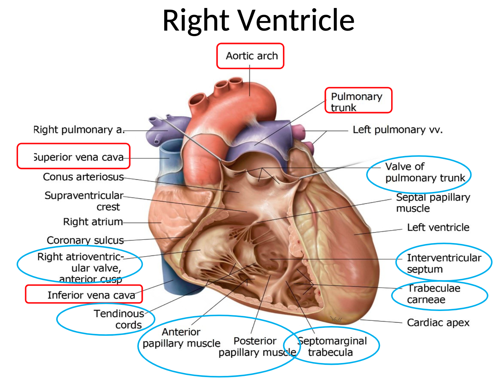
Right Ventricle:
one of the heart's four chambers, located in the lower right portion of the heart below the right atrium, and it pumps oxygen-depleted blood to the lungs for oxygenation
ENDOCARDIUM & MYOCARDIUM
Myocardium is a Cardiac muscle
Endocardium is Inner lining of the heart
Function:
•allow rapid, simultaneous, widespread contraction
•inherent rhythm
•modulated by ANS & hormones
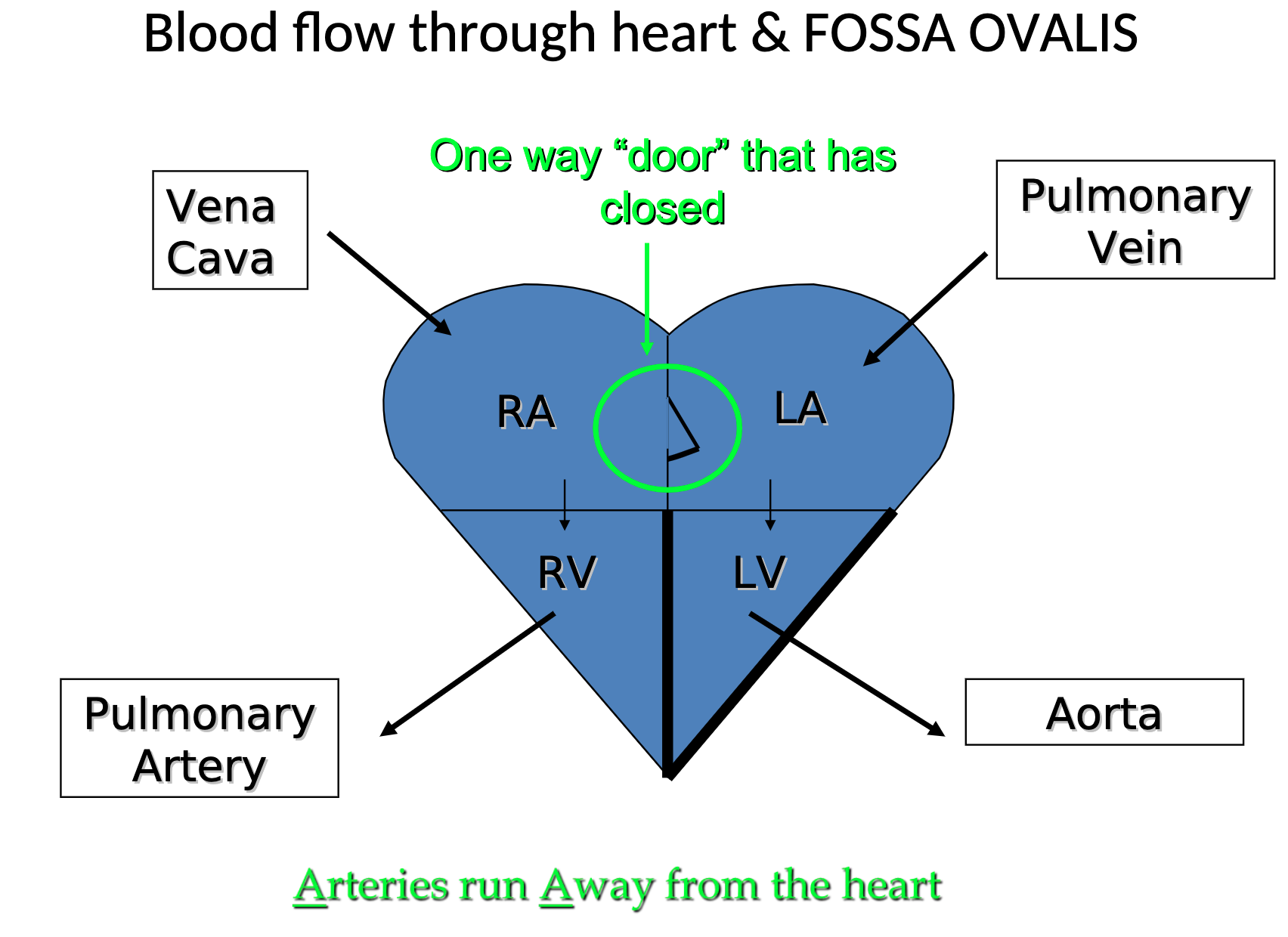
Blood Flow Through the Heart
Circulatory Pathway:
Superior Vena Cava (SVC): Drains upper body blood into the Right Atrium.
Inferior Vena Cava (IVC): Drains lower body blood into the Right Atrium.
Right Atrium → Right Ventricle → Pulmonary Arteries (to lungs for oxygenation).
Pulmonary Veins: Return oxygenated blood from lungs to Left Atrium.
Left Atrium → Left Ventricle → Ascending Aorta (distributes blood to the upper body).
Descending Aorta: Supplies lower body.
Fossa Ovalis: Remnant of the foramen ovale; a one-way door that closes post-birth.
Anatomy of the Heart
Chambers of the Heart:
Right Atrium, Right Ventricle (both serve pulmonary circulation- oxigenating).
Left Atrium, Left Ventricle (both serve systemic circulation - oxygenated to tissue).
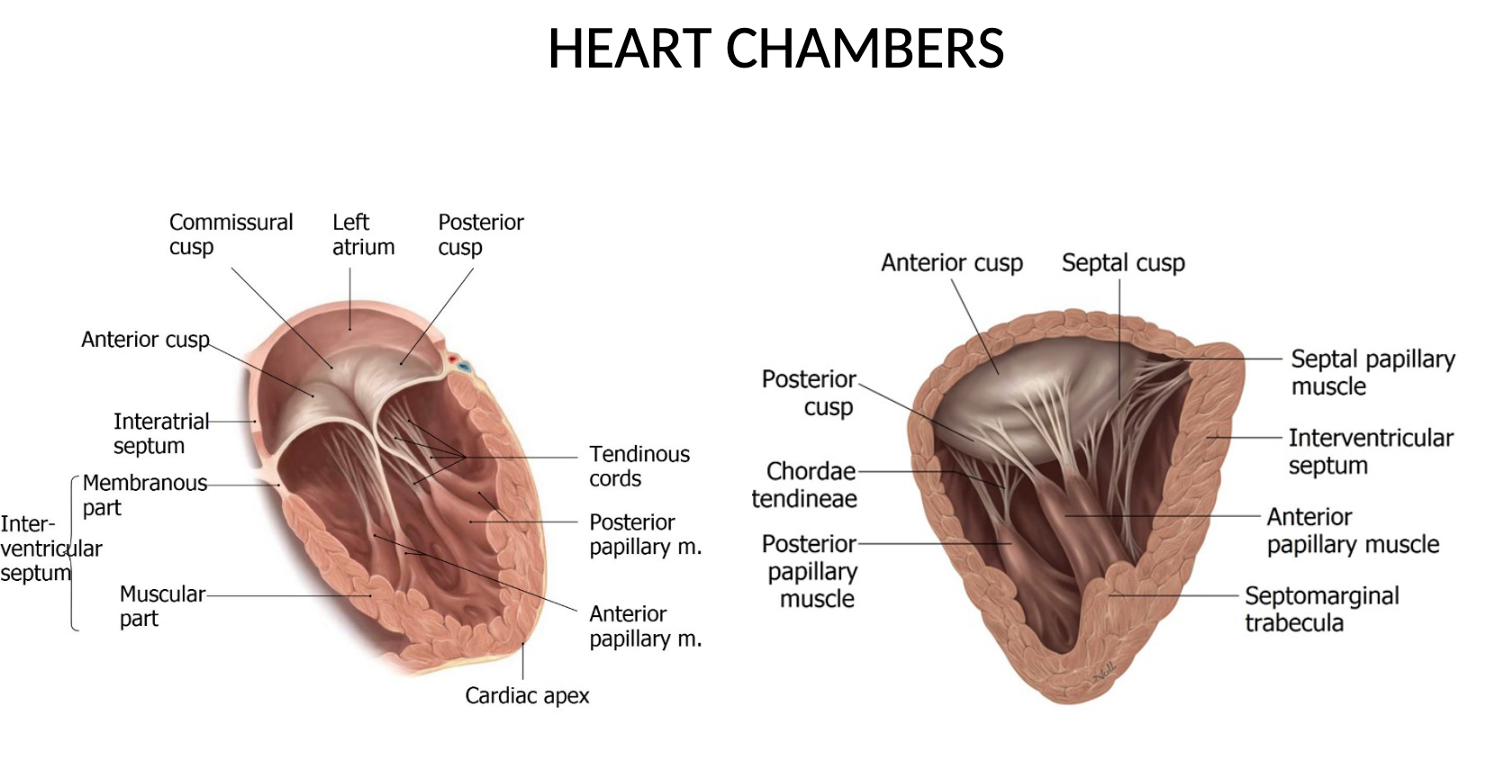
Valves:
Tricuspid Valve (Right)
Bicuspid Valve (Mitral) (Left)
Aortic Valve
Pulmonary Valve
Papillary Muscles and Chordae Tendinae support valve structure during contraction.
Heart Sounds
Systolic sound: First sound, believed to be caused primarily by contraction of the ventricles and by vibrations of the closing AV valves
Diastolic sound: Short, sharp sound; thought to be caused by vibrations of the closing of SL valves
• Heart sounds are clinically significant because they provide information about the functioning of the valves of the heart
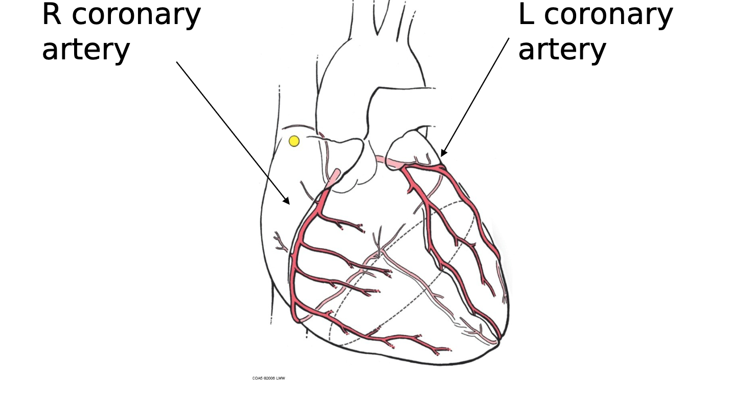
Blood Supply & Drainage of the Heart
Coronary Arteries supply blood:
Right Coronary Artery: Supplies right atrium, ventricle, and nodes.
Left Coronary Artery: Supplies left atrium, ventricle.
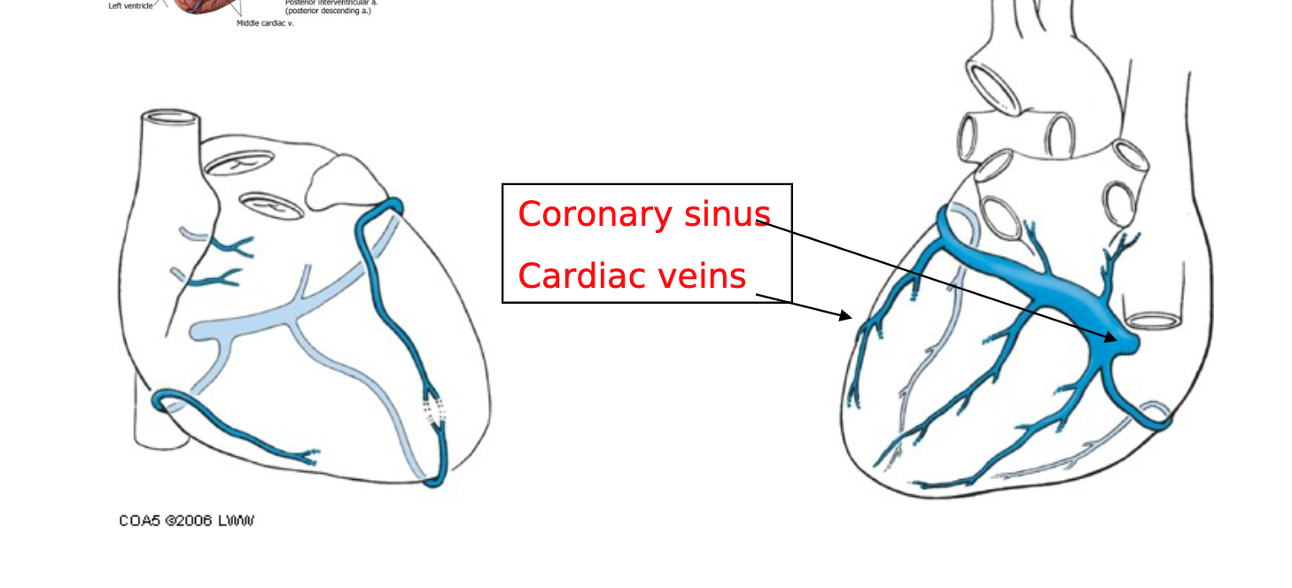
Venous Drainage: Blood is drained mainly via cardiac veins into the Coronary Sinus
Great Cardiac Vein, Middle Cardiac Vein, Small Cardiac Vein. cardiac veins drain mainly into the coronary sinum
Blood flow within the heart itelf: contribution of blood by the R & L coronary a
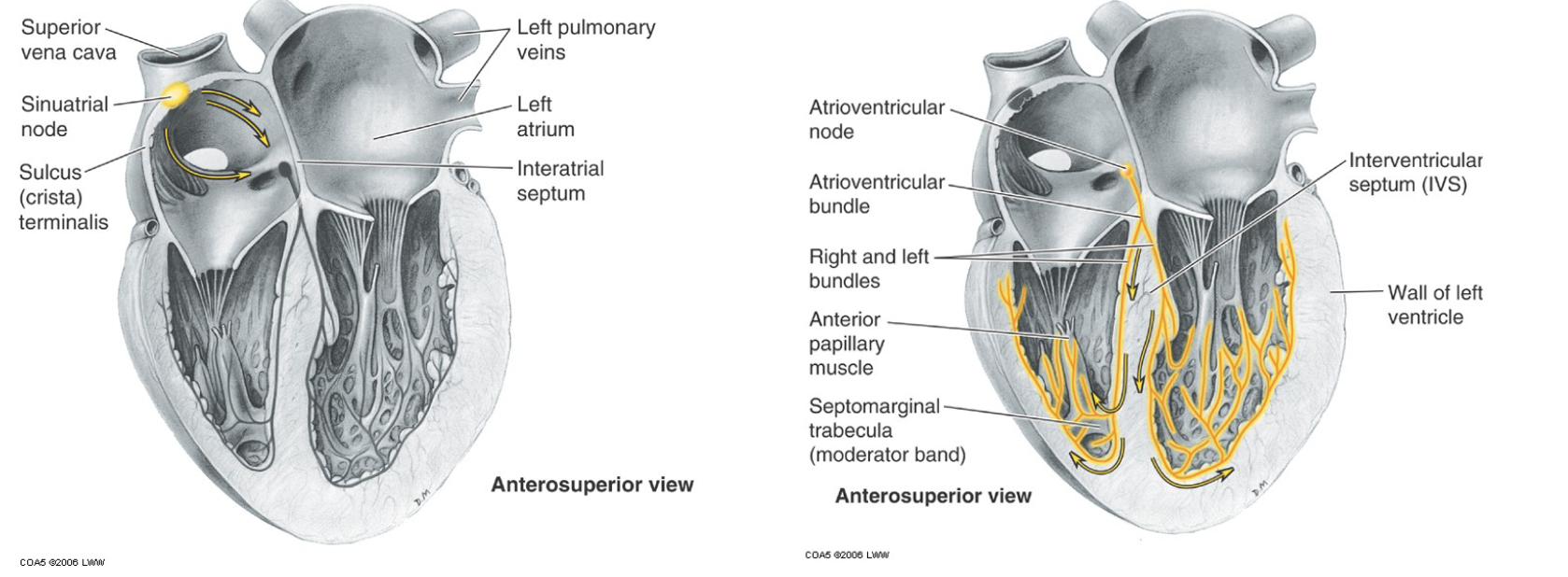
Electrical Conduction Through the Heart
The cardiac conduction system is a network of specialized cells that initiates and coordinates the heart's electrical activity, ensuring the heart beats in a rhythmic and efficient manner.
Conducting System:
Sinoatrial (SA) Node: Acts as pacemaker, generates 70-80 beats/min.
Atrioventricular (AV) Node: Receives impulse from SA node and delays it.
Atrioventricular Bundle (Bundle of His): Path for electrical impulses to travel through ventricles.
Purkinje Fibers: Spread impulses throughout ventricles, causing contraction.
Electrocardiogram (ECG): Visual representation of heart's electrical activity (P, Q, R, S, T waves)
Functions of the Heart
Primary Functions:
Pumps blood throughout the body.
Regulates blood flow and pressure.
Conducts electrical impulses for contraction rhythm.
Heart Sounds:
Systolic Sound: First sound due to ventricular contraction and AV valve closure.
Diastolic Sound: Caused by closure of semilunar valves.
Key Considerations for Examination
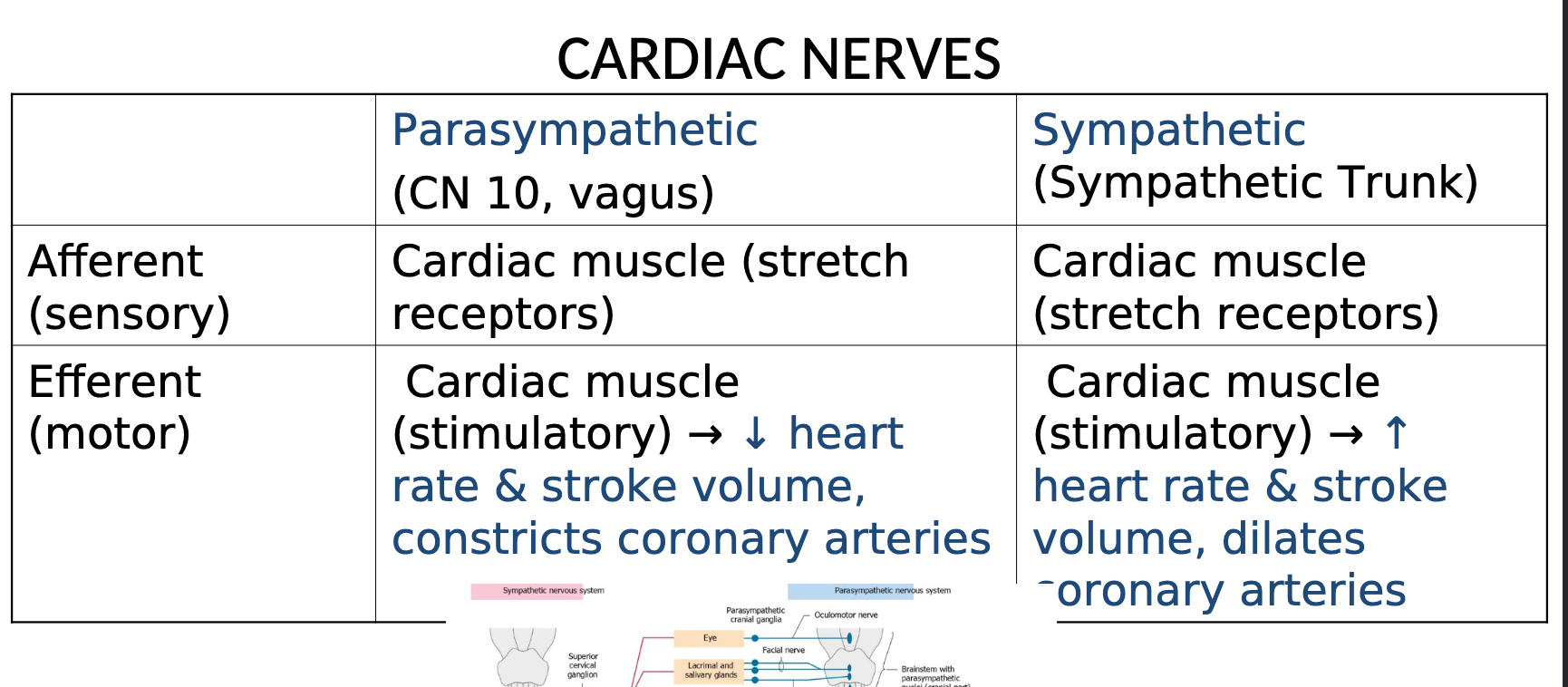
Understanding the labeling of heart diagrams including chambers, valves, great vessels, blood supply, and nerve supply.
Knowledge of blood flow direction and mechanics of valve function.
Familiarity with the structure and function of the heart's conducting system and its components.