Final Exam Review - Medical Interventions || PLTW
1.1
A medical intervention is anything used to treat, prevent, cure, or relieve the symptoms of human suffering
Bioinformatics - the collection, classification, storage, and analysis of biochemical and biological information using computers, can be used to identify disease pathogens
DNA Sequencing
Use of BLAST for DNA sequencing
Comparing a found DNA sequence of a patient to the DNA sequence of a virus or bacteria found in a database

ELISA testing
Enzyme-linked Immunosorbant Array
Takes advantage of the body’s natural immune response (antigens & antibodies)
Found in strep throat tests, pregnancy tests, drug tests, & others
Color change = positive
The greater the color change, the more of a substance is present
Qualitative result - results are observations (color, size, etc.) and not number data.
Compare to a Serial Dilution
Antigens vs. Antibodies

Antigens
Any substance that the body recognizes as foreign
protein, sugar, lipid, etc.
Found on the surface of viruses, bacteria, pollen, & sometimes cancer cells
Antibodies
A defender protein produced by the immune system
identify specific antigens or foreign bodies
ELISA process
The ELISA test begins with a pre-treated tray full of small wells.
pre-coated with antibodies for the pathogen being studied. The serum of patients is then added to these wells.
If the serum contains the bacteria/substance, the antigens on the outside of those cells will be bound to the antibodies in the wells, trapping the antigens on the wells using antigen-antibody interactions. To make this test something with visible results, MORE antibodies are added.
something called a primary antibody is added. The role of this is to latch on to the antigen, forming a platform on which a secondary antigen with an enzyme attached can be added.
this is the next step – adding the second antigen which is linked to an enzyme.
Finally, a substrate is added that the enzyme responds to. The enzyme acts on the substrate and causes a color change
Serial Dilution
A serial dilution is something that is created and used to compare the results of an ELISA to. It involves beginning with a known concentration of antigen (say 100 ng/mL) and diluting it (watering it down).
It involves placing that 100 ng/mL sample in a well, transferring part of it to a new well with a set amount of water.
If you use equal parts water and antigen, 100 becomes 50. In the next well, it’s repeated to reduce the amount of antigen to 25, then 12.5, then 6.25, and so on.
When the antibodies and substrate are added to this a series of colors, from darker to lighter, is created.
These samples have known amounts of antigen.

1.2
Gram (+) v. Gram (-) bacteria
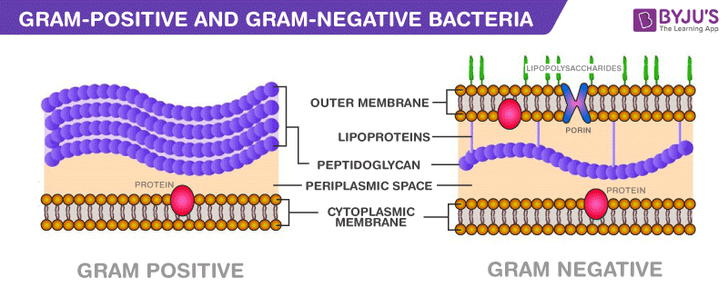
Gram positive bacteria have:
The cell wall contains a thick layer of peptidoglycan and teichoic acids. There is approximately twenty times more peptidoglycan than the Gram negative bacteria.
no outer membrane present.
no porins present.
The Gram-stained cell is purple.
Gram negative bacteria have:
The cell wall contains multiple layers, including a thin layer of peptidoglycan.
The outside layer is called the outer membrane, which is made of a lipid bilayer whose outside is composed of lipopolysaccharides called endotoxins.
The outer membrane serves as a barrier to the passage of most molecules and contains specialized proteins, called porins, which allow certain molecules to pass through the membrane.
The region between the plasma membrane and the outer membrane is called the periplasm and is filled with a gel-like fluid and proteins involved in a variety of cellular activities.
The Gram-stained cell is pinkish-red
Bacteria Structure | Definition |
Nucleoid | Gel-like region within the cytoplasm containing the single, circular, double-stranded DNA molecule. This chromosomal DNA is supercoiled, meaning tightly packed into a twisted form. The DNA contains all of the genetic information necessary for normal functioning of the cell. |
Plasmids | Circular double-stranded DNA molecules. They are typically 0.1% to 10% of the size of the chromosomal DNA and only carry a few to several hundred genes. A single bacterial cell can carry multiple plasmids. Normal functioning of a bacterial cell is not dependent on the genetic information contained in a plasmid, but the DNA often codes for proteins that are advantageous to the cell. For example, plasmids might contain the information coding for the proteins that enable the cell to destroy or be immune to certain antibiotics. Plasmids can be transferred from one bacterial cell to another bacterial cell. |
Ribosomes | Structures involved in protein synthesis. Facilitate joining of amino acids. |
Cell Wall | Rigid barrier that surrounds the cell, keeping the contents from bursting out. Peptidoglycan provides the rigidity for the cell wall. |
Plasma Membrane | Also called cell/cytoplasmic membrane. Semipermeable membrane that surrounds the cytoplasm of the cell. This phospholipid bilayer is embedded with proteins that act as a barrier between the cytoplasm and the outside environment. |
Capsule | A distinct and gelatinous layer, called glycocalyx, enveloping the cell. This layer enables the bacterial cell to adhere to specific surfaces and sometimes protects bacterial cells from human immune systems. |
Flagella | Protein appendages that are anchored in the membrane and protrude out from the surface. The flagella spin like propellers, moving the bacterial cell forward. |
Pili | Filamentous appendages which are similar in structure to flagella, but function in a different manner. Some pili enable the bacterial cell to attach to a specific surface (these pili are called fimbriae). Other pili are involved in conjugation, a mechanism of DNA transfer from one bacterial cell to another (these pili are called sex pilus). |
Endotoxins | Lipopolysaccharide molecules that make-up the outer leaflet of the outer membrane of Gram negative bacteria. Endotoxins are different from exotoxins, which are proteins synthesized by both Gram negative and Gram positive bacteria and function as potent toxins. |
Antibiotics
β-Lactam Antibiotics: Irreversibly inhibit enzymes involved in the final steps of cell wall synthesis. The enzymes inhibited by these drugs mediate the formation of the peptide bridges between adjacent strands of peptidoglycan. These drugs vary in their spectrum of activity; some are more active against Gram positive bacteria; whereas, others are more active against Gram negative bacteria
Tetracyclines: Reversibly bind to the 30S ribosomal subunit, blocking the attachment of tRNA to the ribosome and preventing the continuation of protein synthesis. They are effective against certain Gram positive and Gram negative bacteria.
Fluoroquinolones: Inhibit one or more of a group of enzymes called topoisomerases, which maintain the supercoiling of the chromosomal DNA within the bacterial cells. The inhibition of these enzymes prevents essential cell processes. The fluoroquinolones are active against a wide variety of bacteria, including both Gram positive and Gram negative bacteria.
Sulfonamides: Inhibit the growth of many Gram positive and Gram negative bacteria. They are structurally similar to paraminobenzoic acid (PABA), a substrate in the pathway for folic acid biosynthesis. Because of this similarity, the enzyme that normally binds with PABA preferentially binds with the sulfonamide drugs, resulting in its competitive inhibition. Human cells are not affected by these drugs because they lack this enzyme.
Antibiotic Resistance
Bacteria often will form Antibiotic resistance to commonly used antibiotics.
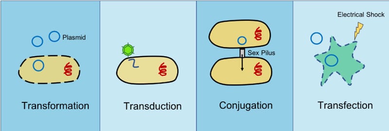
Bacteria mutate via transformation, transduction, or conjugation.