Neuromuscular Junction
The two ions that play a crucial role in propagating an action potential are sodium and potassium.
Ion fluxes depend upon intracellular-extracellular concentration gradients of Na+ and K+ (in reductionist terms…)
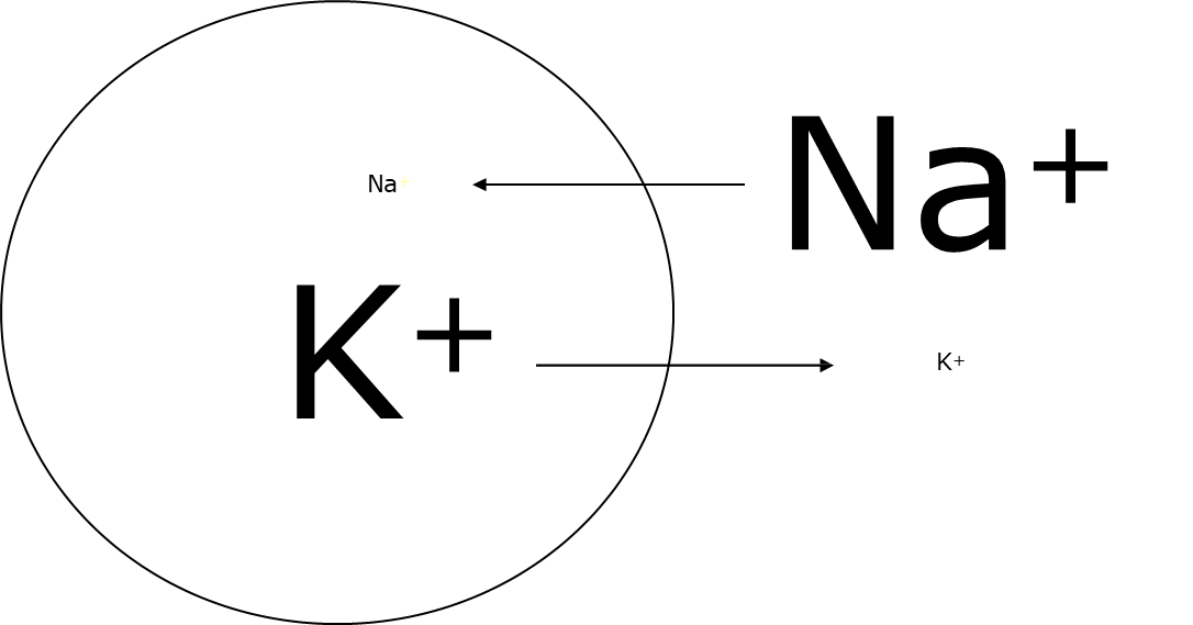
The cell is said to be at rest when there is a higher concentration of sodium ions extracellularly compared to a higher concentration of potassium ions intracellularly.
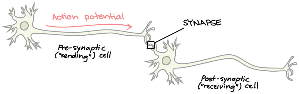
Action potentials propagate down muscle fibres by a wave of depolarisation.
Depolarisation results from an influx of sodium ions moving into the cell from the extracellular fluid down the electrochemical gradient, causing the inside of the cell to become less negative and more positive. This rapid change in membrane potential initiates the release of neurotransmitters at the neuromuscular junction, facilitating communication between the motor neuron and muscle fibre.
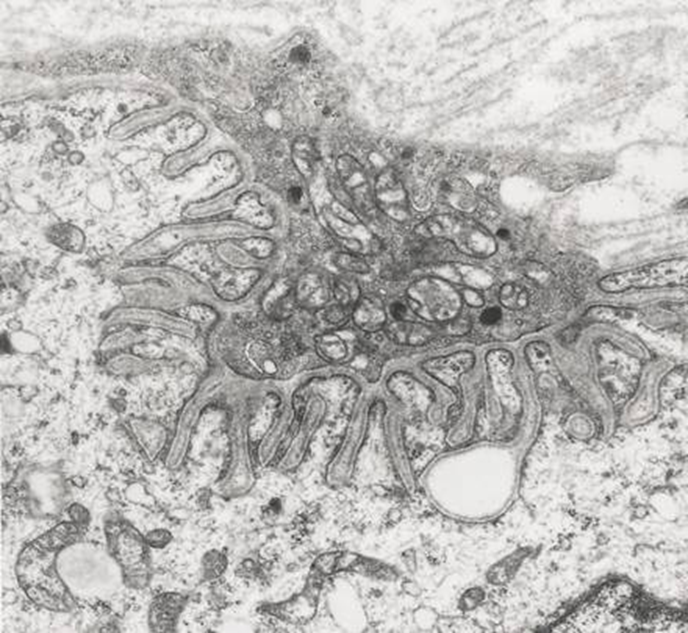
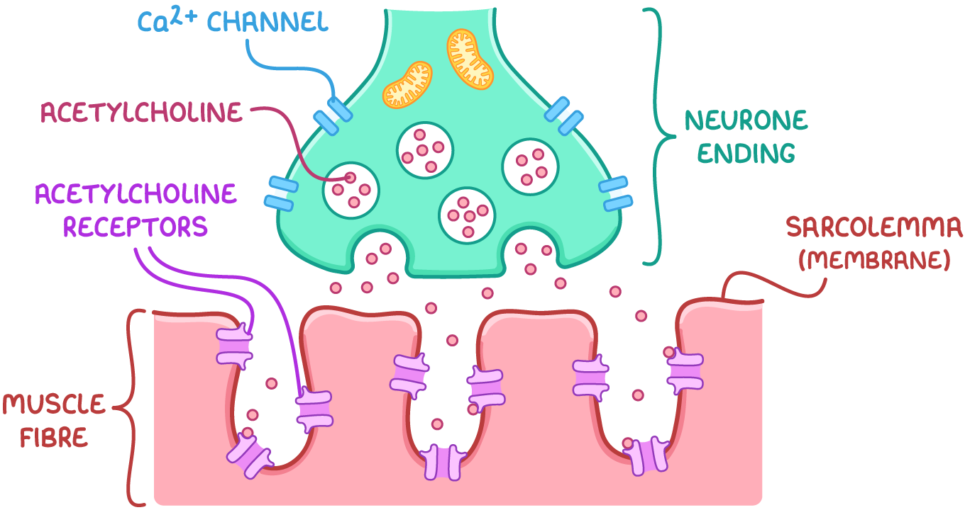
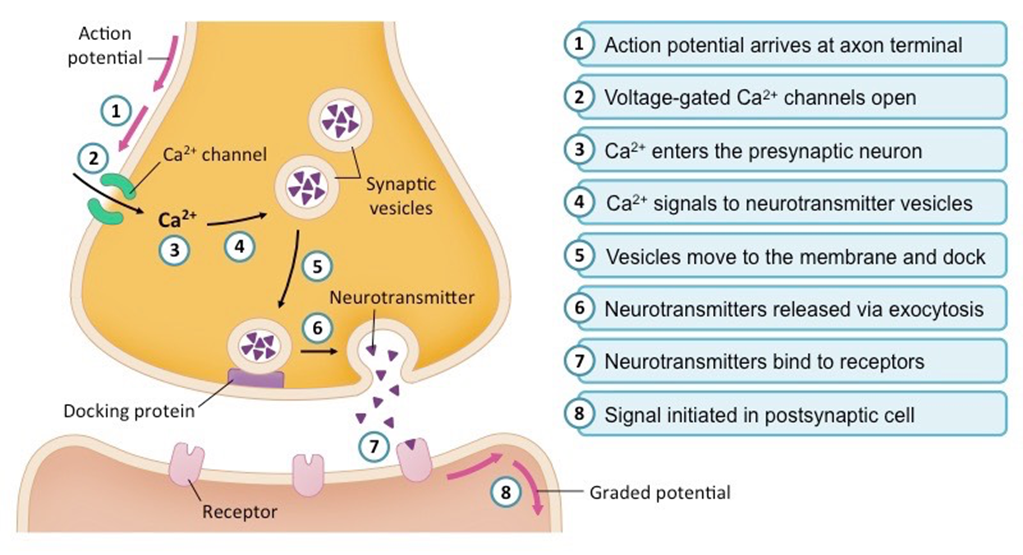
Signals propagate down and end at the axon terminal, in which the neuromuscular junction begins. At this junction, acetylcholine is released into the synaptic cleft, binding to nicotinic receptors on the muscle fibre membrane, which further amplifies the depolarisation and leads to muscle contraction.
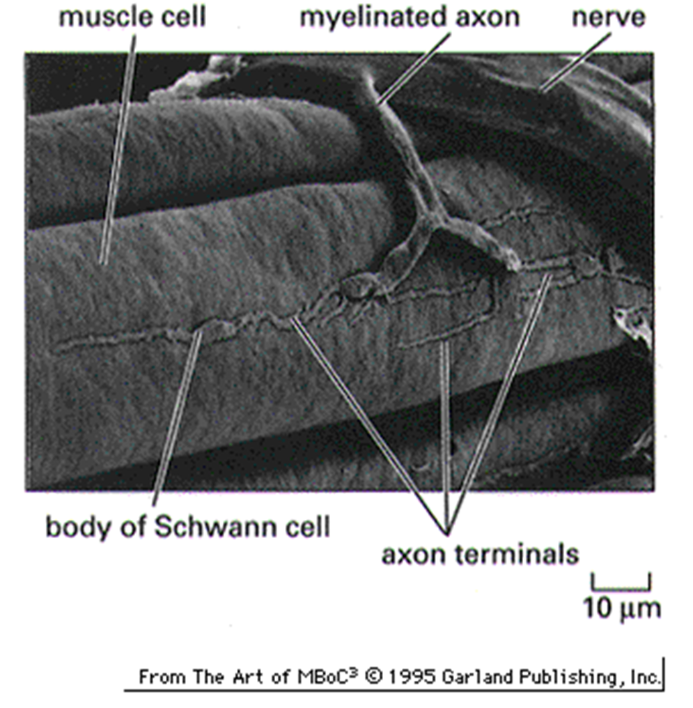
This is a histological image of a neuromuscular junction
The myelin sheath insulates the nerve and speeds up conduction velocity, which is the speed at which an action potential can propagate down.
The sub-neural clefts, which are the villi-like projections on the muscle fibre, help to increase the surface area in which neurotransmitters can bind to their receptors.
The receptors on the postsynaptic cell are called nicotinic acetylcholine receptors. These receptors play a crucial role in muscle contraction by allowing the influx of sodium ions, leading to depolarisation of the muscle membrane.
Acetylcholine release at the neuromuscular junction:
It is dependent upon depolarisation of the presynaptic membrane of the motor neuron
This depolarisation results in Ca2+ ion influx via voltage-dependent Ca2+ channels
The Ca2+ ions promote fusion of the vesicles with the presynaptic membrane
Activation of nAChRs by ACh (2 molecules per receptor) leads to depolarisation of the muscle fibre membrane, via net influx of Na+ ions
Release of a single vesicle of ACh (happens at rest) results in a so-called miniature end-plate potential, whereas release of several (when the motor neuron is activated) causes an end-plate potential or EPP

What are the arrangements of contractile proteins in striated muscle?
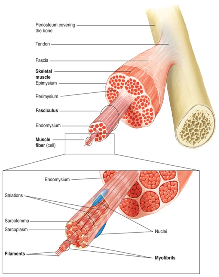
The sarcomere is the lowest functional unit of skeletal muscle that is arranged in parallel. They are separated by Z lines.
Z lines are where actin filaments are anchored and provide structural support, effectively maintaining the organization of the sarcomere during muscle contraction.
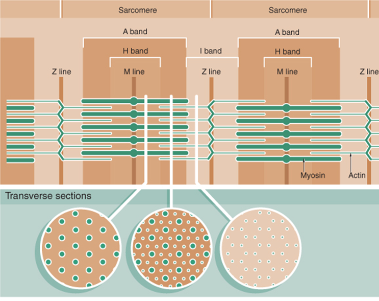
The M line runs straight through the midpoint of the myosin filaments.
What is the skeletal muscle contraction: sliding actin filament contraction?

Excitation-contraction coupling in skeletal muscle:
The events link to muscle excitation (action potential) to muscle contraction (cross-bridge recycling).
The events are:
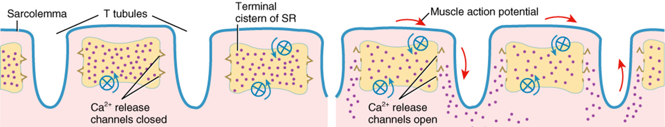
An action potential propagates down the sarcolemma
Transverse tubules conduct AP into the cell’s interior
Calcium ions are then released in channels in the sarcoplasmic reticulum.
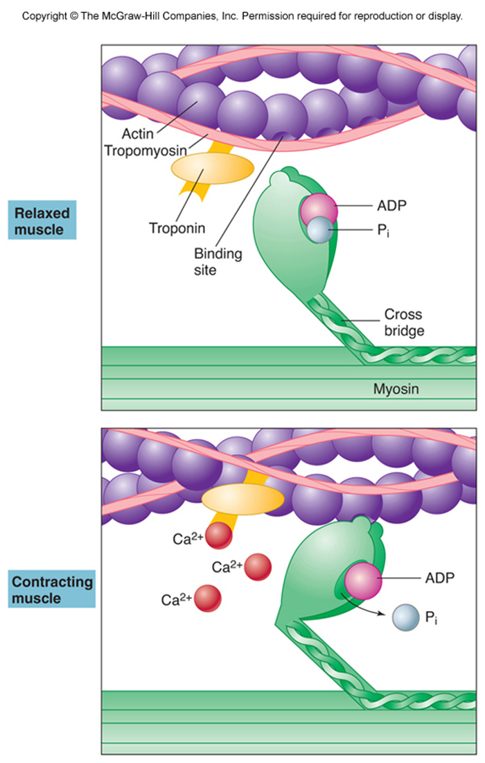
Activation of muscle contraction by action potentials:
Action potentials are induced by the release of calcium ions from the sarcoplasmic reticulum into the sarcoplasm.
When calcium binds to troponin on the actin filament it causes tropomyosin to experience a conformational change.
As a result, tropomyosin exposes. myosin binding sites on actin, enabling myosin to bind to actin.
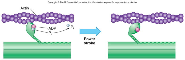
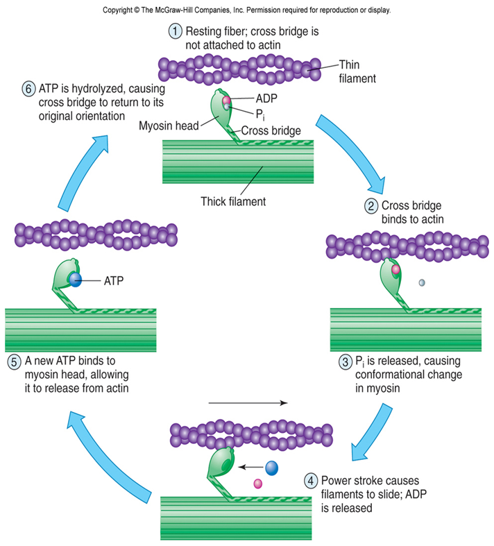
Cross-bridge recycling:
Myosin head binds to actin
This results in a power stroke. The globular heads bend towards the centre of the sarcomere.
The actin filaments are pulled towards the centre of the sarcomere
The cross link is broken, and the head unbends.
Myosin binds to the next actin molecule on its filament.
These steps are repeated, shortening the length of the sarcomere.
What is the relationship between T tubules and the sarcoplasmic reticulum?

The rate at which muscles contract is based on the rate at which action potentials propagate down muscle fibres and the rate at which ATP is converted to ADP and Pi.
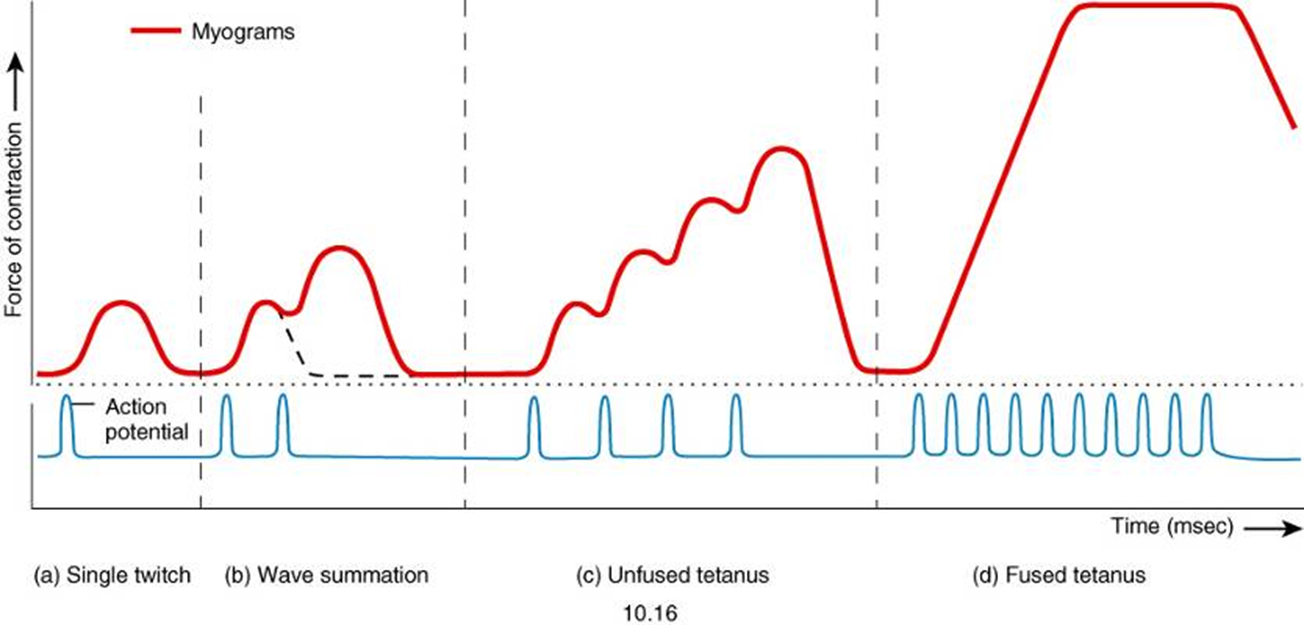
Action potentials and the myogram