Unit 4 - Cell Communication + Cell Cycle
Cell Cycle
Cell Division
Interphase: must occur in order for Mitosis to begin
G1 Phase: cell growth
S Phase: DNA replicated
G2 Phase: continue growth and prepare for Mitosis
If cells are not needed for mitosis, they rest in G0 Phase
Mitosis - process where two daughter cells (cells of same genetic material) are created
Prophase: nuclear envelope breaks down and DNA condenses into chromosomes
Pro-Metaphase: centrosomes begin forming the miotic spindle and attaching chromosomes to it
Metaphase: all chromosomes lined up along miotic spindle via kinetochores
Kinetochore - region on chromosome’s centromere where microtubules can attach to
Anaphase: spindle fibers pull towards opposite poles of the cell, separating the chromosome into 2 sister chromatids
Chromatids - the identical halve of a chromosome
Telophase: miotic spindle disappear, chromosomes decondense into DNA and 2 nuclear envelopes reform at opposite poles of the cell
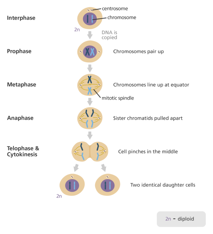
Cytokinesis: a step after Mitosis where cytoplasm and plasma membrane are split into two, fully forming the daughter cells
For plant cells, a cleavage furrow forms, followed by a cell wall to separate the cell into two daughter cells
Cell Cycle Regulation
3 major checkpoints
G1 Checkpoint - checks if cell is ready to undergo mitosis
Nutrients, space, size, growth signals
G2 Checkpoint - checks if DNA is duplicated correctly
M Checkpoint - checks if chromosomes are attached to spindle fibers and can separate
Occurs during metaphase
Growth factors also signal cells to undergo cell division
Paracrine, but can also be autocrine
Leave G0 phase, enter G1
Cell communication
Platelet-Derived Growth Factor (PDGF): protein released from platelets that stimulates cell growth, division, and migration by binding to PDGF receptors and triggering cells to enter S phase
Wound healing, blood vessel formation (angiogenesis), and tissue repair
Regulatory proteins/protein complexes conduct assessments and progression, including:
Kinases: enzymes that activate or inactivate other proteins through phosphorylation
Cyclins: regulator proteins specialized in each phase of cell division
G1 cyclin, G1/S cyclin, S cyclin, M cyclin
Unlike CDKs, they are not always present - instead, they accumulate
At low levels when not needed, peaks at stages where it is needed to initiate transition between phases
EXCEPT for G1, which is mostly high throughout
ex. S cyclin peaks between S phase and G2 phase, initiating the cell to enter G2 phase
Cyclin-Dependent Kinases (CDKs): inactive until it binds to a cyclin
Unlike cyclin, it is always present
A type of kinase
Phosphorylates target proteins specific to each cell division stage
Maturation-Promoting Factor (MPF): type of CDK that allows cell entry into Mitosis
Cyclin in MPF degrades after Mitosis, regulating when cell can perform it
Phosphorylates target proteins that drive events like nuclear envelope breakdown, spindle fiber formation, etc.
Cancer
Cancer - uncontrolled cell division; exhibit:
Immortality
Inducing angiogenesis
Angiogenesis - forming new blood vessels to give cancer cells nutrients
Resisting apoptosis
Mutated tumor-suppressor genes like p53
Sustained proliferative signaling
Continuous intake of growth factors/mutations to genes
Evading growth suppressors
Activating invasion and metastasis
Uncontrolled cell division can lead to both benign and malignant tumors
Benign tumors grow in one spot and don’t spread
Malignant tumors spread to other parts of the body (metastasize)
Metastasizing makes them hard to cure
Cancer is caused by mutations in certain genes:
Growth factor genes
ex. PDGF received despite not being ready for cell division
Receptor genes
Tumor-suppressor genes
Regulatory/stability genes
Genes for signaling pathway molecules
Tumor-Suppressor Genes prevent cancer by telling a cell to stop dividing
2 copies in every individual
Both copies must mutate in order to cause cancer
Some are born with 1 already mutated, making them more prone to cancer because only 1 more needs to mutate
That means cancer can be hereditary
Proto-Oncogenes code for proteins that stimulate cell growth + division
Normal
Oncogenes are the mutated form of Proto-Oncogenes that rapidly increases cell growth/division
Cancer-causing
Stability genes maintain healthy DNA and repair DNA
Responds in the Gene Instability Pathway to repair mistakes in DNA
If faulty, mutations occur more, which can lead to cancer or other diseases
p53: gene that responds to stresses on DNA replication and cell division by producing a crucial tumor suppressor protein, thus performing:
DNA repair (to prevent mutated genes from being inherited), apoptosis (for irreparable cells), halted cell division (senescence to allow time for DNA repair)
RAS proteins: crucial cell signaling products that activate the transcription factors causing cell division
Normally regulated
In cancerous cells, they are mutated, locking them “on” and causing uncontrolled cell growth/division
Normal cells usually silence their Telomerase genes, a gene that adds DNA to chromosomes to prevent them from shortening, thus allowing the cell to divide continuously
Cancer genes activate them, allowing for continuous cell division without the shortening of DNA → immortality
Cancer cells do not exhibit these because of mutations to their genes:
Density-dependent inhibition: if environment has too many cells in one area, the cell will not undergo cell division
Anchorage dependence: cell division can only occur if the cell is attached to a substratum (foundation)
Mutation to this gene is what causes malignant tumors
Mistakes in mitosis can also cause cancer since they lead to abnormal amounts/structures of DNA, ultimately promoting uncontrolled cell growth
BUT less likely to cause cancer than above factors (checkpoints can stop cells from dividing when they notice the mistake in mitosis)
Cell Communication
Cell communication requires:
Ligand - signaling molecule that binds to receptor
Receptors (two main types)
Intracellular receptors: receptor proteins inside the cell
Plasma Membrane receptors: receptors on the plasma membrane
Responses depend on the type of receptor (and thus, the type of ligand because ligands only bind to specific receptors based on their shape)
4 types:
Juxtacrine: two cells touch, either via plasma membranes, ligand presented by cell + receptor of another cell, or receptors on both cells
ex. Helper-T cell + Antigen Presenting Cell
Autocrine: a cell releases signal molecules to itself
ex. cancer cells release their own growth factors for uncontrolled proliferation
Paracrine: a cell releases signal molecules to another cell nearby
ex. Quorum sensing - when bacteria cells send each other signals so they can sense their population density and coordinate group behaviors
Endocrine: a cell releases signal molecules to another cell far away
Signal travels through blood vessels for animal cells and air or tissue for plant cells
ex. the pancreas releases insulin and glucagon (hormones) to regulate blood sugar levels
Responses are triggered via the Signal Transduction Pathway
Reception - a ligand binds to cell receptor
Binding of ligand causes the receptor to undergo conformational change, allowing it to alter its function and activate/block downstream pathways
G Protein-Coupled Receptors (GPCRs):
When a ligand binds to the GPCR, it triggers a GDP from a G Protein to be exchanged with GTP, activating the G Protein
The activated G Protein then activates an enzyme
Activated enzyme triggers transduction (series of responses that lead to the cellular response)
Process ends when GTP → GDP

Ligand-Gated Ion Channels:
Ion channel doesn’t open unless a ligand binds to it
Due to ligand, ions can flow into the cell and trigger cellular response
Ligand can detach and close channel
Receptor Tyrosine Kinases (RTKs):
When ligand binds to RTKs, the two receptors dimerize (come together and activate) and phosphorylate each other
Phosphorylated RTKs act as docking sites for other proteins to activate a cascade of responses
Lipid Hormonal Signaling - lipid hormones travel through the plasma membrane and bind to intracellular receptors
Because they are nonpolar and hydrophobic
Don’t rely on transduction/secondary messengers/ amplifying of signals
Transduction - the overall series of reactions that convert the initial signal from reception into an internal cellular response, often amplifying the signal through phosphorylation cascades
Important role of Second Messengers - intracellular non-protein and hydrophilic molecules that relay and amplify initial signal to target molecules
ex. cAMP, Ca2+
Activate protein kinases
On diagrams - smaller arrow → second messenger → larger arrow → target protein/kinase
Between kinases, never a protein
Kinases phosphorylate other molecules (often other kinases), activating or inhibiting them
Activated by second messengers
Relay and amplify signal
Phosphorylation cascades

Protein Phosphatases deactivate target proteins/kinases by removing their phosphate group
End transduction when initial signal is no longer present
Response - the cellular response resulting from transduction
In lipid hormonal signaling, altered gene expression is a common response
Growth factors trigger cell to grow/divide
Drugs in signal transduction:
Bind to a receptor (like a competitive inhibitor) to stop a ligand from initiating the targeted response
Destroy/disable ligands, enzymes, protein kinases, second messengers, anything involved with transduction
Lower amount of cAMP
Alter the receptor’s shape so it can’t bind to ligands
Can’t dimerize (for RTKs)