Liver Segmental Anatomy Notes
Liver Segmental Anatomy (Couinaud’s Anatomy)
Introduction
Traditionally, the liver is divided into left and right lobes using the falciform ligament. Couinaud’s anatomy divides the liver into eight functional units, each with its own blood supply (portal and hepatic) and biliary drainage.
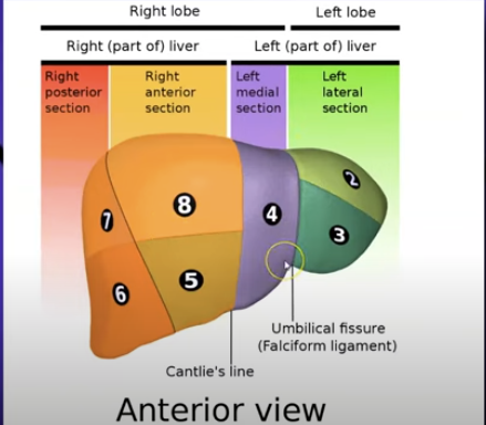
Cantile’s line contains the gallbladder and middle hepatic vein
right hepatic vein separates 6 and 7 from 5 and 8
used the hand pneumonic to make a fist
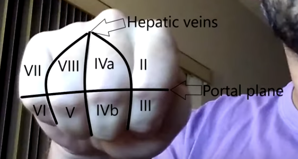
thumb - caudate lobe
knuckles separate liver in the horizontal plane where the portal vein lies that separates from superior and inferior
three lines = three hepatic veins
Each segment is independent, ensuring that damage to one segment does not affect the others. This is particularly important for surgical resections.
The Couinaud system is based on the branching of the portal triad (portal vein, hepatic artery, and bile duct).
Division
Left Hemi Liver:
Segments two, three, and four. These segments are supplied by the left hepatic artery and portal vein branch.
Right Hemi Liver:
Segments five, six, seven, and eight. These segments are supplied by the right hepatic artery and portal vein branch.
Falciform Ligament:
Separates segments two and three from four. It is a peritoneal fold that extends from the anterior abdominal wall to the liver.
Cantlie's Line:
An imaginary line that extends from the gallbladder fossa to the inferior vena cava (IVC). It contains the gallbladder and middle hepatic vein, separating segments four, five, and eight. This line is crucial for surgical planning.
Right Hepatic Vein:
Separates segments seven and six from eight and five. The hepatic veins drain directly into the IVC.
Hand Mnemonic
Use a fist to represent the anterior surface of the liver. This mnemonic helps visualize the spatial arrangement of the liver segments.
Thumb: Caudate lobe (segment one). The caudate lobe receives blood supply from both the right and left hepatic arteries and portal vein branches.
Knuckles: Horizontal plane where the portal vein lies, separating superior from inferior segments. This plane is important for distinguishing between the upper and lower segments on imaging.
Digit Lines: Represent hepatic veins.
Left hepatic vein: Drains segments two, three, and four.
Middle hepatic vein: Drains segments four, five, and eight.
Right hepatic vein: Drains segments six, seven, five, and eight.
Segment assignments:
Thumb: Caudate lobe.
Index finger: Segments two and three.
Middle finger: Segments four a and four b.
Ring finger: Segments five and six.
Pinky finger: Segments seven and eight.
Segment Summary
Falciform ligament separates segments two and three from segment four. This is a key landmark for dividing the left lobe.
Middle hepatic vein separates segments: superior (four a and eight), inferior (four b and five). It serves as an important surgical landmark.
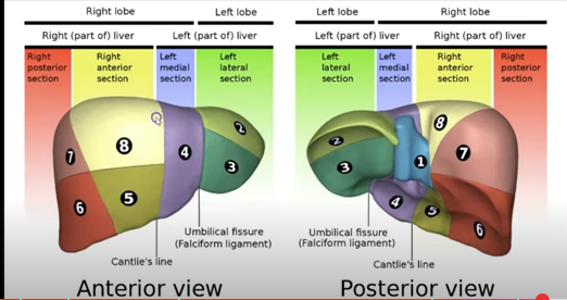
Lateral segments: left lateral (segments two and three), left medial (segment four), right anterior (segments five and eight), right posterior (segments six and seven).
Ultrasound Views
Sagittal
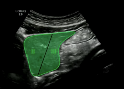
Probe placement: The probe is typically placed in the intercostal spaces to obtain sagittal views of the liver.
Aorta: Superior segment two, inferior segment three. The aorta is a useful landmark for identifying these segments.
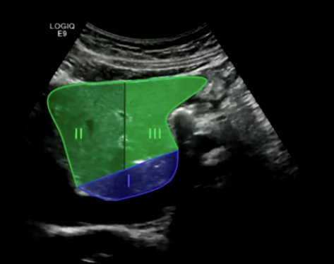
IVC: Segment one (caudate lobe), segment three, and segment two. The IVC is located posterior to the liver.
Gallbladder: Segment four. The gallbladder is located in the gallbladder fossa of segment four.
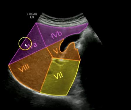
Portal vein plane: Segments four b, five, eight, and seven. This plane is crucial for visualizing the portal vein branches.
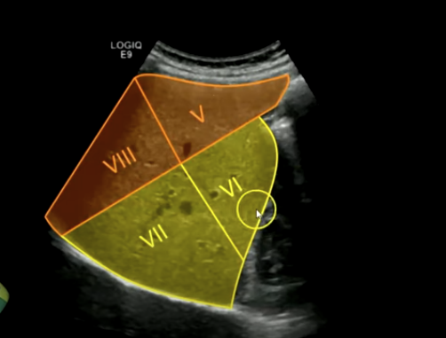
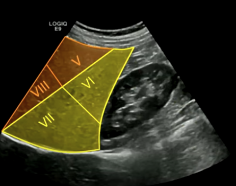
Kidney (lateral view): Segments five, eight, six, and seven. The right kidney is located inferior and lateral to the right lobe of the liver.
Kidney (far lateral): Segments five, six, seven, and eight.
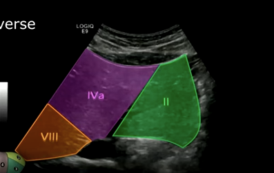
Transverse
Superior segments of the liver: Segment two, segment four a, and segment eight.
Inferior to superior angling (angling upward): The portal vein branch feeding segment three, feeding segment two, the caudate lobe segment one, segment four b, and segment five.
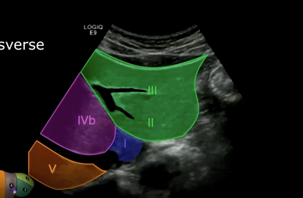
Inferior segments (close to transducer): Segments three and four b.
Posterior segments: Segments two and five and the caudate lobe.
Pancreas (angling upwards): Segments three, caudate lobe, segment five, and segment four b. The pancreas is located posterior and inferior to the liver.
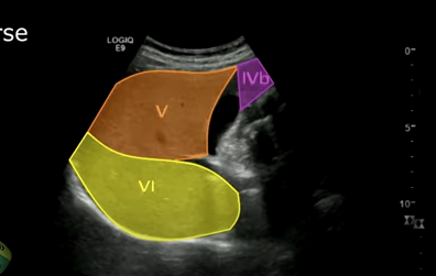
Right lobe (transverse): Gallbladder, segments four b, five, and six.
Superior segment of the right lobe (transverse): Hepatic veins, segments four a, eight, and seven.
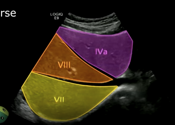
Middle hepatic vein separates four a from eight.
Right hepatic vein separates eight from segment seven.

Quick Recap
Ultrasound: divides the liver into left lobe and right lobe.
Falciform ligament: left lobe, right lobe.
Couinaud’s anatomy: divides into left hemi liver and the right hemi liver.
Cantlie's line (middle hepatic vein, main lower fissure, and gallbladder) separates right from left hemi liver.
Posterior View
Caudate lobe (segment one).
Conclusion
Couinaud’s anatomy is beneficial for surgical applications and improving anatomical knowledge for sonographers. It provides a standardized approach for describing liver anatomy.
Useful for CT, MRIs, and radiologists in mass and tumor localizations. This detailed knowledge aids in accurate diagnosis and treatment planning.