Class 5 Oocyte Meiosis and Meiotic Arrest Factors
Anatomy and Structure of the Oocyte and Follicle
The GV Oocyte Structure:
Germinal Vesicle (GV): The distinct nucleus of the immature oocyte.
Nucleolus: A prominent structure within the GV.
Cytoplasm: The internal cellular matrix of the oocyte.
Plasma Membrane: The outer cellular boundary.
Zona pellucida (ZP): A glycoprotein layer surrounding the plasma membrane of the oocyte.
Cumulus oophorus: A cluster of cells (granulosa cells) that surround the oocyte both in the ovarian follicle and after ovulation.
Corona radiata: The innermost layer of the cumulus cells, directly adjacent to the zona pellucida.
The basement membrane, the granulosa, is peripheral to granulosa cells that developed around the oocyte , providing structural support and separating the follicle from the surrounding stroma of the ovary. There are two levels of the theca, responsible for the secretion of hormonal support of the egg.
Follicular Organization:
Oocyte: The central gamete.
Granulosa Cells: Cells surrounding the oocyte that provide support and produce hormones.
Theca Cells: The outer layer of the follicle that provides structural integrity and produces androgen precursors.
Cumulus-Oocyte Complex (COC): The functional unit comprising the oocyte and its surrounding cumulus cells.
Oogenesis and Meiotic Progression
The Meiotic Timeline:
Prenatal Period: Primordial germ cells migrate to the developing ovary. Following a period of proliferation, they enter Meiosis I.
At Birth: The ovary contains numerous primordial-stage follicles. These consist of small, non-growing oocytes surrounded by a single layer of granulosa cells.
Developmental Progression: Follicles develop step-wise to become preovulatory, Graafian-stage follicles containing a fully grown oocyte.
First Meiotic Division (Prophase I):
Stages of Prophase I:
Leptotene
Zygotene
Pachytene
Diplotene
Dictyate Stage: A prolonged resting phase where the oocyte remains arrested until ovulation (in the adult ovary). The chromosomes remain condensed and undergo genetic recombination later on. The germinal vesicle develops around it and all eggs born with the female remains in the primary oocyte stage until puberty, at which point hormonal signaling initiates their maturation process.
Resumption of Meiosis (Puberty):
Triggered by the mid-cycle surge of gonadotropins.
Diakinesis: The final stage of Prophase I.
Germinal Vesicle Breakdown (GVBD): The dissolution of the oocyte nucleus.
Metaphase I (MI): Chromosomes align at the metaphase plate.
Anaphase I: Homologous chromosomes separate.
Telophase I: The first polar body is extruded. In contrast to regular cytokinesis, the formation of the polar body during oogenesis is asymmetrical, resulting in one larger oocyte and a smaller polar body that typically degenerates.
Polar bodies get their name because they surround the pole of the chromosome
Second Meiotic Division: (Ovulation)
Metaphase II (MII): The oocyte enters the second meiotic division but arrests again at Metaphase II (the ovulated state).
Fertilization: Meiosis II is only completed if fertilization occurs.
Anaphase II and Telophase II: Following sperm entry, the second polar body is extruded.
Pronuclear Egg: The final zygote stage with two pronuclei.
Oocyte Maturation and Meiotic Arrest
Maturation Definition: Oocyte maturation is defined as the progression from the prophase of Meiosis I (GV stage) to Metaphase II (MII).
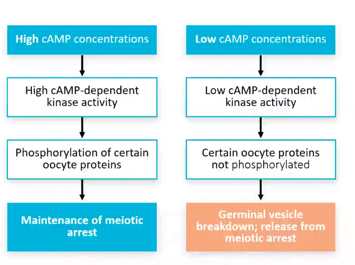
The Role of cAMP in Meiotic Arrest:
High Concentrations: Correlated with high -dependent kinase (Protein Kinase A) activity. This leads to the phosphorylation of specific oocyte proteins that maintain meiotic arrest at the GV stage.
Low Concentrations: Results in low kinase activity. Certain oocyte proteins remain unphosphorylated, leading to Germinal Vesicle Breakdown (GVBD) and release from meiotic arrest.
Key Molecules:
Adenylate Cyclase: Synthesizes from .
Phosphodiesterase (PDE): Breaks down to .
Milrinone: A specific phosphodiesterase inhibitor.
Historical Context:
Pincus & Enzmann (1930s): Asked the question first about meotic arrest in the MI phase: Conducted early experiments on egg activation and parthenogenesis in rabbits. Their results showed that the germinal vesicle and internal enviroment itself keeps the oocyte from developing further.
Pincus (1950s): A key figure in the creation of the birth control pill.
Spontaneous Maturation (Pincus and Enzmann) : Maturation of the ovum can be achieved simply by isolating it from the inhibitory follicular environment (J Exp Med, 1935).
Maturation Promoting Factor (MPF)
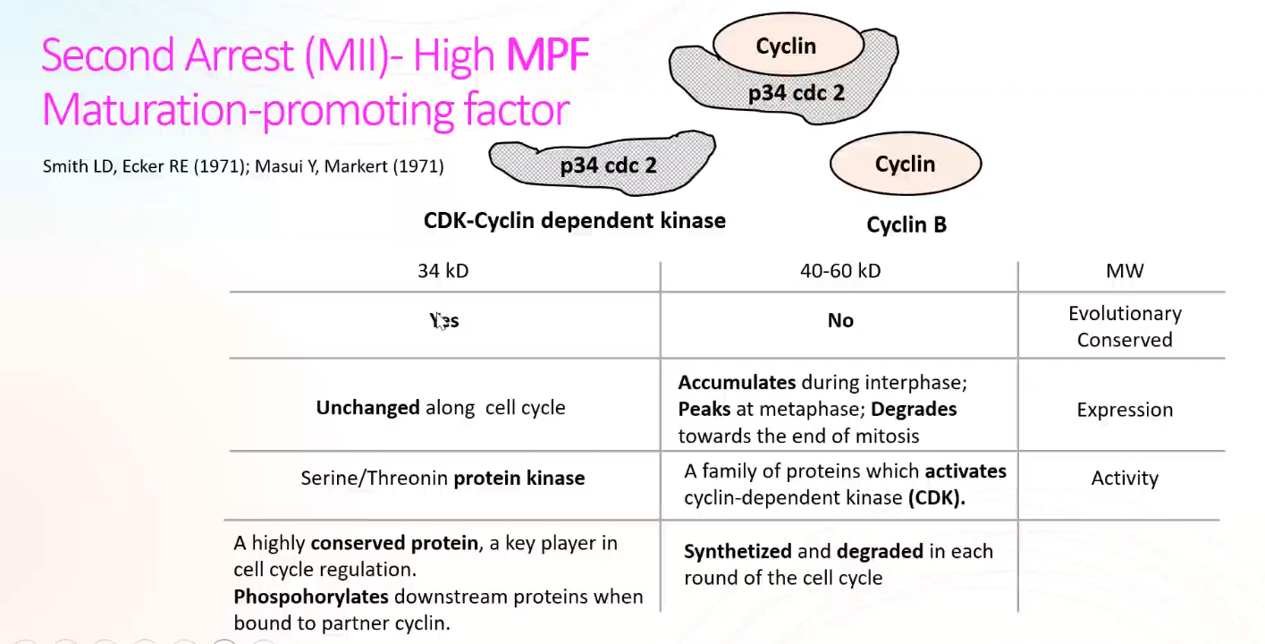
Molecular Composition: MPF is a heterodimer in most cells consisting of:
(CDK - Cyclin Dependent Kinase):
Molecular Weight: Approximately .
Function: Serine/Threonine protein kinase.
Characteristic: Evolurionarily conserved; remains unchanged in concentration throughout the cell cycle.
Activation: Phosphorylates downstream proteins when bound to a partner cyclin.
Cyclin B:
Molecular Weight: .
Function: A family of proteins that activates CDK.
Characteristic: Synthesized and degraded in each round of the cell cycle. Accumulates during interphase, peaks at metaphase, and degrades toward the end of mitosis.
MPF Regulation Cycle:
forms complexes with Cyclin B during S and G2 phases.
Activating Phosphorylation: Occurs on Threonine-161.
Inhibitory Phosphorylation: Occurs on Tyrosine-15 (and Threonine-14 in vertebrates).
G2 to M Transition: Dephosphorylation of Threonine-14 and Tyrosine-15 activates MPF.
Exit from Mitosis: MPF activity is terminated by the proteolytic degradation of Cyclin B.
Cytostatic Factor (CSF) causes Metaphase II Arrest
The Phenomenon: MII oocytes possess a specific activity that maintains arrest at the M phase, preventing cleavage without fertilization.
Identification (Masui and Markert, 1971): Identified a "cytostatic factor" in the cytoplasm that causes the arrest of mitosis and cleavage.
CSF MOA:
Appears during oocyte maturation, reaching its highest level at MII.
Must cause metaphase arrest when microinjected into embryonic cells (blastomeres).
Must be inactivated at fertilization upon egg activation.
The Molecular Identity of CSF:
Mos (c-mos): A MAPK kinase kinase that activates the MAPK pathway.
Experimental Evidence:
knock-out mice are subfertile because oocytes fail to arrest at MII and progress to Metaphase III (MIII).
mRNA induces metaphase arrest in blastomeres.
Anti-sense RNA against prevents oocytes from arresting at MII.
Conclusion was that CSF contains c-mos but not solely composed of it
Complex Nature: CSF is not a single molecule but a regulatory module consisting of Mos-MAPK-Emi2.
Mechanism of Action: This module inhibits the Anaphase-Promoting Complex/Cyclosome (APC/C) and sustains high MPF activity, thereby maintaining MII arrest.
Summary: Experimental Cytoplasmic Injections
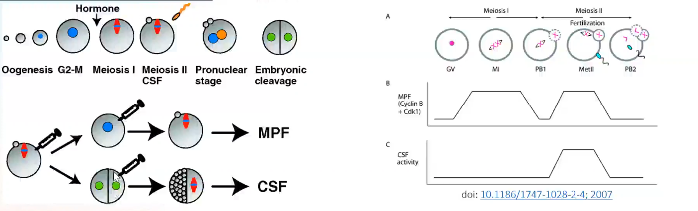
Methodology: Small amounts of cytoplasm from mature oocytes are injected into either immature oocytes or one blastomere of a two-cell frog embryo.
Results:
Immature Oocytes: Resume meiosis (due to MPF activity).
Blastomeres: Injected blastomeres arrest in metaphase (due to CSF activity).
Conclusion: The cytoplasm of mature oocytes contains two distinct biochemical activities (MPF and CSF) that regulate the maturation and arrest process.