Hemodynamic Monitoring
Hemodynamic Monitoring
PACU
Monitoring of blood flow & pressures within the body
Balance between blood flow, resistance & pressures within the body
Focuses on circulation, perfusion & tissue oxygenation
Hemodynamic Monitoring: Key Terms
Stroke Volume (SV)- amount of blood leaving the left ventricle (LV) during each contraction. Normal: 60-100 mL/beat
Cardiac Output (CO)- amount of blood leaving the LV per minute. (HR x SV) Normal: 4-6 L/min. Essential for adequate tissue oxygenation
Cardiac Index (CI)- is the CO/ sq, meter of body surface area. More accurate than CO. Normal 2.5-3 L L/min.
Ejection Fraction (EF)- % of blood ejected from the ventricles with each beat. Normal: 55-70%
Contractility- force of contraction. Frank-Starling law: bigger the stretch = bigger the contraction
Preload- volume of blood in ventricles at the end of diastole
Afterload- the resistance/pressure the ventricle must overcome to eject blood during systole
Importance of Hemodynamic Monitoring
Aids in diagnosis
Assists in guiding therapies & treatments
Evaluates patient response to current therapies & treatments
Indications:
asses heart function, fluid balance, effects of drugs that affect cardiac output
Monitoring perfusion, especially in situations with unstable BP
Types: non-invasive & invasive
Non-invasive Monitoring
Continuous blood pressure (dynamap), pulse ox (continuous SpO2/noninvasive arterial oxygen monitoring, capillary refill, pulses (arterial & peripheral)
Transesophageal ultrasound (TEE)
Invasive Hemodynamic Monitoring
Invasive methods include: arterial lines, jugular central lines, pulmonary artery catheters (AKA Swan Ganz) and Pressure bag & transducer
Pressure bag & transducer
NSS bag, pressure device, tubing w/ transducer, pole mount
Transducer at Phlebostatic Axis
Zeroing & flushing
Arterial lines (A-lines)
Continuous BP monitoring
Via artery: usually radical
Allen’s test
Blood access: labs & ABGs
Indications: hypotension, hypertension, resp. failure, shock, during surgery admin of vasoactive drugs
Risks: hemorrhage, infection, thrombus, NV impairment
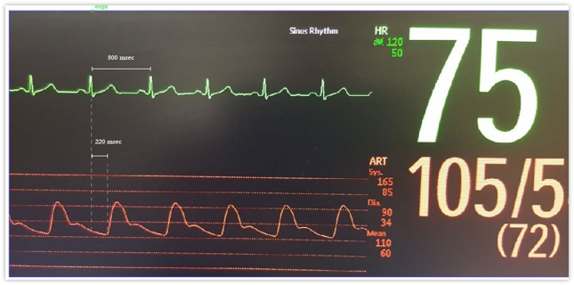
look at pics
A-lines Waveform
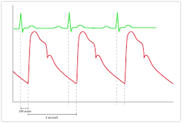
EKG Aline
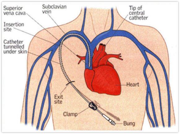
Subclavian Central Venous Catheter (CVC)
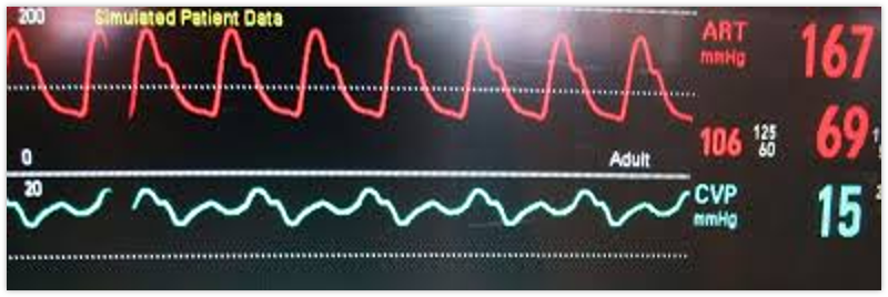
Central Venous Pressure (CVP)
Central line cath
Triple lumen: can infuse blood, IV fluids & meds
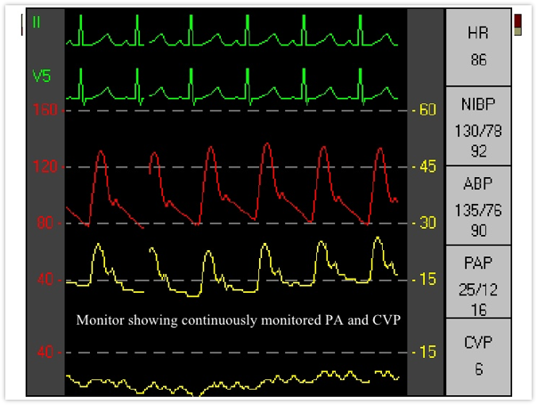
Measure CVP/right atrium pressure
Normal 2-6 mmHg
Low readings indicate hypovolemia
High readings indicate fluid overload
Possible complications: infection (CLABSI). pneumothorax, air emboli, bleeding
Aline & CVP Waveforms
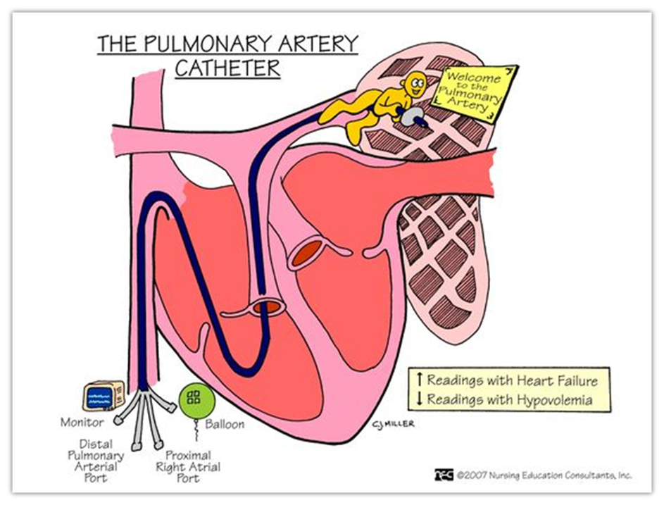
Pulmonary Artery Catheter/Swan Ganz
Used for complicated cardiac, pulmonary intravascular volume problems
Ports for fluids, withdrawal of blood for labs, to measure heart pressures & cardiac output & monitor core temp
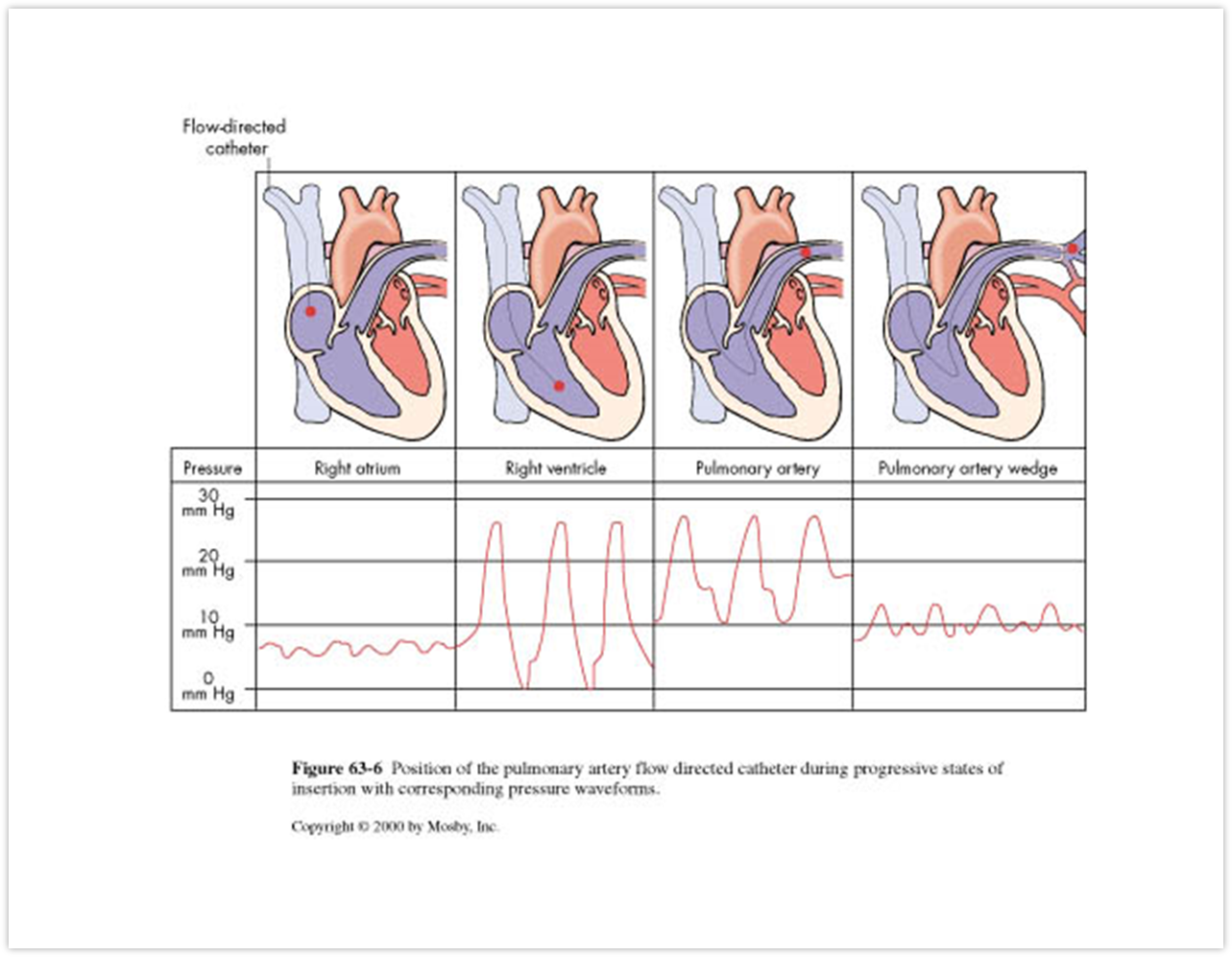
Pulmonary capillary wedge pressure (PCWP) Normal 6-12 mmHg
PCWP increased with fluid overload & HF
PCWP-
balloon inflated & floated through the heart, it wedges into the pulm artery & blood flow is temp. stopped
measures the pressures in front of the balloon, the left side of the heart
Increased pressures can indicate: LV failure, ischemia, pericarditis, mitral valve disease, fluid overload
Decreased pressures can indicate: hypovolemia, shock, vasodilating drugs
Nursing Management in Hemodynamic Monitoring
Assess patient baseline and as needed
General appearance, LOC, skin color/temp, vitals, resp status, peripheral circulation, urine output, changes in pt condition and/or numbers
Monitor & compare invasive measures with non-invasive
Monitor sites of invasive lines for:
s/s infection, dressings/drainage, catheter placement, use of lines (measuring/capped/IVF), changes in readings
Reminder
that while advanced monitoring is great technology, you must still look at your patient!