Major Vessels
Pulmonary/Systemic Circuits
pulmonary circuit
pulmonary trunk (from right ventricle) → pulmonary arteries → lungs
blood deoxygenated
gas exchange occurs within lungs
pulmonary veins to left atrium of heart
blood oxygenated
systematic circuit
blood leaves left ventricle → aorta (ascending/descending)
blood enters right atrium ← vena cava (SVC/IVC)
Arterial Receptors
sensory structures in walls of major aa.
monitor BP and blood chemistry
transmit informaton to the brainstem
regulates HR, BV diameter, and respiration
baroreceptors in carotid and aoritc sinuses
monitor BP from iinternal carotid aa. and aorta
transmit via glossopharyngeal and vagus nn.
baroflex - keeps BP steady by rapidly adjusting cardiac output to match arterial BP
chemoreceptors
carotid bodies
monitor blood chemistry
transmit signals via glossopharynealgeal nerve
adjust respiratory rate to stabilize pH, CO2, and O2
aoritc bodies
similar to carotid bodies
innervated by vagus nerve
Arteries
major aa. include:
aorta and its branches
arch, thoracic aorta, and abdominal aorta
head/neck aa.
circle of willis
appendicular aa.
Aorta (& Branches)
aortic arch
branchiocephalic trunk
right common carotid a. (right of head)
righ subclavian a. (right shoulder/UL)
left common carotid a. (left of head)
left subclavian a. (left shoulder/UL)
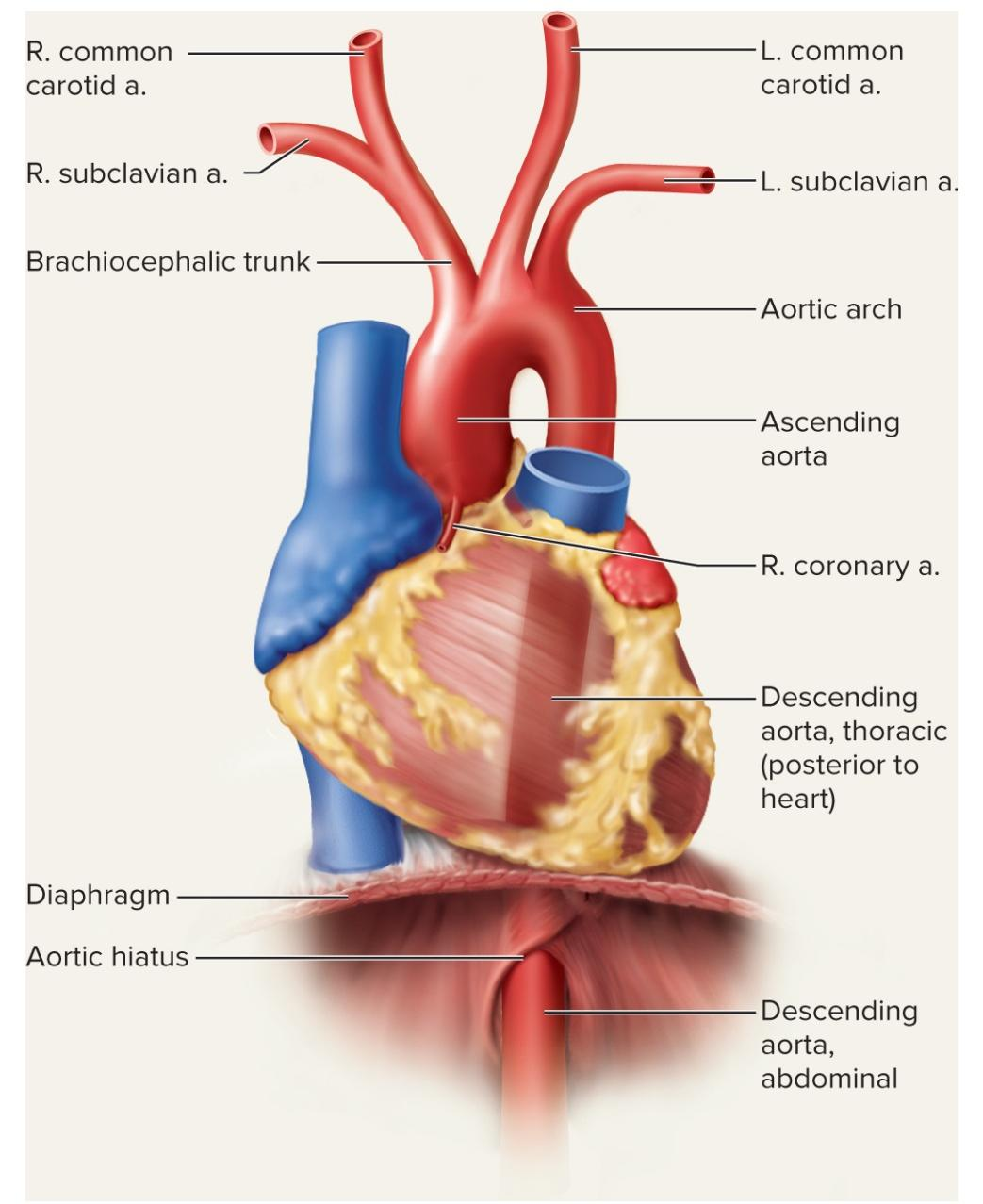
ascending aorta
right/left coroary aa.
descending aorta
thoracic aorta and abdominal aorta
arteries associated with esophagus, lungs, liver, spleen, kidneys, intestines, testes/ovaries, and various muscles
Head and Neck
common carotids
internal carotid a.
external carotid a.
vertebral aa.
from subclavian aa.
come together to form Basilar a.
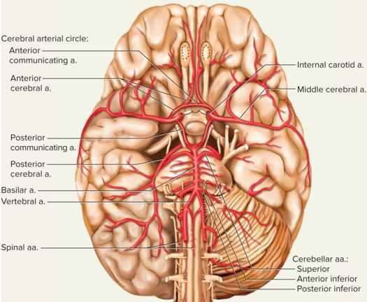
circle of willis
Circle of willis
internl carotid aa.
vertebral aa.
basilar a.
cerebellar aa.
pontine aa.
cerebral aa.
communicating aa.
Thorax and Upper Limb
subclavian aa.
axillary aa.
brachial aa.
radial aa.
deep/superficial palmar arches
ulnar aa.
interosseous aa.
deep/superficial palmar arches
Abdominopelvic
celiac trunk
common hepatic a.
splenic a.
left gastric a.
superior mesenteric a.
renal aa.
gondal aa.
ovarian/testicular aa.
inferior mesenteric a.
common iliac aa.
internal iliac a.
Lower Limb
common iliac aa.
internal iliac aa.
external iliac aa. → femoral aa.
deep femoral
popliteal aa.
posterior tibial aa.
fibular aa.
anterior tibial aa.
dorsal pedal aa.
Venous Sinuses
some veins expand to form venous sinuses
thin walls, large lumens, and no smooth muscle
not capable of vasomotor responses
e.g.dural venous sinus and coronary sinus
Major Veins
superior vena cava
jugular, vertebral, axillary vv. drain into the subclavin vv., which then drain into the brachiocephalic vv. → SVC
inferior vena cava
iliac, hepatic, renal, and gonadal vv. drain here
Head and Neck
dural sinuses
internal jugular vv.
exxternal jugular vv.
Dural Sinuses
drain blood from brain into intenal jugular vv.
also drain CSF via arachnoid granulations
5 unpaired sinuses: superior sagittal, inferior sagittal, straight, occipital, and intercavernous
confluence of the sinuses
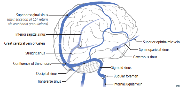
5 paired sinuses: transverse, sigmoid, cavernous, superior petrosal, and inferior petrosal
confluence of the sinuses
Thorax
blood from tissues and organs in the thorax drained by the intercostal veins and azygos vein
azygos v. formed by right lumbar v. and right subcostal v.
azygos vein system drains to the SVC
hemiazygos and accessory hemiazygos vv. drain into azygos v.
Upper Limbs
deep and superficial veins
hand vv. drain into forearm
deep/superficial venous palmar arches/network
radial, ulnar, cephalic, and basilic
forearm drains to arm
basilic, radial, and ulnar vv. drain to the axillary vv.
median cubital vv.
between cephalic and basilic
arm to subclavian vv. →brachiocephalic vv. → SVC
Abdomen
veins drain into the IVC
internal iliac and external iliac vv.
drain into the common iliac vv.
gonadal vv.
ovarian vv.
testicular vv.
renal vv.
hepatic vv.
phrenic vv.
Lower Limbs
foot drains to crural region → drains to leg
dorsal vv. superficial and drain to small saphenous vv. → great saphenous vv.
great saphenous vv. drains to femoral vv.
plantar vv. deep and drain to fibular, anterior tibial, and posterior tibial vv.
popliteal vv. → femoral vv.
femoral vv. drain into the external iliac vv. → IVC
FUN VESSEL FACTS
if all vessles from one body were laid end-to-end, they would stretch over 60,000 miles
could circle the earth twice (80% from capillaries)
veins appear blue through skin due to light reflection, but venous blood is actually dark red
Galen (2nd century) was the first to describe circulation
thrught venous blood was made in the liver and that there were 2 one-way systems for blood
theory held until william henry corrected in the 1600s
superficial veins play a role in thermoregulation
dilate to release heat; constrict to conserve heat
great saphenous vein = longest in body