CN I + CN XI + Cervical plexus
Learning objectives
Overview of the brain
Describe somatomotor and somatosensory pathways in the brain
Define the nerve components on CN I, XI, and the cervical plexus
Describe the pathways of each of these nerves
Describe the functions associated with each of these nerves
Describe select clinical correlations associated with each nerve + testing in clinic
Overview of the Brain
Gyri = folds
Postcentral gyrus: Sensory area
Precentral gyrus: Motor area
Sulci = grooves
Central sulcus: Divides frontal and parietal lobes
Lateral sulcus: Divides parietal and temporal lobes
Parietod-occipital sulcus: Divides parietal and occipital lobes
Fissures = clefts
Structures within the diencephalon (center area of the brain)
Epithalamus: Emotion, sleep wake cycles, mood
Thalamus: Relay point for sensory and motor info (somatic signals)
Hypothalamus: Coordination center for homeostasis (autonomic signals)
Cereborospinal Fluid Circulation
Function of CSF: Buffer zone to protect brain while the head moves
Secreted by epithelial cells in choroid plexus in ventricles of the brain
Circulates through ventricles and into subarachnoid space around spinal cord
Total volume = 90-150 ml
400-500 ml produced per day → Excess and used CSF absorbed into venous system
Hydrocephalus: Increased CSF volume leading to increased intercranial pressure
If sutures haven’t fused (infants) → large head (emergency)
Arachnoid Granulations
Protrusions of arachnoid mater and subarachnoid space into superior sagittal sinus or plexus around it, including lateral lakes.
In humans, not covered by dura
Function: Transport CSF into venous sinus system
Regulate CSF volume to prevent increased intercranial pressure
Newborn distribution: Microscopic (need less CSF because brains are smaller and they have other means to remove CSF)
Increase in size and distribution → widespread by 3 yrs old
Adult distribution: Concentrated around sagittal sinus
Skull Fracture and CSF Leak
Skull fractures, especially in the frontal nasal area, can cause CSF to leak (clear and watery)
Emergency situation because don’t want bacterial exposure
Clinical presentation: CSG rhinorrhea
Requires defect in dura and pathway from nasal sinus into nasal cavity
Cribiform plate → Nasal cavity
Somatomotor and Somatosensory Pathways to Brain
Skeletal motor signals (from brain) begin in primary motor cortex (precentral gyrus)
Somatosensory signals (to brain) are processed in the primary somatosensory cortex (postcentral gyrus)
Both somatomotor and somatosensory signals relay through the thalamus
Homunculus (motor or sensory): A map of corresponding regions in each gyrus distorted by concentration
Somatomotor
Upper motor neurons (UMN): Integrates all excitatory and inhibitory signals from cortex → translates to a movement signal.
3 tracts of somatomotor signal
Anterior corticospinal tract: Voluntary mvmt of trunk muscles
Lateral cortocospinal tract: Voluntary mvmt of limb muscles
Corticonuclear fibers: Motor signals to cranial nerves
Lower motor neurons (LMN): Transmits signal to muscle
3 types of LMN (general breakdown = somatomotor and visceromotor)
Somatic motor neurons
Special visceral efferent brachial motor neurons
General visceral motor neurons
Axons of UMN relay through thalamus → spinal cord → synapse directly onto LMN → Signal transmitted to muscle
Somatosensory
Info gets conveyed up to the brain from primary sensory neurons in peripheral ganglia (outside CNS) → through thalamus → into post-central gyrus
Ex/ Pathway when you touch something with your hands
Sensory ending in skin → Splanchnic n. → Sympathetic trunk → White communicating ramus → Ventral ramus → Spinal n. → DRG → Dorsal horn → Peripheral ganglia → Thalamus → Post-central gyrus
Hypothalamus
Control autonomic signals → Communicate with spinal cord through hypothalamospinal tract
Origin symp signals: Lateral horns
Cranial Nerve I: Olfactory Nerve
Fiber components: Special sensory (smell)/Special visceral afferent only
Transmit smell to brain from receptors in olfactory epithelium
No peripheral ganglia
Pathway: Cribiform plate (of ethmoid bone) → Brain
Unmyelinated processes of the olfactory cells (olfactory axon) pass through the cribiform plate in bundles (to olfactory bulb)
Division within the brain:
Lateral olfactory stria: Primary olfactory cortex
Medial olfactory stria: Autonomic response to olfaction (ex. salivation)
Distribution of nerves: Lateral wall and midline (cover superior section)
Pathology/Disease
Anosmia: Inability to detect smell
Hyposmnia: Reduced ability to smell
Smell distortion: Change in smell
Lesions or tumors, trauma to the cribiform plates (sever olfactory tracts), distortion can be caused by migraine or epilepsy
Affect processing
Testing: Strong stimulus presented to one nostril at a time and patient asked to sniff
Stimulus must be adjacent to nostril
Cranial Nerve XI: Accessory Nerve - Trapezius and SCM
Fiber components: Somatomotor ONLY
Carries axons of LMN within the ventral horns of C1-C5
Pathway: Ventral horn of C1-C5 → Spinal root → Through foramen magnum → Jugular foramen → Muscle
Accessory n. formed by connections with dorsal root
Testing:
SCM - Place hand on side of face and ask patient to rotate against hand (test both sides)
Trapezius - Place hand on shoulders and ask patient to shrug (push up against hands)
Sternocleidomastoid (SCM) - Within investing layer
Originate: Mastoid process and lateral superior nuchal line
Attachment: Manubrium of sternum and medial 1/3 of clavicle
Innervation: CN XI
Function:
Unilateral - mediolateral head rotation
Bilateral (posterior fibers) - head extension
Bilateral (anterior fibers) - head flexion
Trapezius - Within Investing layer
Origin: Median nuchal line, spinous process
Attachment: Scapula and clavicle (ends)
Innervation: CN XI
Function:
Upper fibers:
Bilateral - head extension
Unilateral - lateral head flexion
Elevate and rotate scapula
Middle fibers - retract and rotate scapula
Lower fibers - depress and rotate scapula
Cervical plexus
Plexus: Mesh of intersecting nerves
4 main plexuses from spinal nerves (somatoplexus): Cervical, brachial, lumbosacral, coccygeal
Autonomic plexuses: celiac plexus-organs, myenteric plexus-GI tract, submucosal plexus-intestinal mucosa, pharyngeal plexus of CN X-palate, pharynx, cardiac plexus-heart
Cervical plexus origin: Arises with contributions from C1-C5 ventral rami
C5-T1 also contribute to brachial plexus (SM to anterior and middle scalene + levator scapulae)
Fiber components: Somatosensory and somatomotor fibers
Somatosensory branches: lesser occipital, greater auricular, transverse cervical, supraclavicular, phrenic
Somatomotor branches: ansa cervicalis, nerve to thyroid, phrenic
Branches of cervical plexus
To meninges - C1 (sensory)
Lesser occipital n. - C2 (sensory)
Greater auricular n. - C2-3 (sensory)
Transverse cervical n. - C2-3 (sensory)
Supraclavicular n. - C3-4 (sensory)
Phrenic n. - C3-5 (mixed)
Inferior root of ansa cervicalis - C2-3 (motor)
Superior root of ansa cervicalis - C1 (motor)
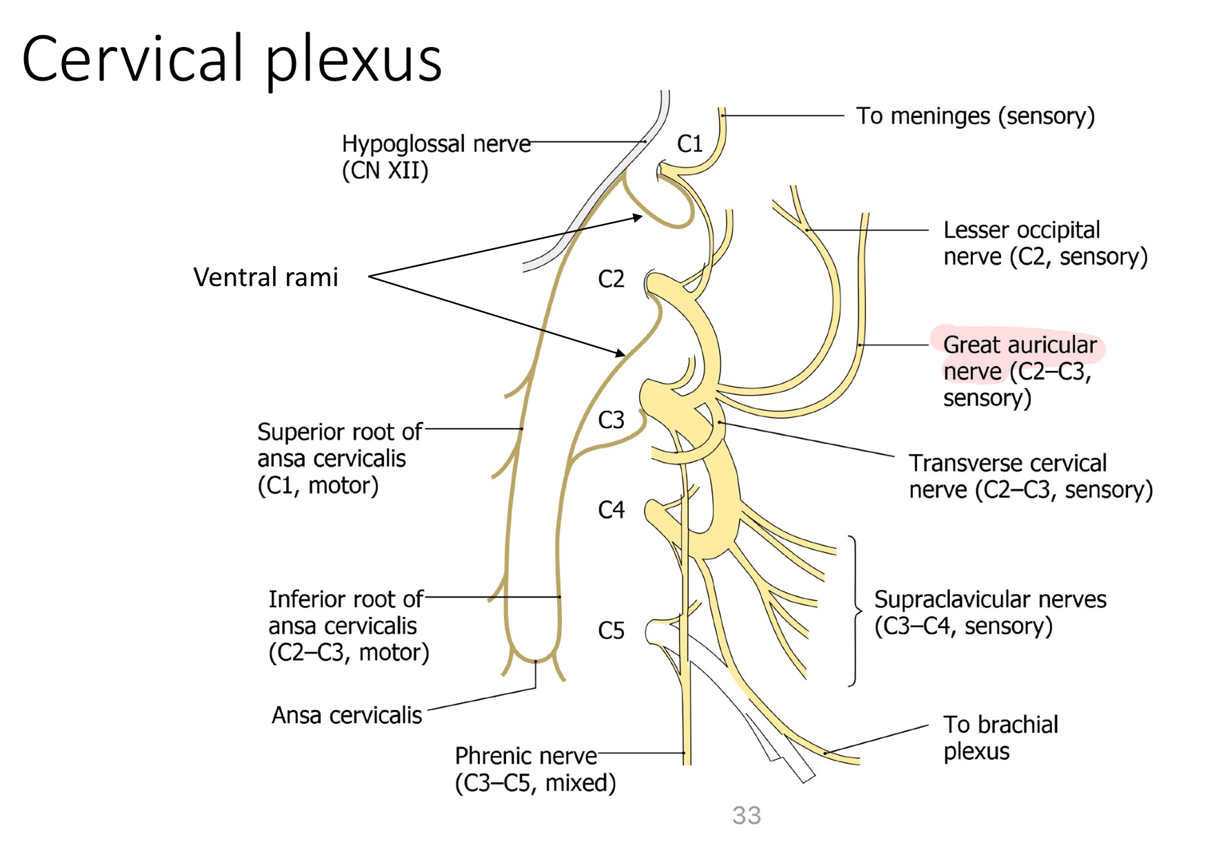
Ansa cervicalis (superior root + inferior root)
Innervation: Somatomotor to infrahyoid muscles (except thyroyoid)
Motor portion of the cervical plexus
Superior root: hitchhiking or relaying of the cranial nerves - axons that don’t originate on a CN but join the CN for a short period of time to get to a target
C1 fibers hitchhike on hypoglossal to reach thyrohyoid
Nerves for somatosensation in the neck
Lesser occipital n. → SS to back of head
Most posterior
Transverse cervical n. → SS to anterior neck
Anterior neck
Supraclavicular n. → SS to superior clavicle (inferior neck)
Inferior
Greater auricular n. → SS around ear and angle of mandible
Ear/gonion
Puncta nervosa (Erb’s point): Location for anesthetic-midpoint on the posterior side of SCM muscle
Phrenic nerve
C3-5 “keeps the diaphragm alive”
SS and SM to diaphragm
Location: On top of anterior scalene
Referred Pain - Viscerosensory pain
Pain is perceived in a location distant from the actual site of injury or pathology
Follows sympathetic pathway except for 1 exception (signal goes to dorsal horn)
Examples:
Angina (heart pain): sensation to left upper limb
Signal mixed around T1-2 area
Viscerosensory