Nervous System (7 & 8)
LO1: Describe the structure and function of neurons and glial cells of the CNS and PNS, and the functional organisation of these cells in the nervous system
LO2: Describe the anatomy of the meninges and their role in protecting the CNS
LO3: Describe the anatomy of the blood-brain barrier and its role in protecting the CNS
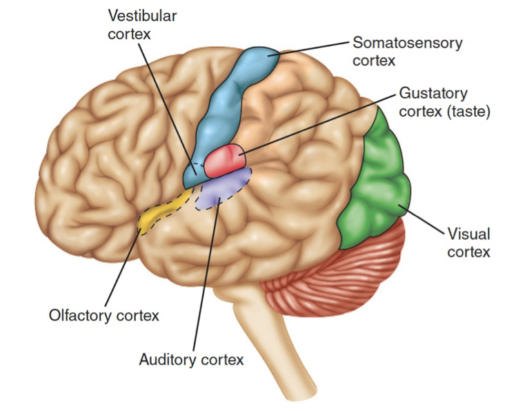
LO4: Describe the major anatomical regions and function of the cerebral hemispheres, cerebellum and brainstem
LO5: Describe the major anatomical regions and function of the spinal cord and spinal nerves
LO6: Describe the subdivisions of the peripheral nervous system and their function
LO7: Describe the features of the neuron anatomy that relate to the generation and propagation of an action potential
LO8: Describe how cells communicate and how this relates to the structure and function of a chemical synapses
LO9: Understand the nature and role of neurotransmitters in neural communication and how drugs can be used to mimic or block neurotransmitter function
The Structure of the Nervous System
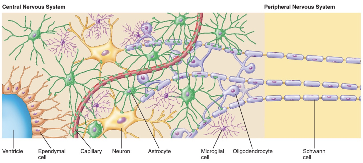
Cells in the Nervous System
Neuroglia are non-neuronal cells that support the functions of neuron’s
Astrocytes: Transport nutrients such as glucose to neuron’s
Oligodendrocytes: Produce myelin in the CNS
Schwann cells: Produce myelin in the PNS
Microglia: Immune cells of the CNS because they phagocytose debris after injury or pathogens to prevent infection.
Ependymal cells: Line ventricles and central canal of spinal cord to produce CSF to cushion and provide the brain with nutrients.
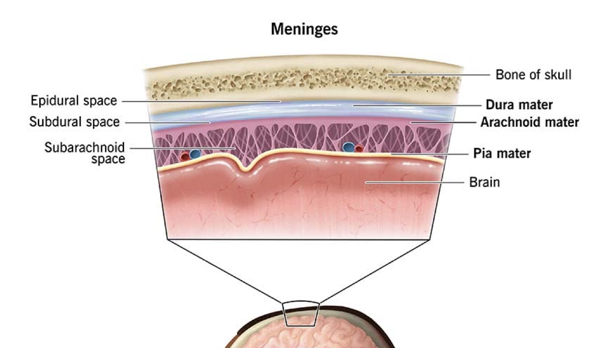
Meninges
First layer of protection is provided by the skull and vertebral column
Beneath the bone there are three layers of connective tissue containing CSF
Dura matter: Outer, tough, fibrous connective tissue attached to the cranium.
Arachnoid matter: Middle layer, attached to dura matter with a space between it and the pia matter called the subarachnoid space for CSF.
Pia matter: Inner thin layer of connective tissue attached to brain.
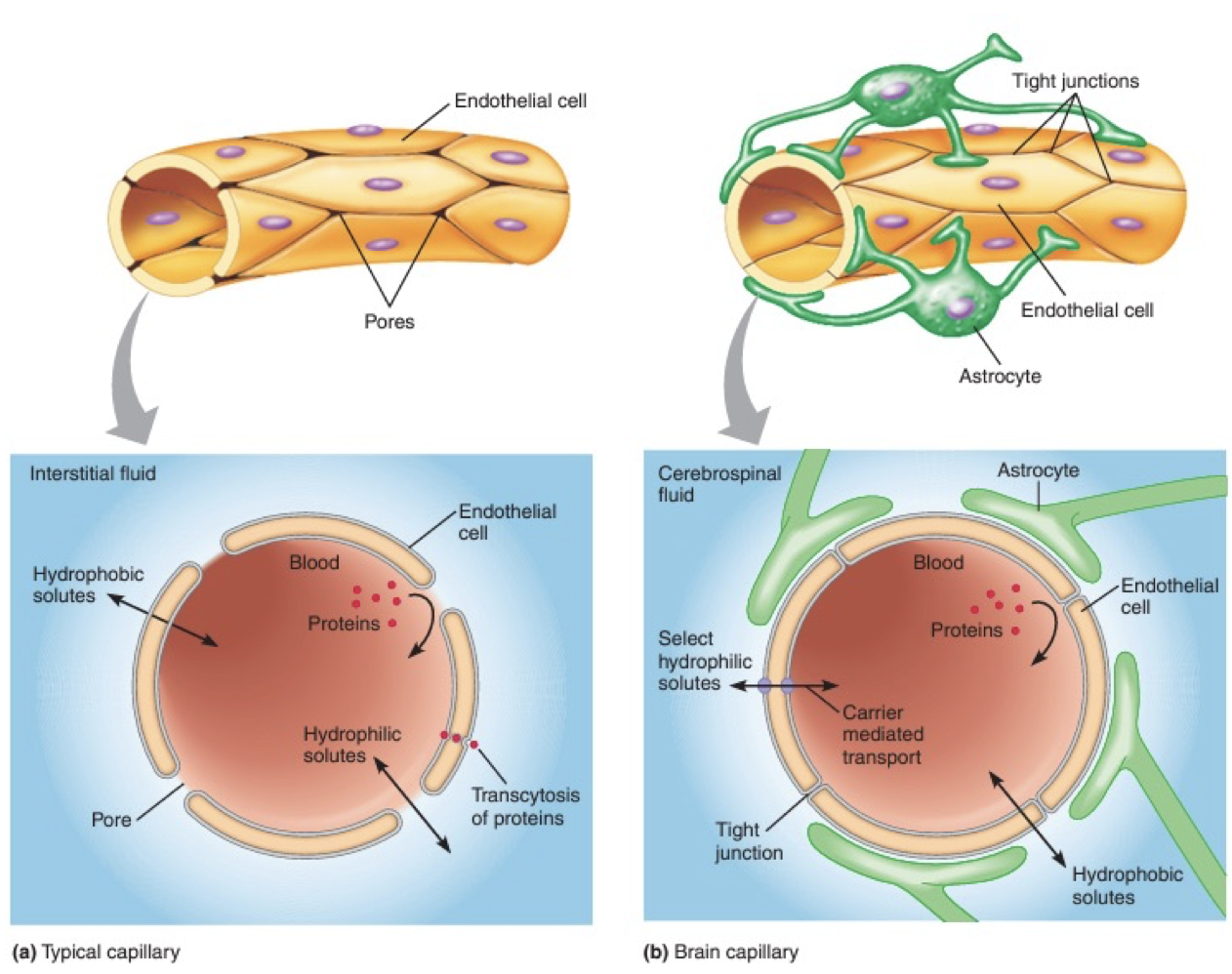
Blood Brain Barrier
Brain receives 15% of blood supply due to neuron’s high metabolic rate (20% of our oxygen and 50% glucose)
Brain cannot store glucose like the liver
Capillaries are made up of a single layer of endothelial cells.
Movement of molecules usually occurs through small pores in the endothelia cells that made up the capillary walls
In the CNS they are held tightly together to restrict the movement of molecules and protect the CNS from harmful things in the blood creating the blood brain barrier.
Also limits the movement of important molecules like glucose so they must be transported across, making it selectively permeable.
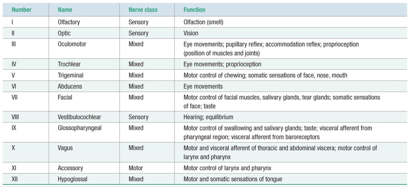
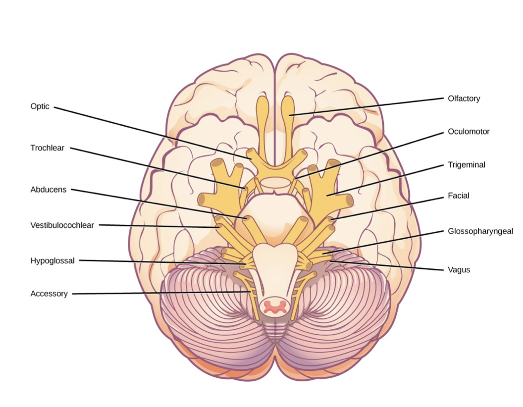
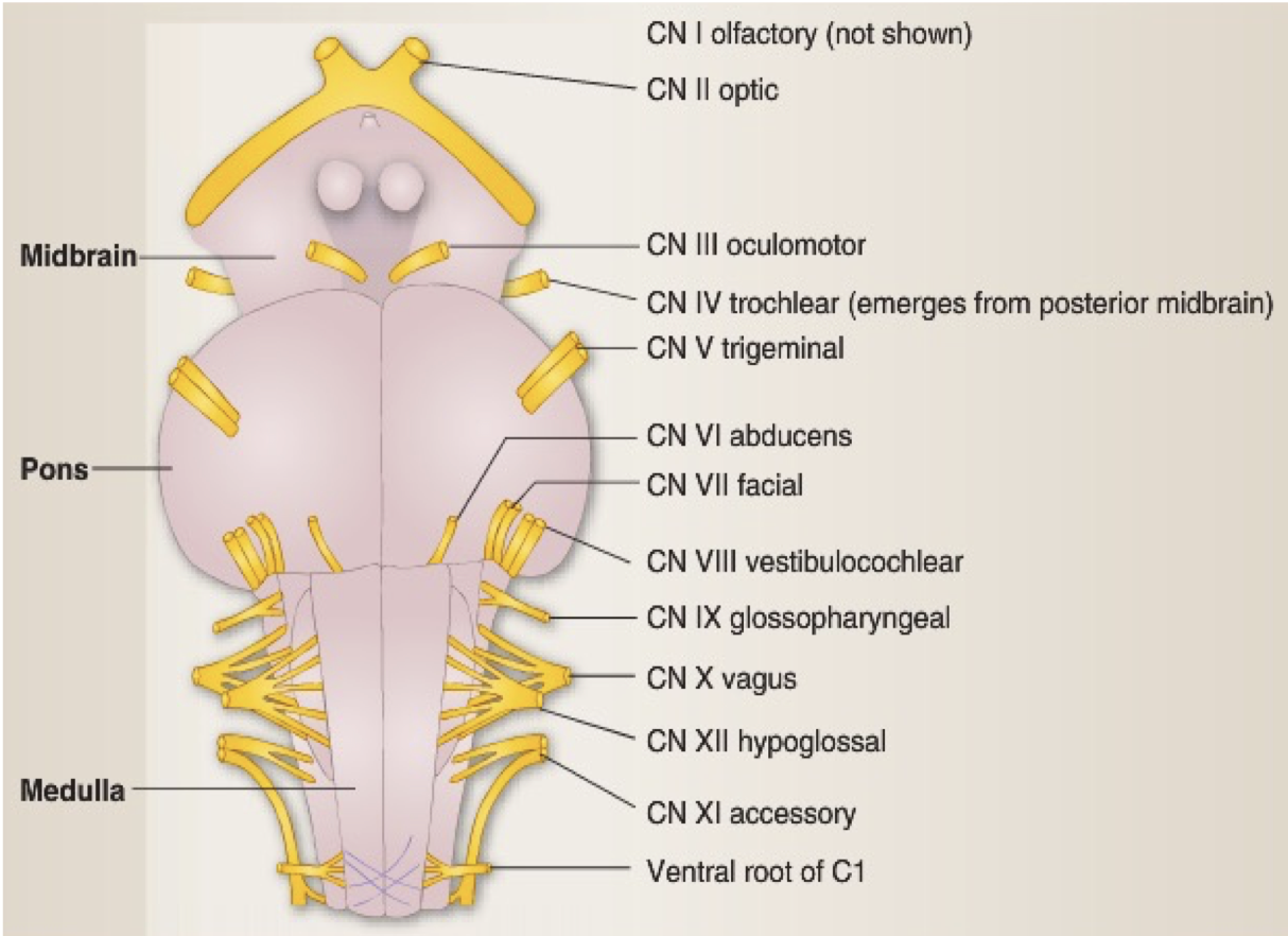
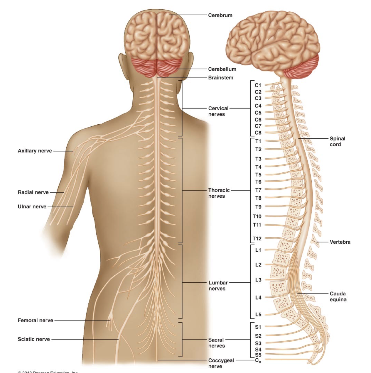
Cranial Nerves
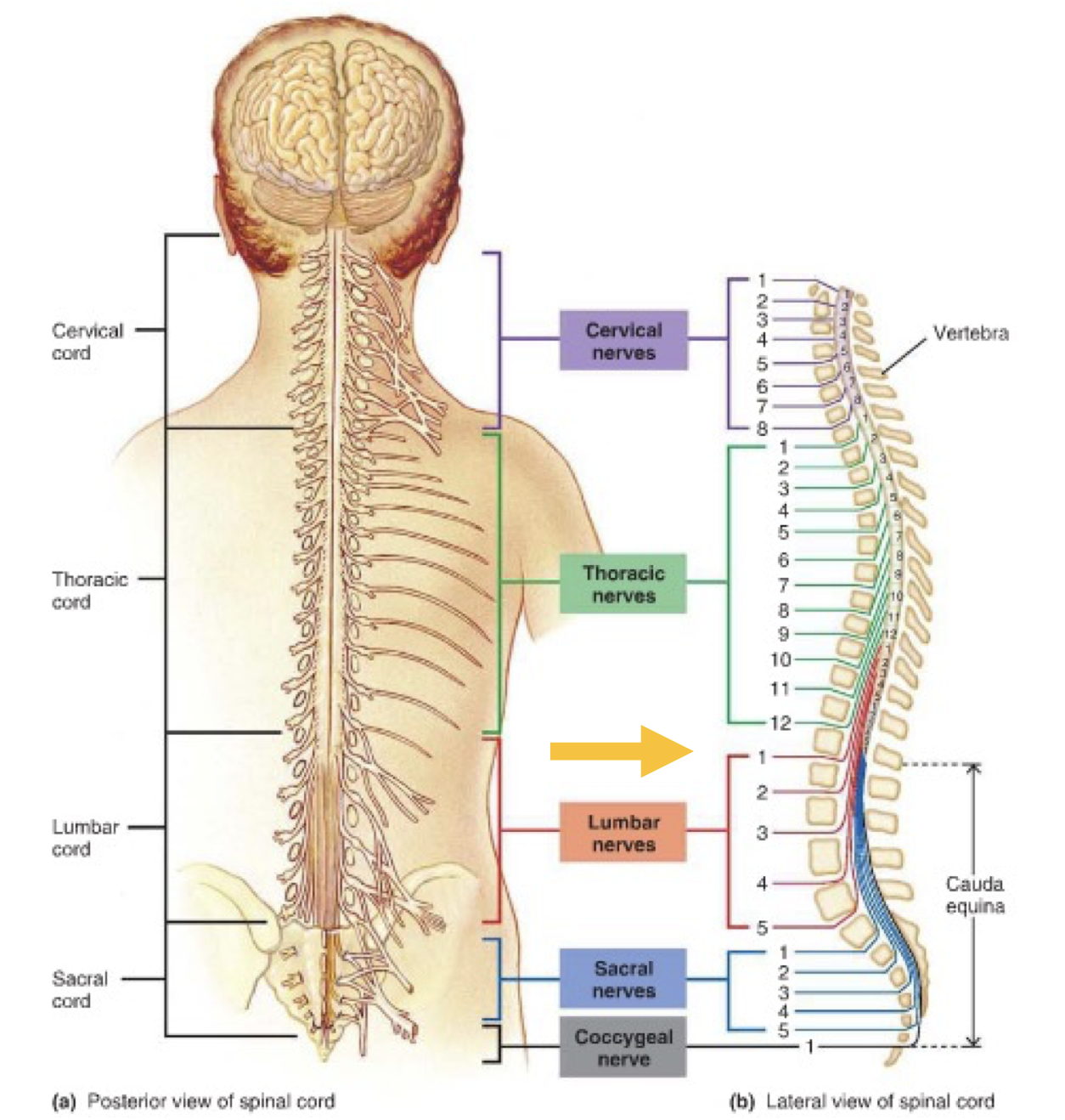
Spinal Cord
Connects CNS to PNS
40 - 50 cm long and 2 cm at the widest point (mid thoracic)
Medulla oblongata to 2/3’s down the vertebral column
Remain nerves continue down column as cauda equina
31 neural segments give rise to 31 pairs of spinal
nerves.
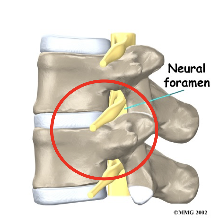
Emerge from the vertebral column through an opening
called neural foramen between adjacent vertebrae.
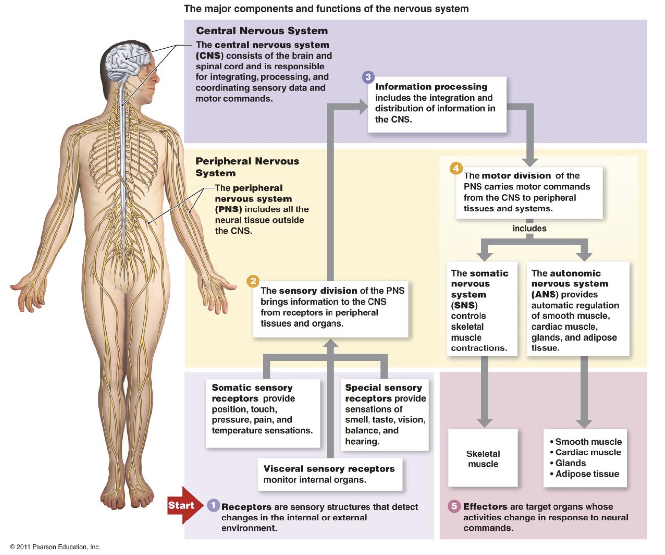
CNS: Brain and spinal chord
PNS: Spinal and peripheral nerves
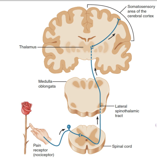
Sensory division (PNS to CNS):
Somatic receptors: Position, touch, pressure, pain and temp
Specialised receptors: Smell, taste, vision, balance, and hearing
Visceral receptors: Internal organs
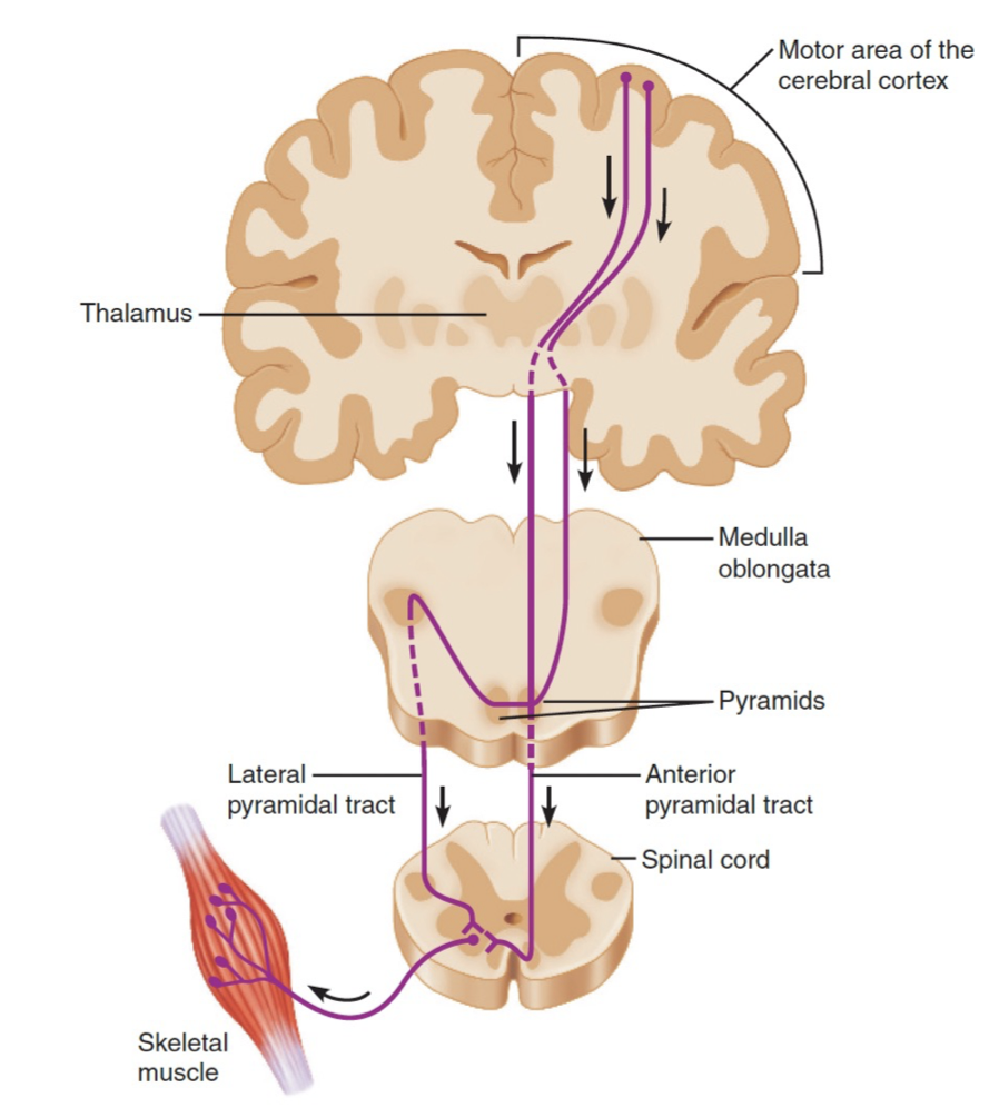
Motor division (CNS to PNS):
Somatic nervous sytem: Skeletal muscles (voluntary)
Autonomic nervous system: Smooth muscle, cardiac muscle, glands, adipose tissue (involuntary)
Sympathetic: Fight or flight
Parasympathetic: Rest and digest. Relaxation.
Enteric Nervous System: Complex network of neuron’s that governs gastrointestinal system (second brain for peristalsis and enzyme secretion)
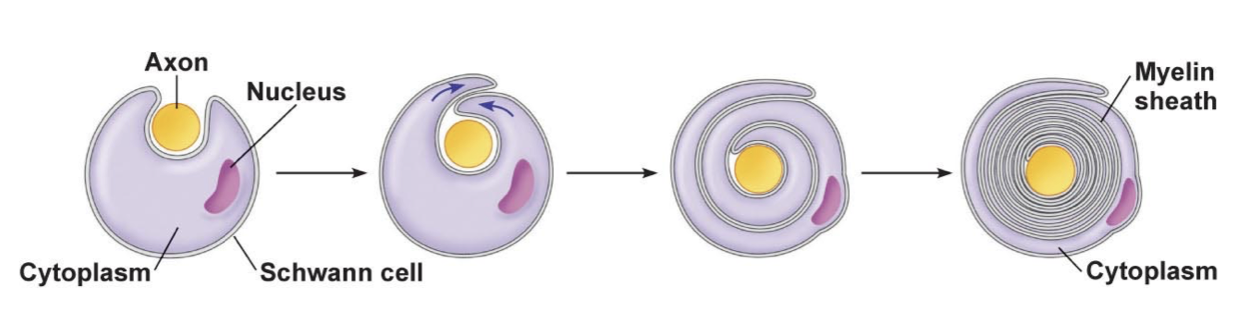
Myelin Sheath
Schwann cell or oligodendrocyte wraps around the axon
Cytoplasm is gradually squeezed out
50 - 150 stacked layers of membrane
Myelin is made of lipids which is a poor conductor of electricity insulating the axon.
Action potentials regenerate at the nodes of ranvier where the is no axon sheath.
Communication of Cells
Juxtacrine signalling
Membrane bound proteins interact with each other and ligands in the extracellular matrix to communicate with ajacent cells.
Signals travel via hydrophobic membrane channels/pores (gap junctions) between cells.
Paracrine signalling
Local communication between cells close together
Release of neurotransmitters from one cell which diffuse to the next cell
Autocrine signalling
Chemical signals released by a cell bind to the receptors on the same cell
Endocrine signalling
Chemical signals released by endocrine cells into the circulatory system to communicate with distant target organs.
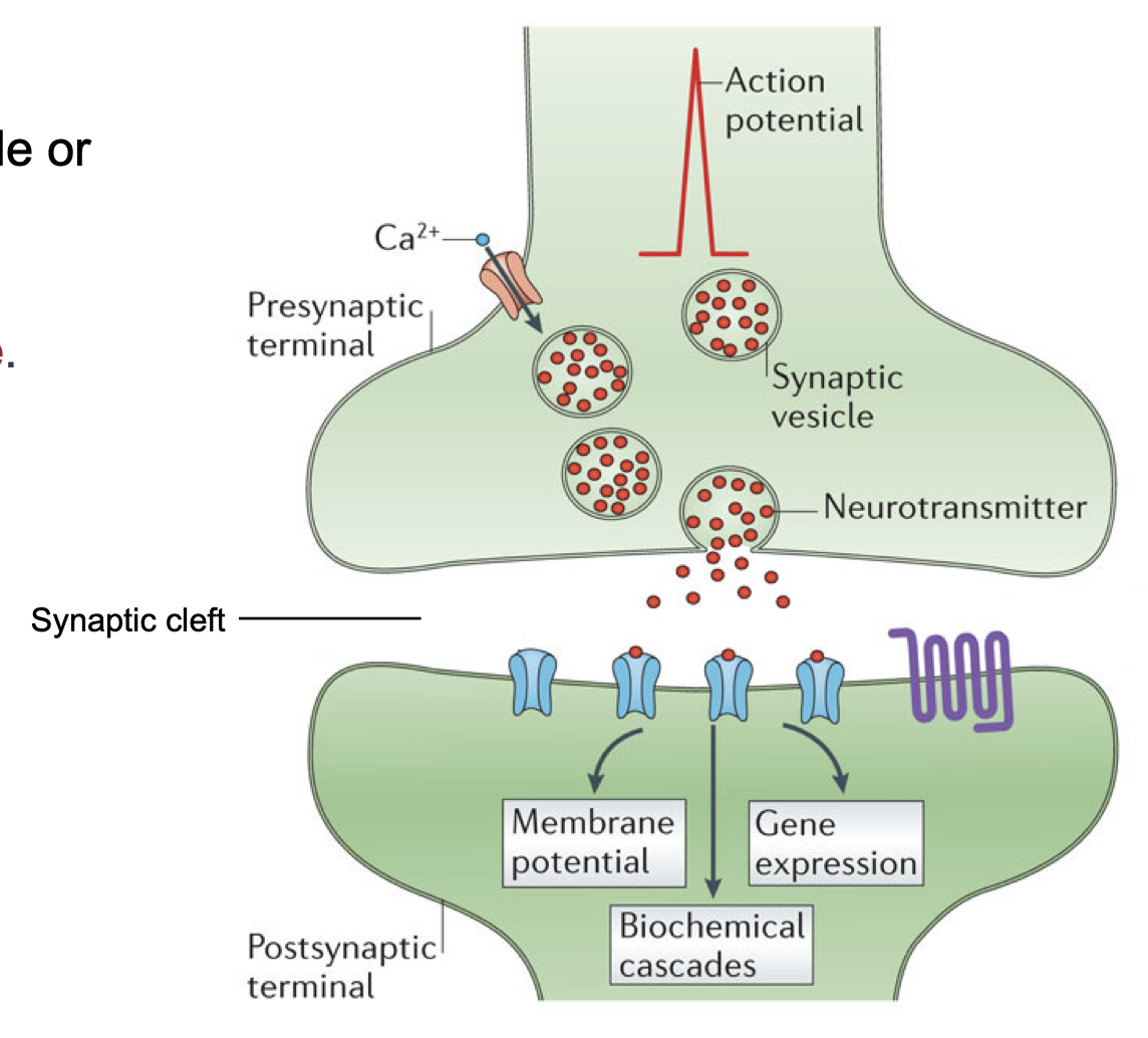
Chemical Synapses
Action potential arrives at the terminal
Triggers calcium (Ca2+) to enter the presynaptic cell
Ca2+ triggers exocytosis of neurotransmitter
Neurotransmitter diffuses across synaptic cleft and binds to receptors on postsynaptic cell
Response triggered in postsynaptic cell
Response terminated by removing neurotransmitter from synaptic cleft
Neurotransmitters
To be a neurotransmitter a substance must be
Present (and usually synthesised) within the presynaptic neuron
Released in a regulated fashion (usually via exocytosis from synaptic vesicles) following stimulation of the pre-synaptic neuron
Receptors for the substance must be present on the post-synaptic target cell
Mechanisms must be present to remove or inactivate the substance
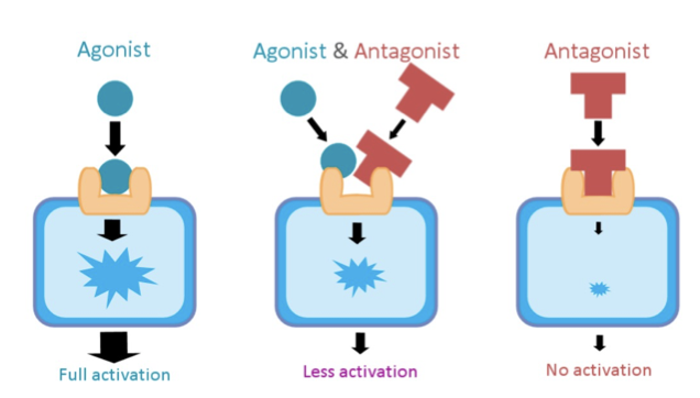
Exogenous Neurotransmitters
Agonist: Similar to an endogenous (natural produced in body) neurotransmitter that it binds to receptor on the pre/post synaptic neuron's and activates them e.g. morphine & heroin (opiates) mimicking the effect of natural endorphins.
Antagonists: Substance that is similar enough that it binds and blocks the receptor but doesn’t activate it e.g. curare binds and blocks muscle receptors causing muscle paralysis