Session 8 a - Visual Coding
Visual Coding Biopsychology 1
General Principles of Perception
The brain interprets signals from the retina based on the type of nerve:
Johannes Muller’s Law of Specific Nerve Energies:
Visual nerve = vision
Auditory nerve = sound
Olfactory nerve(sense of smell) = odors
Anatomy of the Eye
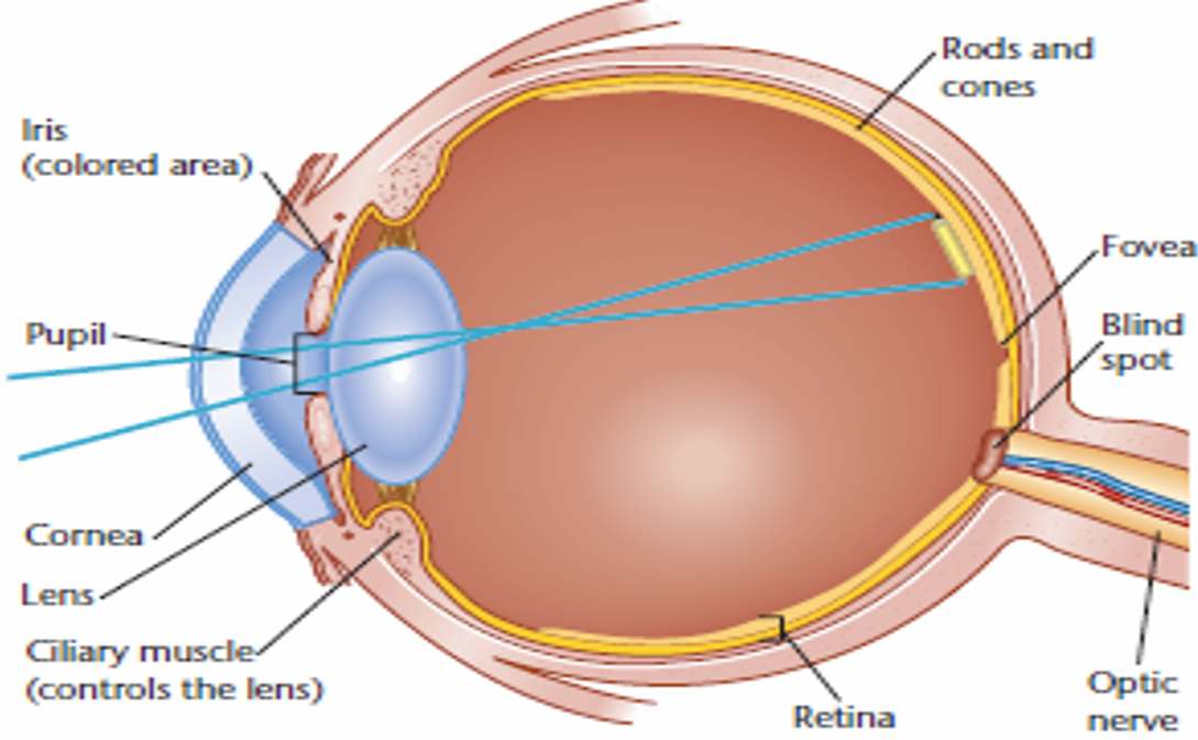
-Photoreceptor cells=specialized cells in the retina of the eye that detect light and convert it into electrical signals that can be processed by the brain, allowing us to see(rods & cones)
Components of the Eye:
Rods=photoreceptor cells in the retina that helps us see in low/dim light but do not detect color
Cones=photoreceptor cells in the retina that helps us see colors and detailed vision in bright light
Iris (colored part)=muscular ring that controls the size of the pupil(regulates how much light enters the eyes)
Fovea=responsible for the sharpest and most focused part of your vision.
Blind Spot=area of the retina without any photoreceptor cells thus cant process vision
Pupil=lens help focus light on the retina
Cornea=Focuses light as it first enters the eye, helping direct it toward the lens
Lens=Focuses light helping direct it toward the retina
Ciliary Muscle=change the shape of the lens for focusing
Retina=captures light and sends signals to the brain to create vision
An image is inverted on the retina; the optic nerve exits on the nasal side.
Visual Receptors: Rods and Cones
-periphery=outer edge of something
-retina’s periphery=outer edges of the retina, the light-sensitive layer at the back of your eye. responsible for motion detection and peripheral vision but doesnt have detailed vision that the center of the retina (fovea) does.
Rods: Located in the retina's periphery; sensitive to faint light.
Cones: Many located in the fovea; essential for color vision in bright light.
Rods:Cones ratio = 20:1 in the retina. Cones provide 90% of the brain's visual input.
Variation in optic nerve axons and visual cortex size is often genetic.
Photopigments release energy when struck by light.
Visual System of the Vertebrate
-bipolar cells=so bipolar cells are neurons in the retina that receive signals from the photoreceptor cells (rods and cones) and sends signals to the ganglion cells.
-Amacrine cells=specialized neurons found in the retina that make the visual signals more clear
Process of visual information:
Messages travel from receptors to bipolar cells.
↓
Bipolar cells send information to ganglion cells.
↓
Ganglion cells' signals combine and go to the brain.
↓
Amacrine cells contribute to shape, movement, lighting changes, and color detection.
Visual Characteristics
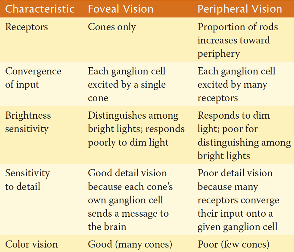
Foveal Vision:
Receptors: Only cones.
Good detail and color vision.
Peripheral Vision:
Proportion of rods increases; poor detail and color discrimination.
Each ganglion cell receives input from multiple cone receptors.
Color Vision
Visible light is a type of energy that moves in waves, and our eyes can only see waves within 400 nm to 700 nm range.
Trichromatic (Young-Helmholtz) Theory: Color perception based on three kinds of cones sensitive to different light wavelengths:
S-cone(short wave length)=sensitive to blue light
M-cone(medium wave length)=sensitive to green light
L-cone(long wave length)=sensitive to red light
Opponent Process Theory: Describes color by organizing it into opposing pairs on a scale:
Red vs. Green, Yellow vs. Blue, White vs. Black.
“Fatigue response” leads to seeing the opposite color to keep it balance
Color Vision Deficiency:
Caused by genetic factors affecting cone development.
Red-green deficiency: occurs due to when the X chromosome made the pigment, the red cones and green cones are using the same pigment instead of different ones. Thus they cant differenciate between the two
Visual Experiments
Eye Pressure Experiment:
Rub your eyes and observe seeing spots/flashes of light in darkness.
Explanation: Mechanical pressure excites visual receptors, perceived as light.
Negative Color Afterimage Experiment:
Stare at a point for 1 minute under bright light, then look at a white surface to see the negative color afterimage.
Afterimage Experiment:
Observe the colors after staring at a colored object and viewing a white page.
Retinex Theory:
Integrates information from the retina and cortex for brightness and color determination.
Highlights color constancy despite lighting changes.
Retinex Theory Experiment:
Close focus on the center of an object and observe changes.
Additional Information
Blind Spot:
Definition: Point where the optic nerve exits, having no receptors.
Fovea and Retinal Periphery:
Fovea: Specialized for acute and detailed vision. No blood vessels or ganglion axons interfere. Has many receptors connected to a single bipolar cell.
Peripheral Retina: More receptors converge to bipolar and ganglion cells. Increased sensitivity to dim light.