Integumentary System
Consists of:
skin (cutaneous membrane)
skin appendages- sweat glands, oil glands, hair, nails


Functions:
insulates and cushions deeper body organs
protects the body from mechanical damage, chemical damage, thermal damage, chemical damage, thermal damage, UV, radiation, microbes, desiccation (drying out)
aids in loss of retention of body heat as controlled by the nervous system (thermoregulation)
aids in excretion of urea and uric acid
synthesizes Vitamin D
2 kinds of tissue compose the skin
epidermis
dermis
Hypodermis (subcutaneous layer)
anchors skin to underlying organs
not technically part of the integumentary system
composed mostly of adipose tissue
serves as a shock absorber and insulates deeper tissues
Epidermis- outer layer
capable of being hard and tough
stratified squamous epithelium
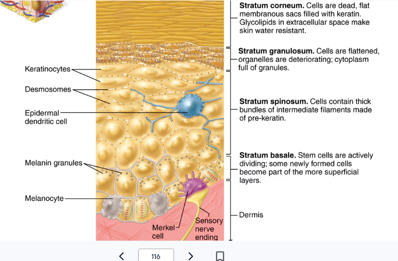
Keratinocytes (the most common cell) produce a fibrous protein called Keratin
Avascular
composed of 5 layers (strata) deepest to top layers:
Stratum Basale
Stratum Spinosum
Stratum Granulosum
Stratum Lucidum (thick, hairless skin only)
Stratum Corneum
Stratum Basale (stratum germinativum)
deepest layer of epidermis
lies next to dermis
wavy borderline with the dermis anchors the two together
cells undergoing mitosis
daughter cells are pushed upward to become the more superficial layers
Stratum Spinosum
cells become increasingly flatter and more keratinized
Stratum Granulosum
filled with keratin; cells begin to die after SG
Stratum Lucidum
formed from dead cells of the deeper strata
occurs only in thick, hairless skin of the palms of the hand and soles of the feet
Stratum Corneum
outermost layer of epidermis
shingle-like dead cells are filled with keratin (protective protein prevents water loss from skin)
Epidermal Dendritic Cells
alert and activate immune cells to a threat (bacterial or viral invasion)
Merkel Cells
associated with sensory nerve endings
serve as touch receptors called Merkel discs
Dermis
connective tissue
underlies the epidermis
2 layers
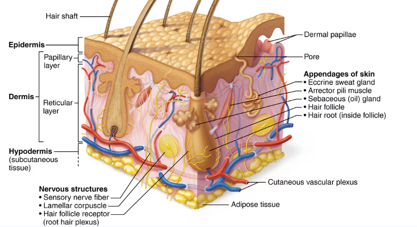
Papillary layer (upper dermal region) contain projections called dermal papillae
indents the epidermis above
many projections contain capillary loops, and others house pain and touch receptors
on palm and sole surfaces, papillae increase friction and gripping ability
fingerprints are identifying films of sweat
Reticular layer (deepest skin layer)
blood vessels
sweat and oil glands
deep pressure receptors (lamellar corpuscles)
other dermal features
cutaenous sensory receptors
phagocytes
collagen and elastic fibers
blood vessels
Skin color
3 pigments contribute to skin color
Melanin
yellow, reddish brown, or black pigments
pigment produced by melanocytes
Melanocytes mostly in the stratum basale of the epidermis
accumulates in membrane-bound granules called melanosomes
amount of melanin produced depends upon genetics and exposure to sunlight
eumelanin (darker) and pheomelanin (Red/pink)
Carotene
orange-yellow pigment from some vegetables
Hemoglobin
red coloring from blood cells in dermal capillaries
oxygen content determines the extent of red coloring
Skin color (not permanent)
Redness (erythema)- due to embarrassment, inflammation, hypertension, fever, or allergy
Pallor (blanching)- due to emotional stress (such as fear), anemia, low blood pressure, impaired blood flow to an area
Jaundice (yellow cast)- indicates a liver disorder
Bruises (black and blue marks)- hematomas
Cyanosis (blue/grey)- severe lack of oxygen
Appendages of the skin
cutaneous glands are all exocrine glands
sebaceous glands (oil glands)
located all over skin except for palms and soles
most have ducts that empty into hair follicles; others open directly onto skin surface
glands are activated at puberty
produce sebum (oil)
makes skin soft and moist
prevents hair from becoming brittle
kills bacteria
increased amounts due to androgens in adolescents
sweat glands
hair and nail follicles
nails
sweat (sudoriferous) glands
produce sweat
widely distributed in skin
2 types
Eccrine (everywhere) glands- cool you
open via duct to sweat pores on the skin’s surface
produce acidic sweat (water, salts, vitamin C, traces of metabolic waste)
function in body temperature regulation (thermoregulation)
Apocrine glands- show up at puberty
ducts empty into hair follicles in the armpit and genitals
begins to function at puberty
release sweat that also contains fatty acids and proteins (milky or yellowish color)
play a minimal role in body temperature regulation
active during pain, sex, stress
Homeostatic Imbalance of Sebaceous Gland
acne= infection of the gland
blackhead= dried blackened sebum; blocked gland
whitehead= same, not blackened
seborrhea= cradle cap in infants; overactive glands
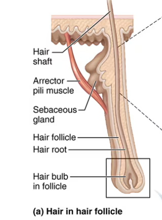
Hair
produced by hair follicle
root is enclosed in the follicle
shaft projects from the surface of the scalp or skin
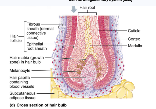
consists of hard keratinized epithelial cells
melanocytes provide pigment for hair color
hair grows in the matrix (mitosis) of the hair bulb in stratum basale
Associated hair structures
hair follicle
dermal region provides a blood supply to the hair bulb (deepest part of the follicle)
Arrector pili muscle connects to the hair follicle to pull hairs upright when we are cold or frightened
If the hair shaft is oval, the hair will be smooth, silky, wavy.
If the hair shaft is flat and ribbonlike, the hair is curly or kinky.
Nails
heavily keratinized, scalelike modifications of the epidermis
stratum basale extends beneath the nail bed, which is responsible for growth
lack of pigment makes nails colorless
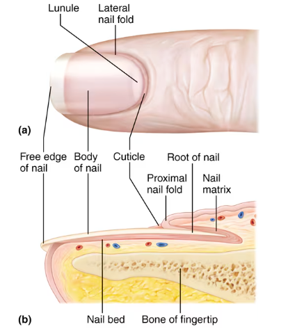
Parts of a nail
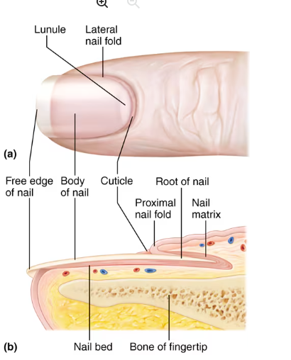
free edge- end of nail, tip
stratum basale beneath nail is nail bed
Body is the visible attached portion
Nail folds are skin folds that overlap the edges of the nail; the cuticle is the proximal edge
Root of nail is embedded in skin
growth of the nail occurs from thickened proximal nail matrix
Infections to know
Athlete’s Foot- caused by fungal infection, itchy red peeling skin between the toes
Boils (furuncles) and carbuncles- caused by inflamation of hair follicles, carbuncles are clusters of boils caused by bacteria
Cold Sores (fever blisters)- caused by human herpesvirus 1, blisters itch and sting
Contact dermatitis- caused by exposure to chemicals that provoke allergic responses, itching redness and swelling of the skin
Impetigo- caused by bacterial infection, pink fluid-filled raised lesions around mouth/nose
Psoriasis- triggered by trauma, infection, hormonal changes, or stress, red epidermal lesions covered with dry silvery scales that itch burn crack or sometimes bleed
Burns
tissue damage and cell death caused by heat, electricity, UV radiation, or chemicals
associated dangers
protein denaturation and cell death
dehydration and electrolyte imbalance
circulatory shock
result in loss of body fluids and infection from the invasion of bacteria
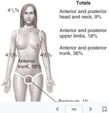
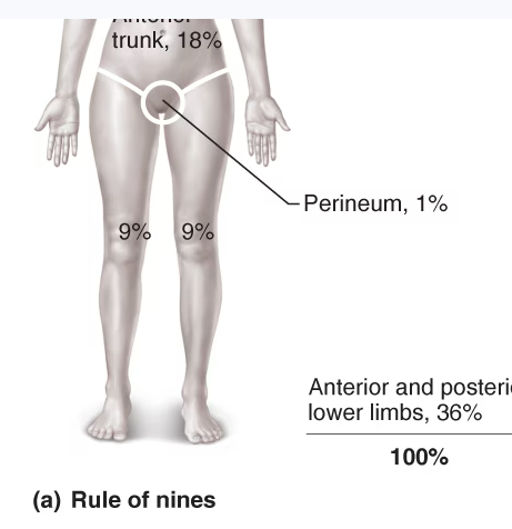
extent of a burn is estimated using the rule of nines
body is divided into 11 areas for quick estimation
each area represents about 9% of total body surface area
the area surrounding the genitals (the perineum) epresents 1% of body surface area
1st degree burn (superifical burn) (sunburn)
only epidermis is damaged
skin is red and swollen
2nd degree burn (partial-thickness burn)
epidermis and superficial part of dermis are damaged
skin is painful, red, and blistered
regrowth of the epithelium can occur
3rd degree burn (full-thickness burn)
destroys epidermis and dermis; burned area is painless
requires skin grafts, as regeneration is not possible
burned area is blanched (gray-white) or black
4th degree burn (full-thickness burn)
extends into deeper tissues (nones, muscle, tendons)
appears dry and leathery
requires surgery and grafting
may require amputation
Criteria for deeming burns critical (if any one is met):
over 30% of body has second degree burns
over 10% of the body has third or fourth degree burns
third or fourth degree burns of the face, hands, or feet, or genitals
burns affect the airways
circumferential (around the body or limb) burns have occured
Skin Cancer
most common form of cancer in humans in the US
most important risk factor is overexposure to ultraviolet (UV) radiation in sunlight and tanning beds
most common types
Basal cell carcinoma- least malignant and most common type of skin cancer, arises from cells in stratum basale that are altered so that they can no longer make keratin, lesions appear as shiny, dome-shaped nodules that develop a central ulcer
Squamous cell carcinoma- believed to be induced by UV exposure, arises from cells of stratum spinosum, lesions appear as scaly reddened papules that gradually form shallow ulcers, early removal allows a good chance of cure, metastasizes to lymph nodes if not removed
Malignant melanoma- most deadly of skin cancers, but accounts for only 5 percent of skin cancers, arises from melanocytes, metastasizes rapidly to lymph and blood vessels, detection uses ABCDE rule for recognizing melanoma
A= asymetry (two sides of pigmented mole do not match)
B= border irregularity (borders of mole are not smooth)
C= color (different colors in pigmented areas)
D= diameter (spot is larger than 6mm in diameter)
E= evolution (one or more of the ABC characteristics is evolving)
Cancer can be classified two ways
Beneign means the neoplasm (tumor) has not spread
Malignant means the neoplasm has invaded other body areas
Review
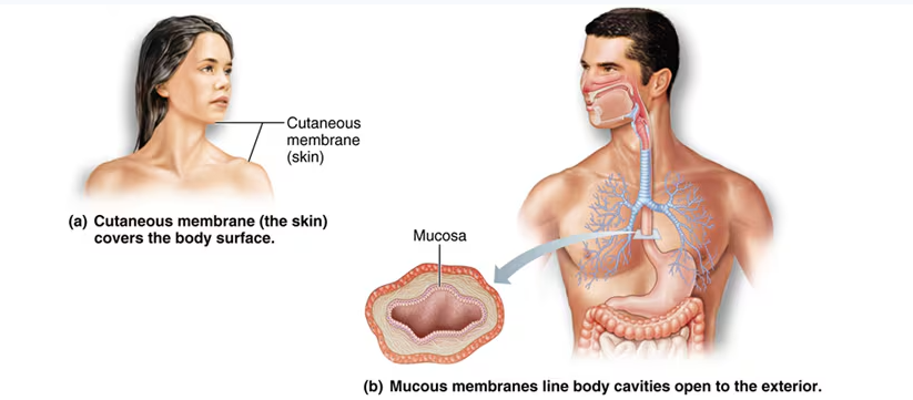
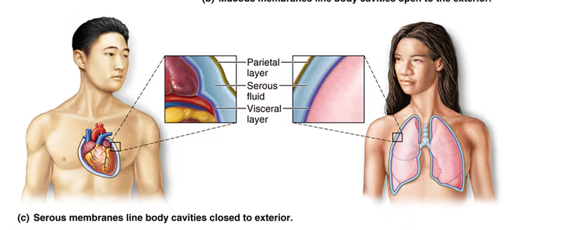
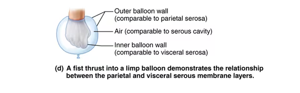
There are four types of membranes. What are they? (Know diagrams)
Cutaneous (most superficial, skin), serous (visceral and parietal), mucuous (moist), synovial (joints)
Can you list five integument functions?
Mechanical damage
Chemical damage
Thermal damage
Ultraviolet radiation
Microbes
Dessicration
Thermoregulation
Aids in excretion of urea and uric acid
Synthesizes Vitamin D
The layers of the epidermis are (come lets get sun burned):
S. Basale
S. Spinosum
S. Granulosum
S. Lucidum
S. Corneum
Melanin: cells, layer, color, granules
Cells- melanocytes
Layer- s. basale of the epidermis
Color- yellow to brown to black
Granules- melanosomes
Influenced by genetics and sunlight
Layers of the dermis:
upper papillary
reticular papillary
Three pigments contribute to skin color:
Melanin
Carotene
Hemoglobin
List 5 emergency skin colors:
Erythema (red)
Pallor (white)
Jaundice (yellow)
Cyanosis (blue)
Hematoma (purple)
What are two types of exocrine glands in this system?
Sebaceous (oil)
Sudoriferous (sweat)
How do the 2 types of sudoriferous glands vary (know location)?
Eccrine is cool (everywhere), apocrine smells (at puberty)
Hair:
Produced by hair follicle
Root is enclosed in the follicle
Shaft (part you shave)
Keratin (pigment)
Melaocytes
S. Basale
What are five major regions of the nails?
Free edge
Body
Nail
Cuticle
Root of nail
Growth
Impetigo
Bacterial infection; pink, fluid-filled raised lesions around mouth/nose
Contact Dermatitis
Itching, redness, and swelling of the skin caused by exposure to chemicals that provoke allergic responses
Basal Cell Carcinoma
Skin cancer that arses from cells in stratum basale that are altered so that they can no longer make keratin
Squamous Cell Carcinoma
Believed to be induced by UV exposure, this cancer arises from cells of stratum spinosum
Diagrams to Know