Nociception (Pain) Study Notes
Nociception
Lecture by Dr. Mariana Vargas-Caballero
Course Overview
Two primary lectures on:
General Overview of Sensory Systems
Nociception
Learning Outcomes
Defining Key Concepts:
Define pain and associated terms related to nociception.
Describe the afferent pathways mediating pain and temperature sensation.
Explain physiological mechanisms required for the integration of painful stimuli.
Discuss descending modulation of nociceptive pathways.
Sensory Systems Framework
Sensory processing integrates environmental stimuli with behavioral responses.
Components of Sensory Systems:
Central Nervous System (CNS):
Composed of the brain and spinal cord, it processes sensory and motor information.
Peripheral Nervous System (PNS):
Composed of cranial and spinal nerves, it functions in transmitting sensory information to the CNS and motor commands to effectors.
Sensory receptors are located at the body's surface and interior, including sensory ganglia and nerves.
Motor Components:
Visceral Motor System:
Includes sympathetic, parasympathetic, and enteric divisions controlling smooth, cardiac muscles, and glands.
Somatic Motor System:
Composed of motor nerves that control skeletal (striated) muscles.
Somatosensory Functional Components
First Order Sensory Neurons: Location and Function
Cell bodies for somatosensory fibers reside in the dorsal root ganglia (DRG) for body fibers.
For facial sensations, somas are present in the trigeminal ganglion.
Features two axon branches:
Peripheral branch: extending to the periphery (PNS)
Central branch: projecting to the spinal cord (CNS)
Structure of First Order Neurons:
Neurons are classified as pseudounipolar, displaying two axonal branches.
Mechanosensory Pathways
First Order Neuron Pathway:
DRG neurons integrate sensory signals, entering via the dorsal root and joining the dorsal column pathway.
Pain and temperature fibers create connections upon entry into the spinal cord, which can serve for clinical diagnostic purposes based on dissociated sensory loss.
Somatic Sensory System Visuals and Figures
Visuals represent organization showing various regions in the brain associated with sensory processing.
Synaptic Pathways:
First-order neurons synapse on second-order neurons in the medulla and thalamus before reaching the somatic sensory cortex.
Ascending pathways are detailed for mechanosensation, pain, and temperature.
Major Classes of Somatic Sensory Receptors
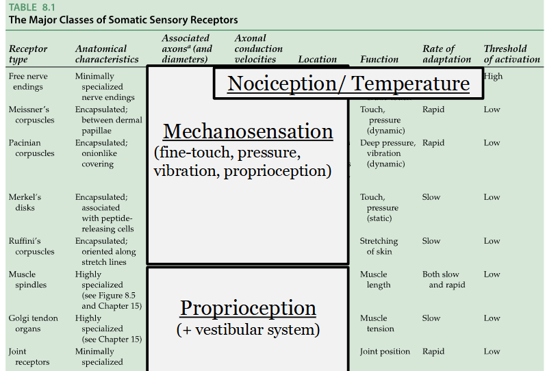
Table 8.1: Summarizes anatomical characteristics and functions of sensory receptors, including:
Free Nerve Endings:
Minimally specialized, associated with nociception and temperature.
Conductive velocities vary based on fiber type:
Aβ fibers (35-90 m/s) for touch, Aδ fibers (5-40 m/s) for sharp pain, and C fibers (0.5-2 m/s) for dull pain.
Encapsulated Receptors:
Mechanoreceptors like Meissner’s corpuscles (for touch), Pacinian corpuscles (for vibration), and Ruffini endings (for skin stretch).
Pain and Nociception Definitions
Pain:
Defined as an unpleasant sensory and emotional experience associated with actual or potential tissue damage (International Association for the Study of Pain).
Nociception: Process of encoding harmful or potentially harmful stimuli, involving peripheral tissues, spinal cord, and multiple brain areas.
Types of Nociceptors
Nociceptive Afferents:
Located near blood vessels and in various tissues (e.g., skin, muscles).
Activated by noxious mechanical, thermal, and chemical stimuli.
Categorization of Nociceptors:
Aδ Fibers: Fast pain, myelinated, localized sharp pain.
C Fibers: Slow pain, unmyelinated, associated with burning and aching sensations.
Signal Transduction Mechanisms
Process of sensory transduction involves several steps from weak to strong stimuli leading to the generation of action potentials.
Ions (e.g., Ca²⁺, Na⁺) flow through stretch-sensitive cation channels, leading to the depolarization of the cell.
Pain Measurement Scales
Numeric Pain Rating Scale: Range from 0 (no pain) to 10 (worst possible pain).
Wong-Baker FACES Pain Rating Scale: Visual representation to help individuals express pain levels, emphasizing emotional aspects of pain perception.
Modulation of Pain Signals
First Order Neurons: Activate to transmit noxious signals to the spinal cord.
Modulation Sites:
Dorsal horn of the spinal cord serves as an initial processing point.
Descending Pathways:
These pathways can inhibit nociceptive signals and mediate pain response altered by psychological factors or endogenous opioids (e.g., enkephalin).
Clinical Implications of Pain Management
Painkillers include:
Opioid Analgesics: Morphine, codeine, acting on opioid receptors.
Non-Steroidal Anti-Inflammatory Drugs (NSAIDs): Target inflammation and pain management pathways.
Local Anesthetics: Block sodium channels and prevent action potential propagation.
Referred Pain and Allodynia
Referred Pain: Pain perceived in a different area from the original source of nociception (e.g., heart attacks felt in the arm).
Allodynia: Pain response to stimuli that normally do not provoke pain, indicating further dysfunction in nociceptive pathways.
Conclusion
The complex organization and processing of nociception are vital for understanding both normal sensory function and pathophysiological states. The modulation of pain involves intricate neural networks capable of adaptation and change depending on the context of the stimuli and the psychological state of the individual.
Nociception
Lecture by Dr. Mariana Vargas-Caballero
Course Overview
Detailed examination of sensory systems with a focus on:
General Principles of Sensory Transduction and Coding
Nociception: The physiological and psychological processing of pain.
Learning Outcomes
Defining Key Concepts:
Distinguish between pain (the perception) and nociception (the physiological signaling).
Map the afferent pathways (Anterolateral System) mediating pain and temperature sensation from the periphery to the cortex.
Explain the physiological mechanisms of signal transduction at the receptor level.
Analyze the descending modulation systems that allow the brain to suppress or amplify incoming pain signals.
Sensory Systems Framework
Sensory processing involves the integration of external and internal environmental stimuli to drive adaptive behavioral responses.
Central Nervous System (CNS):
Brain and Spinal Cord: The primary sites for sensory integration and high-level processing.
Peripheral Nervous System (PNS):
Afferent Division: Transmits sensory information toward the CNS. Includes sensory ganglia (Dorsal Root Ganglia and Trigeminal Ganglion).
Efferent Division: Carries motor commands from the CNS to effectors.
Somatic Motor System: Controls voluntary skeletal muscle contraction.
Visceral Motor System (Autonomic): Controls involuntary functions (Sympathetic, Parasympathetic, Enteric) acting on smooth muscle, cardiac muscle, and glands.
Somatosensory Functional Components
First Order Sensory Neurons:
These are pseudounipolar neurons possessing a single process that divides into two branches:
Peripheral branch: Distal endings contain receptors in skin, muscle, or viscera.
Central branch: Enters the spinal cord via the dorsal roots or the brainstem via cranial nerves.
Location:
Body: Cell bodies reside in the Dorsal Root Ganglia (DRG).
Face: Cell bodies reside in the Trigeminal Ganglion (Cranial Nerve V).
Mechanosensory vs. Nociceptive Pathways
Mechanosensery (Touch/Proprioception): Typically follows the Dorsal Column-Medial Lemniscal Pathway. First-order neurons ascend ipsilaterally (same side) and synapse in the medulla.
Nociceptive (Pain/Temperature): Follows the Anterolateral System (Spinothalamic Tract).
First-order neurons synapse immediately in the Dorsal Horn of the spinal cord (Substantia Gelatinosa).
Second-order neurons decussate (cross the midline) at the level of entry before ascending to the thalamus.
Clinical Significance: Lesions to one side of the spinal cord result in "Dissociated Sensory Loss" (loss of touch on the ipsilateral side and loss of pain on the contralateral side).
Major Classes of Somatic Sensory Receptors
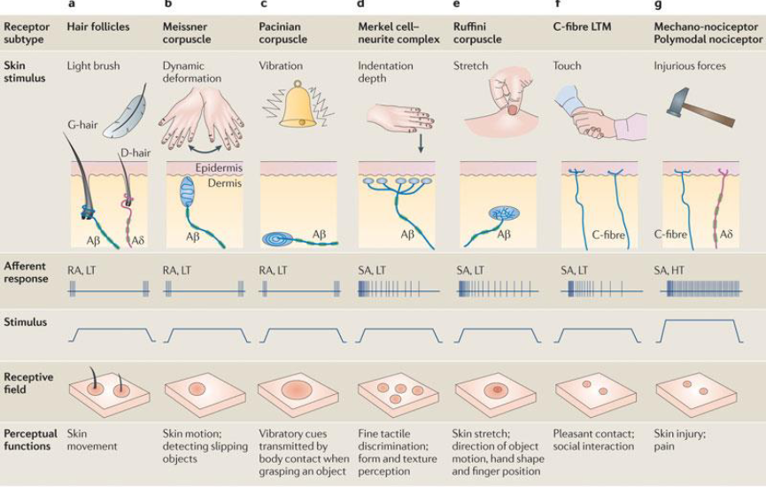
Free Nerve Endings: Non-encapsulated endings responsible for sensing pain (nociceptors) and temperature (thermoreceptors).
Fiber Classification and Velocity:
A fibers: Large, myelinated (). Primarily for touch and pressure.
A fibers: Small, lightly myelinated (). Mediate "First Pain"—sharp, prickling, and well-localized.
C fibers: Smallest, unmyelinated (). Mediate "Second Pain"—dull, aching, burning, and poorly localized.
Encapsulated Mechanoreceptors:
Meissner's Corpuscles: Rapidly adapting; sensitive to light touch and low-frequency vibration.
Pacinian Corpuscles: Rapidly adapting; sensitive to high-frequency vibration.
Ruffini Endings: Slowly adapting; sensitive to skin stretch.
Signal Transduction and Coding
Molecular Transducers: Specific ion channels at the nerve endings convert stimuli into electrical signals.
TRPV1 (Vanilloid Receptor): Responds to noxious heat (>), $H^+$ ions (acid), and capsaicin (the heat in chili peppers).
Mechanotransduction: Stretch-sensitive cation channels ( and ) open in response to mechanical deformation, causing depolarization (Generator Potential).
If the depolarization reaches a specific threshold, Voltage-Gated Sodium Channels trigger action potentials to propagate signals to the CNS.
The Gate Control Theory
Proposed by Melzack and Wall, suggesting that non-painful input (A activity) can "close the gate" to painful input (C fiber activity) in the dorsal horn.
This occurs via inhibitory interneurons in the Substantia Gelatinosa that suppress the transmission of pain signals from second-order neurons.
Modulation of Pain
Peripheral Sensitization: Inflammatory soup (histamine, prostaglandins, bradykinin) lowers the threshold of nociceptors following tissue injury.
Central Sensitization: Continuous bombardment of the dorsal horn by C-fiber activity leads to "wind-up," where neurons become hyper-excitable.
Descending Modulation:
Higher brain centers (e.g., Periaqueductal Gray - PAG) send descending projections to the spinal cord.
Release of endogenous opioids (Enkephalins, Endorphins) inhibits the release of Substance P and Glutamate from primary afferents, reducing pain perception.
Clinical Phenomena
Referred Pain: Occurs because visceral and somatic nociceptors often converge on the same second-order neurons in the spinal cord. The brain misinterprets the source of the pain (e.g., myocardial infarction felt in the left arm).
Allodynia: Condition where normally non-painful stimuli (like a light touch) evoke intense pain, often due to central sensitization.
Hyperalgesia: An exaggerated response to a stimulus that is normally painful.
Pain Management
NSAIDs: (e.g., Ibuprofen, Aspirin) Inhibit Cyclooxygenase (COX) enzymes, reducing prostaglandin synthesis.
Opioids: Bind to mu-opioid receptors in the CNS to mimic endogenous descending inhibition.
Local Anesthetics: (e.g., Lidocaine) Reversibly block voltage-gated channels, preventing the propagation of action potentials.