GI path
GI Physiology & Pathophysiology
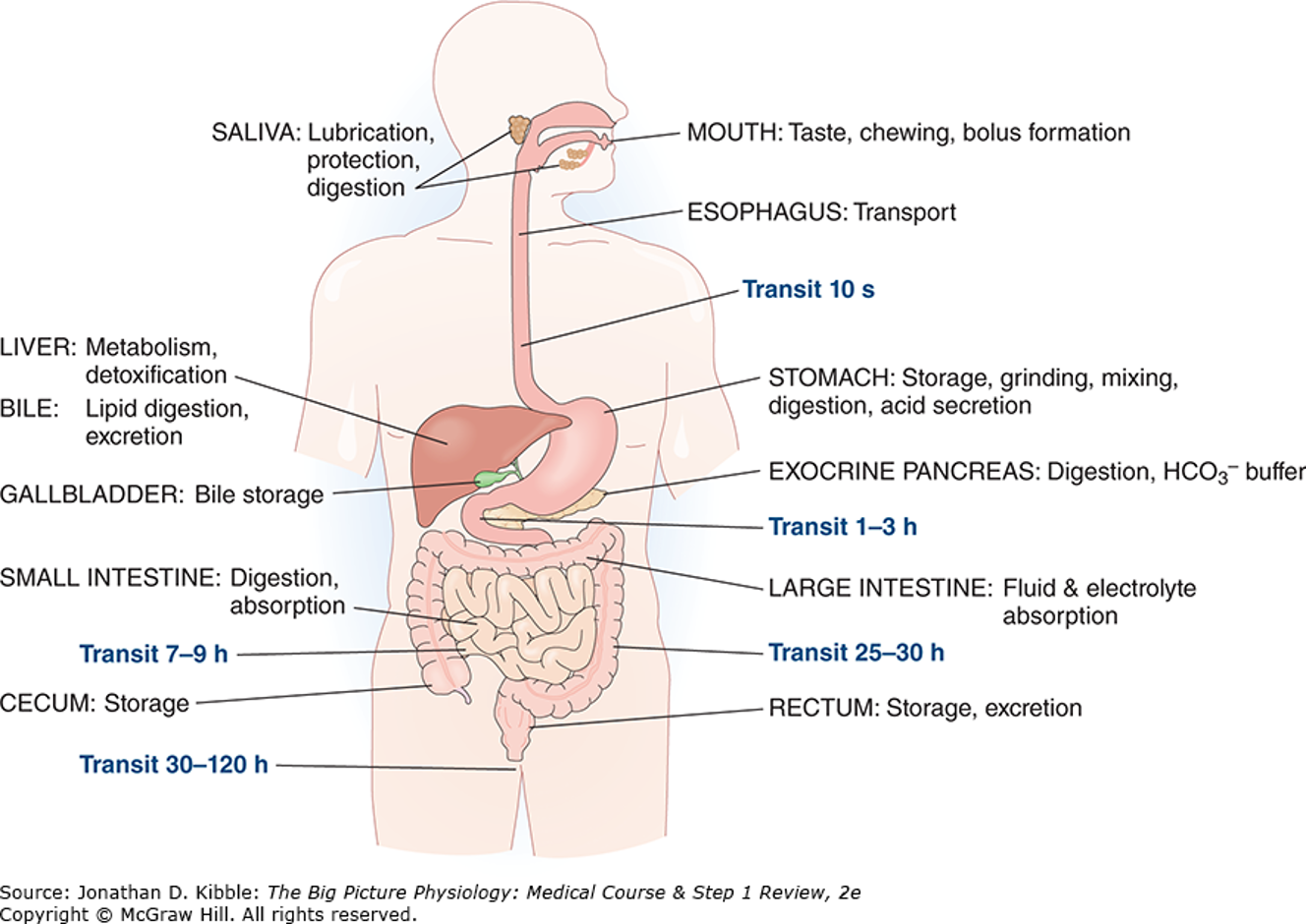
Identify the structure of the GI tract.
The GI tract is a continuous tube that carries food from the mouth to the anus.
GI Tract | Function |
|---|---|
Mouth | Entry of food |
Esophagus | Moves food to stomach |
Stomach | Begins digestion |
Small intestine | Major site of digestion and nutrient absorption |
Large intestine | Absorbs water and forms feces |
Accessory Glands
These organs release secretions that help digestion.
Salivary glands
Liver
Gallbladder
Pancreas
Describe the layers of the GI tract and the components that make up each layer:
Mucosa
The innermost layer consists of a single layer of epithelium stretching from the stomach to the anus.
It is folded to increase the surface area and invaginated to form tubular exocrine glands.
It contains endocrine cells within the epithelial lining that are responsible for releasing GI hormones.
The mucosa is supported by two distinct sub-layers:
Lamina propria: A layer of connective tissue that houses capillaries and nerve endings.
Muscularis mucosae: A thin layer of smooth muscle

Submucosa:
A layer of connective tissue that houses the major blood and lymphatic vessels serving the GI tract. (Immunuty)
It contains the Submucosal (Meissner) Nerve Plexus, which consists of ganglion cells that coordinate intestinal absorption and secretion
Muscularis externa:
Made up of two smooth muscle layers: an inner circular layer and an outer longitudinal layer.
It is responsible for mixing and moving food along the GI tract.
It houses the Myenteric (Auerbach) Nerve plexus, which lies directly between the two muscle layers
Serosa:
A thin connective tissue layer that is continuous with the peritoneal mesentery
It acts as the transit point where major structures enter and leave the GI tract, including blood vessels, extrinsic nerves, and the ducts of large accessory exocrine glands

Discuss the major physiologic processes that occur in the GI tract:
Digestion:
This is the mechanical and chemical process of converting the nutrients found in food into smaller products that the body can readily absorb
Secretion:
Secretion: To aid in the digestion process, exocrine glands and epithelial cells release a large volume of fluid into the lumen of the GI tract
Motility:
This refers to the movement of luminal contents and secretions all the way from the mouth to the anus. It is achieved through the coordinated contractions of smooth muscle within the GI tract walls.
Absorption:
This is the process where the products of digestion—spicifically amino acids, small peptides, monosaccharides and fatty acids are taken into the body. All significant absorption of nutrients occurs in the small intestine
Defense:
Protects the body from microbial pathogens while allowing immunological tolerance for antigenic substances.
Innate immunity utilizes physical and chemical barriers like mucus, secretions, and tight junctions.
Adaptive immunity involves specialized tissues like Mucosa-associated lymphoid tissue (MALT) and Peyer's patches.
Regulation of fluid and electrolyte balance:
The small intestine receives 8 to 9 liters of fluid per day and secretes 1 liter of electrolytes.
Secretion and absorption work closely together to maintain homeostasis, with most of the fluid ultimately being absorbed
Excretion:
The elimination of waste products from the body in the feces.
Excreted materials include undigested food particles, bacteria, and heavy metals such as copper and iron derived from bile

Describe the methods of physiologic regulation of the GI tract by neural, hormonal, and paracrine control.
Neural Control
The Enteric Nervous System (ENS) is a division of the autonomic nervous system located within the wall of the GI tract.
It is arranged into the myenteric and submucosal nerve plexuses and is responsible for much of the moment-to-moment control of gut motility and secretion.
The ENS can function independently of the autonomic nervous system, but it is strongly modulated by it, as well as by endocrine signals and immune modulators.
The ENS utilizes neurotransmitters such as acetylcholine (which stimulates secretion and motility), ATP and nitric oxide (which are inhibitory), and gut-brain peptides like vasoactive intestinal peptide (VIP).
The gut-brain neural axis links the ENS and the central nervous system via parasympathetic and sympathetic nerves.
Parasympathetic innervation (via the vagus nerve and the S2-S4 sacral spinal outflow) generally increases GI activity.
Sympathetic innervation generally decreases motility and secretion while increasing sphincter tone.
Endocrine Control
Endocrine control relies on gastrointestinal hormones, which are polypeptides produced and secreted by specialized enteroendocrine cells.
These chemical transmitters are involved in regulating GI motility, secretion, absorption, and food intake.
True hormones are secreted into the bloodstream and act on distant sites; the primary examples are gastrin, secretin, and cholecystokinin (CCK).
Gastrin is produced by G cells in the stomach and acts to increase H+ secretion and stomach motility.
Cholecystokinin (CCK) is produced by I cells in the duodenum and jejunum; it increases gallbladder contraction and pancreatic enzyme secretion while decreasing gastric emptying.
Secretin is produced by S cells in the duodenum; it increases pancreatic HCO3- secretion and decreases gastric H+ secretion.
Other hormones include Ghrelin (increases food intake and growth hormone), Motilin (stimulates contraction in the fasted state), Guanylin (decreases intestinal Na+ absorption), and Glucagon-like peptide-1 (increases insulin secretion).
Paracrine Control
Paracrine control occurs when a hormone diffuses locally to affect target cells, rather than being transported through the blood.
Serotonin is released from Enterochromaffin (EC) cells in the intestinal mucosa in response to gut wall distension; it causes an excitatory response that increases motility and secretion.
Somatostatin is released from D cells in the GI tract and pancreas; it acts as a potent inhibitor that blocks pancreatic and gastric secretion, relaxes the stomach and gallbladder, and decreases nutrient absorption in the small intestine.
Histamine is released by Enterochromaffin-like (ECL) cells in the stomach; it acts directly on oxyntic cells in the stomach to stimulate H+ secretion

Describe the structure, electrophysiology, and mechanical properties of GI smooth muscle.
Structure
The vast majority of the GI tract is composed of smooth muscle, with the exception of the pharynx, a portion of the esophagus, and the external anal sphincter, which are made of striated muscle.
This smooth muscle is arranged into two principal layers within the muscularis externa: an inner circular layer and an outer longitudinal layer.
The thickness of these muscle layers varies depending on the specific region of the GI tract
Electrophysiology
Gap junctions: These connections are found between individual muscle cells and allow signals to pass directly from cell to cell, ensuring that the muscle contracts efficiently in bundles.
Interstitial cells of Cajal: This is an extensive network of stellate cells located within the muscle layers that are closely associated with both the smooth muscle cells and enteric neurons.
These cells function as a pacemaker region. They spontaneously activate a pacemaker current and propagate "slow waves" via electrotonic conduction to the smooth muscle, which leads to depolarization and the activation of L-type Ca2+ channels to initiate contraction
Mechanical Properties
Tonic contractions: These are sustained contractions typical of sphincters, which act as one-way valves to facilitate forward flow and prevent the retrograde movement of contents.
Peristaltic contractions: These are moving waves of contraction designed to propel food (digesta) forward along the GI tract. Peristalsis involves smooth muscle contracting proximally (behind the food) and undergoing receptive relaxation distally (ahead of the food)
Segmental contractions: These involve narrow contracted segments alternating with relaxed segments. This type of movement does not propel food forward but instead allows for the thorough mixing of luminal contents with digestive secretions.

Describe the following GI disorders:
Disorders of motility
Disorders of motility
These occur when the nerves, muscles, and signaling pathways of the GI tract fail to coordinate the normal movement of food, fluid, gas, and stool.
Common symptoms include nausea, abdominal pain, dysphagia, bloating, and constipation.
Examples include achalasia, gastroparesis, chronic intestinal pseudo-obstruction, and irritable bowel syndrome (IBS)
Disorders of secretion:
Disorders of secretion
These involve the GI tract secreting too much or too little fluid, electrolytes, acid, bile, mucus, enzymes, or hormones.
Secretory diarrhea can result from infectious, inflammatory, medication, or postsurgical states.
Gastric acid hypersecretion is seen in conditions like Zollinger-Ellison syndrome.
Reduced acid secretion occurs in pernicious anemia.
Pancreatic insufficiency is also classified as a secretion disorder
Disorders of digestion and absorption:
Disorders of digestion and absorption
Maldigestion: Occurs when nutrients are not broken down properly in the gut lumen, such as in cases of pancreatic insufficiency and lactase deficiency.
Malabsorption: Occurs when nutrients are digested but cannot be transported or absorbed by the body, as seen in Celiac disease, Crohn's disease, and short bowel syndrome.
Malassimilation: A broad term encompassing both maldigestion and malabsorption, which often presents with clinical symptoms like chronic diarrhea, steatorrhea, and weight loss
GI manifestations of systemic disease:
GI manifestations of systemic disease
Diabetes: Presents with gastroparesis, nausea/vomiting, constipation/diarrhea, and DKA-related abdominal pain.
Chronic Kidney Disease (CKD) / End-Stage Renal Disease (ESRD): Can cause uremic nausea/anorexia, GI bleeding, constipation, and carries a risk for pancreatitis.
Cirrhosis: Leads to variceal bleeds, ascites/spontaneous bacterial peritonitis (SBP), encephalopathy, and portal hypertensive gastropathy.
Heart failure: Can cause congestive hepatopathy, bowel edema/anorexia, and carries a risk for ischemic colitis.
Systemic Lupus Erythematosus (SLE) / Vasculitis: Presents with mesenteric vasculitis, pancreatitis, serositis, and pseudo-obstruction.
Cancer / Chemotherapy / Immunotherapy: Associated with neutropenic enterocolitis, obstruction, mucositis, and immune colitis/hepatitis.
Systemic sclerosis: Causes esophageal dysmotility/GERD, gastroparesis, pseudo-obstruction, and small intestinal bacterial overgrowth (SIBO).
Neurologic disease: Manifests as dysphagia/aspiration, constipation, ileus, and neurogenic bowel.
Endocrine / metabolic disorders: Includes thyroid-related diarrhea/constipation, adrenal crisis pain, and hypercalcemia-related constipation.
HIV / immunocompromise: Associated with infectious diarrhea, esophagitis, CMV colitis, biliary disease, and lymphoma/Kaposi sarcoma (KS).
Chronic opioid use: Leads to constipation, ileus, narcotic bowel, nausea/vomiting, and biliary spasm.
Alcohol use disorder: Can cause gastritis/bleeding, pancreatitis, hepatitis/cirrhosis, malnutrition, and withdrawal vomiting
