Dental Anatomy and Occlusion
Lecture 1: oral cavity, dental anatomy and morphology nomenclature
maxilla: upper jaw that doesnt move
mandible: lower jaw, only freely moving part of the skull
labial: The lips
palatine raphe is the seam that runs along the hard palate
glossal is tongue
pharyngeal is throuat
retromolar pad the pad behind last molar
pterygomandibular raphae for injections
Lingual is tongue
Frenum soft tissue attachment
diastema is a gap between teeth
tubercle, soft tissue, tiny projections
buccal refers to cheek
mucosa is the pink tissue lining the cheek
vestibule is the dead space
buccal vestibule
between the space of teeth and cheek is the buccal corridor
narrow buccal corridor is a wide smile (Preferred)
wide buccal corridor is a narrow smile
gingical sulcus is the space popcorn kernal gets stuck here
1-3 mm is good
gingival margin is where the teeth and gum meet, arch
alveolar: teeth attached to the bone
Periodontal ligament: what holds the tooth in the head
the tooth is suspended by ligaments
attach root of tooth to bone
alveolar crest fibers
horizontal fibers
oblique fibers
actively resisting the forces of chewing
apical fibers
located in the bottom third of the root
interradicular: located only between roots of multirooted teeth, not every tooth has it,
Transseptal: exists between teeth, connects one tooth to another
Tissues of tooth
Enamel: protective external surface layer
highly calcified
cemento is the enamel of the root surface, covers the root surface
Pulp
exists in the root canal
it’s alive,
dentil-producing cells (odontoblasts) produce dentin throughout the life of a tooth
cementoenamel junction
runs around the waist of the tooth
junction between enamel cocvering and
surfaces of teeth
Outer surfaces
facial surface
surface towards teh face
buccal surface
facial surface of posterior cheek
premolars and molars
Labial surface
facial surface of anterior teeth (lips)
Towards the gums
Gingival
towards the
Cervical
Marginal ridge
occur on the outside of teeth
Mesial and distal
Mamelons and perikymata
mamelons, small bulges of tubercles on the incisal edges of newly erupted permanent incisors
Perikymata
numerous, minute horizontal ridges on the enamel of newly erupted permanent teeth
wear away from toothbrushing, eating, abrasion things
Valleys
Sulcus
broad v-shaped depression on the occlusal surface of each posterior teeth running mesiodistally between the buccal and lingual cusps
Groove
channel running at the very base of the sulcus,
Central
separates
crests of curvature and height of contour are the same thing
Embrasures
spaces around each contact area
lingual embrasure is the largest
Furcations
w
Lecture 2: Anterior Teeth
Taper: gradual narrowing
concave: having an outline or surface that curves inward
convex:
proximal view means either mesial or distal
certical line is the curve
cervical line: mesial and distal surface, curved incisaly
The Incisors: Function
cut food
enable articulate speech
incisor root has a distal defelction, slight bent at the tip of the root
all inciso
maxillary central incisor root doesn’t have distal defelction
maxillary central incisor has the poorest root to crown ratio
maxillary lateral incisor root has a distal defelction at the apex
mandibular lateral incisor
Canines
the mandibular canine often considered the longest crown in the mouth ( along with the maxillary central incisor)
mesial aspect of crown is flat and continuous with the mesial aspect of the root, one continuous line
Posterior Teeth
The Posteriors: General Classification
All posterior teeth have cusps
all have transverse ridges
occlusally means higher up
Maxillary 1st premolar
buccal cusp tip offset to the distal, everything else is mesial.
only premolar which as 2 roots
incisocervically refers to just the crown, from the incisal surface to the gingival margin (cervical third)
Maxillary 2nd premolar
the smaller the central groove, the wider the mesial and distal marginal ridge.
Mandibular 1st premolar
distal marginal ridge is more occlusally oriented than the mesial marginal ridge,
tilt noticeably toward the lingual surface at the cervix
contact and marginal ridge are more occulsal on distal
Mandibular 2nd Premolar
cup tips are more rounded
cusp tips are offset to mesial
Types
4 lobes: U or H shaped Groove
5 Lobes: Y shaped groove (most common)
one giant buccal cusp and 2 smaller lingual cusps (mesial lingual is bigger an distal bucal)
They do not have transverse ridge
more special in mesial half of first premolar
maxillary second premolar
5 lobes = 3 cusps
4 lobes = 2 cusps
mandibular first molar, which as 5 cusps
develops from 5 lobes
The Molars Lecture
the root trunk: gets longer as you travel posteriorly
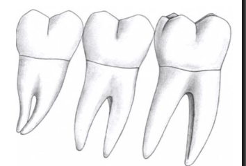
Maxillary molars
3 roots: trifurcation
mesil buccal, distal buccal, and palatal
wider buccal-lingual than mesial distal
Mandibular molars
pentagonal in shape
wider mesiodistal than buccallingual
2 roots
longest root to crown ratio
Maxillary 1st molar
widest mesiodistally in maxillary arch
widest facial lingually in the dentition
Cusp of carabelli seen on its lingual surface of Mesial lingual cusp
does not develop from lobe
Mandibular 1st molar
widest faciolingually in the mandibular arch (the widest of all ficiolingual is the maxillary first molar
mesial root is longest of any molar with greatese faciolingual dimension of any tooth
maxillary third molars (typically not asked, but good to know)
are the shortest of all permanent teeth
Root Anatomy and Pulp Morphology
root furcation = space between the roots
bifurcation = 2 roots
trifurcation = 3 roots
F
Pulp cavity = pulp chamber + root canals
orfice is the opening/path from pulp chamber to root (pulp) canals
Pulp horns
incisors generally have 3 pulp horns
exception is the maxillary (peg) lateral, 1 pulp horn
cusped teeth (canines, premoals, molars) have one pulp horn under each functional cusp
pronounced in the teeth of younger people
Accessory (lateral) canal, chemical irrigation used to reach (disinfecting the area)
most often located in the apical third of the root
Types of Canal configurations
Type 1 just one canal
Type 2: two canals that combine in to one apically, single root
type 3: two separate canals
type 4: single canal, but
Anterior Teeth
all have one root, except the mandibular canine, which is most likely to have 2 roots (facial and lingual)
Premolars
Maxillary first premolars
if it has 3 roots, the third is usually
Molars
Maxillary moalrs
most often have 3 roots and 4 canals
two canals in the mesiobuccal root
more likley in maxillary first molar
Third molars: usually have 3 canals
considerable variation in root formation
Triangles are shown to account for the root horns when accessing the roots.
we see oval access, a canine has one pulp horn
ovals in premolars, when there are two cusps, its in the buccal lingual formation
Principles of Digital Dentistry
Intraoral scanners Advantages
reduce patient discomfort
time efficient
can “add” to a scan, no need to redo
eliminates shipping time to lab
Enhanced diagnostics
better visualization and planning
early detection of dental issues
Eliminates impression materials and stone models
allows better communication with lab
Accuracy
9 microns
no material distortion
Versatility
Pt education
Addititive manuafacturing
you take an intraoral scan you and send it to a 3d scanner that will add material in small increments that will give you the object ( 3d model, etc.)
Subtractive Manufacturing
take digital impression, subtracts the parts you don’t want , grinds around
Intraoral scanners work
project light to area being scanned and the light will bounce off the object and hit back at the sensor (capture reflections by imaging sensors
Mirrors are widely used to increase the measuing area of a single view (multiple angles)
Operative Dentistry
Dental Caries
preventable
chronic infectious disease
biofilm-mediated
Cavity (hole)
used to describe a caries lesion that had progressed to the point that there was a breach in the surface integrity of the tooth
Drill, use Prepare
Fill, use Restore
GV Black noticed carious lesions were not random
caries repeated developed in predictable anatomical locations
Pit-and-fissures lesion in places hard to clean
Smooth-surface lesions occured proximally and cervically
GV black caries classification
Class 1: Pit and fissure
Class 2: proximal surface of posterior teeth, always has 2 distinctions
smooth surface caries
two distinctions mesial/distal and occlusal
mesial occlusal, distal occlusal, medial occlusal distal
happens at/below contact point
Class 3: proximal surfaces of anterior teeth
does not involve incisal edge
smooth surface caires
Classify based on mesial/distal, and has lingual/facial
Class 4: same as class 3 but includes the incisal edge, commonly seen in fractures
Class 5: gingival third of the crown, facial or lingual surface of all teeth
Class 6: incisal edge of anterior teeth or the occlusal cusp tips of posterior teeth
not common
all classes are smooth surface caries except for class 1
The cavity preparation became specifically engineered with certain mechanical properties
cavosurface finish: finishing enamel walls and margins
“Extension beyound prevention” prepare (drill) beyond the decay to prevent spread of caries.
for GV Black: Preparation = disease + tooth anatomy + material requirements + prevention
Cariology: study of caries as a disease, not simply a defect requiring surgical removal
Classic GV Black
prep with properties to hold and maintain the restoration
“extension for prevention”
Modified GV Black
similar shape but preserve as much natural tooth structure as possible
prevents tooth fracture
Contemporary
remove decay and restore with a resin-based restoration (i.e. composite)
Modern Operative Dentistry: Tooth preparation
1. conserve as much healthy tooth structure as possible
2. remove all defects while simultaneously providing protection of the pulp-dentin complex
3. form the tooth preparation
4. allow for esthetic placement of a restorative material where indicated
FActors that dictate preparation include:
iatrogenic damage: damaging harm to teeth, surrounding tissues accidentally
9 steps of cavity preparations
1. Outline Form
2. Resistance Form (primary)
the shape and placement of the preparation of the walls and floors, comes from smooth flat floors that helps resistance
line angles should be rounded and not 90 degrees
forces oritned parallel to long axis
•3. Retention Form (primary)
to retain your restoration, hold it in place
result of tipping forces or lifting (forces oriented at an angle to the long axis)
Comes from walls
Amalgam macro-mechanical retentsion, dont make the walls straight, make it cone shaped
compositive can be put of straight walls
• 4. Convenience Form
is the shape or form that provides adequate observation, accessibility, and ease in the preparation, caries removal, and restoration of the tooth
5. Removal of Decay
6. Pulpal Protection (if needed)
dentin provides little protection
avoid over desiccating and over drying the dentin
7. Secondary resistance and retention form
come from other things that are not the floor or wall
slots and pins
• 8. External wall (cavosurface) finishing
cavosurface finishing, (margination), and finishing external preparation walls all mean the same thing
allows the creation
unsupported enamel is weak, make sure the enamel is covered by dentin
make sure all enamel walls that form a 90 degree exit angle with the cavosurface
• 9. Debridement
cleaning of the tooth preparation using air/water syring
Handpieces (not drill)
High speed
must always be used with water
friction grip burs
Slow speed
for caries excavation and finishing
latch grip burs (or friction grip burs)
forward- caries excavation
reverse - smoothing and polishing
motor, contra angle, nose cone
Carbide Burs
have cutting etch
round is for excavating decay
come in high speed and slow speed
1556 high speed
RA 56 low speed
Diamond Burs
used to cut teeth
comes in different grits
coarse grits used to remove bulks
fine grits for finishing and crown preparations
Operative Dentistry 2
3.8mm sink halfway your about at 2mm
class 2
broken gingival contact
that is where the decay happens
at or below the contact point
open facial and lingual embrasures 0.25mm
measured with tine of explorer
axial depth is the distance between gingival floor and axial wall
premolar 1mm
molar 1.5mm
axial wall should be convex or follows contour of gingical cavo-surface margin or straight
axial pulpal line angle should be rounded (line that forms when axial wall and pulpal floor meet
broken gingival contact is when the two adjacent teeth are no longer touching
you should be able to pass your explorer
The proximal box is centered on contact area because the contact area is centered
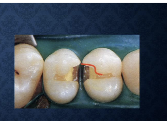
S curve = extreme facial contact in the maxillary premolars, MO surface
Class 2 Tools and Procedures
Interproximal guard is the curvy guard
Class 3
facial or lingual, lingual 90% of the time
outline form is a rectangular shape
mesial and distally you don’t cross the marginal ridge
you wnat your preperation to interuupt the triangle (in order to know that you have broken gingival contact)
unsupported enamel on facial wall is acceptable since susceptability for fracture is low
you want everything at 90°
330 bur (pear shaped)
Class V (5)
shape of the outline form is directly proportional to the mesio-distal diameter of the tooth, with the outline following the contour of the cementoenamel junction
prep will be in the middle 2 quarters (middle half)
in the incisal half of prep, 1.5mm (because the incisal half is thicker than the gingival half)
gingival half prep needs to 1mm
a bevel is a softening of the prepared cavosurface margin (softening to 45°)
exposing more enamel for bonding
Only bevel for composite restoration
Only bevel on enamel
Diamond burs
Don’t bevel for Amalgam
1556 carbide bur
Rubber Dam
mandatory for all bonded restoration because bonded restorations do not cure in the presence of moistures
Isolation strategies
Anterior isolation (class 3, 4, 5)
first premolar to first molar; use 2 clamps
2 pieces of foss in each hole and tie to ligate clamps to the rubber dam frame
Other methods of controlling the operating field
drugs
antisialogogues: not routinely used for operative procedures
epinephrin
dri-angles (bibulous paper)
Cotton rolls
vacuum devices
Place the rubber clamp below the height of contour to prevent it from flying out
the bow should be faced towards the posterior, face the throat/back of the mouth
bring marker and mirror for friday as well as rubber day stuff
tuesday: learners blocks, white yellow red (on tuesday)
bring handpieces and all of your burs