GPHY
WEEK 7. MUSCULAR SYSTEM |
|---|
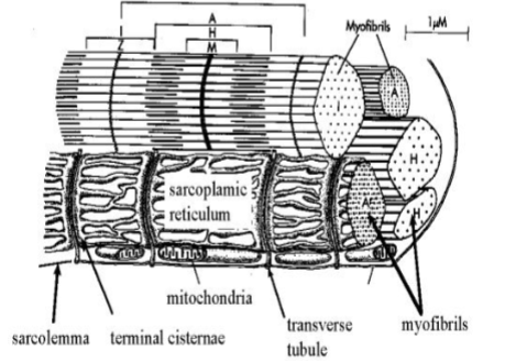
Parts of the Muscle Tissue
- Sarcolemma – cell membrane
- Sarcoplasm – cytoplasm of the muscle cells
- Sarcosome – granules in the cytoplasm
- Myofibrils – fine threadlike structures in the sarcoplasm
- Sarcoplasmic reticulum – endoplasmic reticulum
- Sarcomere – structural and functional unit of skeletal muscle
Connective tissue covering
- Endomysium - A thin layer of connective tissue that surrounds individual muscle fibers (muscle cells) within a muscle.
- Perimysium - A thicker connective tissue layer that surrounds bundles of muscle fibers called fascicles within a muscle
- Epimysium - A dense layer of connective tissue that surrounds the entire muscle, providing structural support and protection.
–
Myofibrils
- Light bands – isotropic bands ( I bands)
- Dark bands – anisotropic bands (A bands )
- Z line – bisects the I band
- H band ( Heller band ) – lighter mid portion of the A band
- M line ( Mittelscheibe line ) – bisects the H band
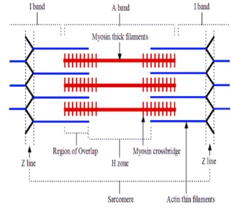
Muscle Filaments (Myofilaments)
- Thick filaments – occupy the middle zone of sarcomere
- Thin filaments – occupy the peripheral zones of the sarcomere
–
Proteins in Muscle Filaments
- Actin – principal protein component of thin filaments
- Troponin and tropomyosin – arranged along both sides of the actin filament
- Myosin – make up the thick filaments.
–
Neuromuscular Junction
- Presynaptic terminal - End of a neuron's axon releasing neurotransmitters.
- Synaptic vesicles - Small sacs in the presynaptic terminal storing neurotransmitters
- Synaptic cleft - Tiny gap between neurons where neurotransmitters diffuse.
- Postsynaptic membrane - Membrane of the receiving neuron or cell where neurotransmitters
- bind, triggering cellular responses.
–
Neurotransmitters
- an action potential travels along an axon membrane to a neuromuscular junction
- calcium channels open and calcium enters the presynaptic terminal
- acetylcholine is released from presynaptic vesicles
- acetylcholine stimulates Na channels on the postsynaptic membrane to open
- Na diffuses in the muscle fiber
- Action potential travels along the sarcolemma and t-tubules
- Sarcoplasmic reticulum releases Ca
- On the actin, Ca binds to troponin. Myosin attachment sites are exposed
- Myosin heads bend causing the actin to slide past the myosin.
- Contraction occur
–
Types of Muscle Tissue
- Skeletal muscle - associated with the skeletal system.
- usually attached to bones
-striated and voluntary
- Cardiac muscle – found in the heart
-striated and voluntary
- Smooth muscle - found in the walls of visceral organs like blood vessels digestive tract, respiratory tract, urinary and reproductive organs.
- non-striated and involuntary.
–
Parts of Skeletal Muscle
- Origin –stationary attachment of muscle usually attached to bones
- Insertion – more movable attachment of the muscle
- Belly- fleshy part of the muscle
–
MUSCLE GROUPS ACCORDING TO THEIR PRIMARY ACTIONS
- Prime mover or agonist- muscle whose contraction is chiefly responsible for producing a particular movement.
- e.g. orbicularis oculi is the prime mover in closing eyes
- Antagonist – oppose action of the agonist
- e.g. levator palpebrae superioris which opens the eye is the antagonist of the orbicularis oculi which closes the eye.
- Synergists- contraction assists prime movers in performing action to reduce excess and unnecessary motion.
- e.g. latissimus dorsi is a back muscle that extends , adducts and medially rotates arms. Teres major is a synergist helping latissimus dorsi in starting its actions when the arm is at full flexion.
–
BASES OF NOMENCLATURE OF MUSCLES
Muscles are named according to:
- Shape - trapezius, rhomboideus, deltoid, quadratus
- Size - maximus, minimus,major, minor
- Location - frontalis, occipitalis, branchi, femoris, abdominis
- Direction of fibers - rectus, oblique, transversus
- Points of attachment – sternocleidomastoid, hyoglossus
- Position - superior, inferior, external, internal
- Action - flexor, extensor, supinator, pronator
- Structure - semitendinosus, semimembranosus
- Number of bellies - digastric
- Miscellaneous – Sartorius , (tailor’s muscle)
- Combination-
- e.g. external oblique (position and direction of fibers)
–
AXIAL MUSCLES
A. Muscles of Head
- Muscles of facial expression
- Mouth
- - buccinators (trumpeteer’s muscle)
- orbicularis oris (purses lips)
- risorius (for sardonic smile )
- Depressor anguli oris (triangulis )
- zygomaticus major (for sweet smile) (laughing muscle)
- Eye
- corrugator supercilii (draws eyebrow medially)
- orbicularis oculi (closes eye)
- Nose
- procerus (wrinkles root of nose)
- nasalis (compresses nose as n sniffing)
- Scalp
- epicranius
- moves scalp forward and backward
- made up of the following
- frontalis (for transverse wrinkles in forehead when one is Surprised)
- Occipitalis
- Mouth
- Extraocular muscle - move eyeball in different directions
- Recti muscles - superior rectus inferior rectus , medial rectus, lateral rectus
- Oblique muscles - superior oblique and inferior oblique
- Levator palpebral superioris - opens eyes
- Muscles of mastification - muscles used in chewing
- Masseter
- Temporalis
- Medial pterygoid
- Lateral pterygoid – protects mandible
- Muscles of the tongue
- Intrinsic muscles - form shape & body of tongue
- Extrinsic muscles - attached to other structures aside from tongue
e.g. genioglossus
- protrudes tongue
- Muscles of the middle ear- contract when there is loud sound to weaken the vibration of the ossicles
- Stapedius
- weakens the vibration of stapes
- Tensor tympani
- weakens the vibration of malleus
- Muscles of soft palate
- tensor veli palatini - elevate soft palate during swallowing
- levator veli palatini
- Muscles of pharynx – muscles of swallowing
e.g. constrictor muscles- superior, middle and inferior constrictors.
B. MUSCLES OF NECK
- Superficial muscle
- platysma (only muscle in superficial facial of neck)
- Lateral neck muscles
- e.g. sternocleidomastoid
- Suprahyoid muscles- elevate hyoid bone and depress mandible
- Digastric
- Mylohyoid
- Infrahyoid muscle (STRAP MUSCLES)- depress hyoid bone and elevate
- Larynx
- Laryngeal muscles
- Intrinsic
- Cricothyroid
- tenses vocal fold
- Posterior cricoarytenoid
- abduct vocal fold
- Cricothyroid
- Extrinsic
- Sternothyroid
- Thyrohyoid
- Intrinsic
C. MUSCLES OF THE THORACIC WALL
- mainly used for respiration
- External intercostal
- Internal intercostal
- Transversus thoracis
D. DIAPHRAGM
- Separates thoracic cavity from abdominal cavity
- Most important muscle of respiration supplied by phrenic nerve
E. MUSCLES OF ABDOMINAL WALL
- Antero- lateral abdominal muscles - flex trunk and increase abdominal cavity pressure used in activities like defecation , micturition
1. External intercostal
2. Internal intercostal
3. Transversus thoracis
POSTERIOR ABDOMINAL MUSCLES
e.g. psoas major/ minor
F. MUSCLES M OF PELVIS
- Piriforms- found at posteripr wall of pelvic cavity
- Obturator internus – found at lateral wall of pelvic cavity
- Levator ani and Coccygeus form the pelvic diaphragm to support pelvic organs separating pelvic cavity and perineum
G. PERINEAL MUSCLES
- Bulbospongiosus and Ischiocavernosus cover the root of clitoris or penis
H. MUSCLES OF BACK
- Superficial- actually belong to muscles of the upper extremities
- Trapezius
- Latissimus dorsi
- Levator scapula, Rhomboid minor and Rhomboid major attached to medial border of scapula
- Deep (Postvertebral muscles)
- intrinsic muscles of the back
- Erector spinae (sacropinalis) - extends vertebral column
–
APPENDICULAR MUSCLES
- MUSCLES OF UPPER EXTREMITY
- Muscles connecting upper limb to thoracic wall (superficial anterior thoracic muscles)
- Pectoralis major – found underneath mammary gland
- Pectoralis minor- underneath pectoralis major
- Serratus anterior- found in lateral thoracic wall
- Scapulohumeral muscles
a. Deltoid – chief abductor
b. Rotator cuff muscles
e.g. Supraspinatus – initiates abduction of arm infraspinatus
Teres minor
- Arm Muscles
a. Anterior compartment
- common action: flex forearm
- Biceps branchi powerful supinator and flexor of forearm
- Brachialis – chief flexor of the forearm
b. Posterior compartment
- action : extends forearm. there is only one muscles here - tricep brachii
- Forearm muscles
- Anterior forearm
- predominant action of this group : flex wrist and flex digits
Superficial group:
- Pronator teres
- Flexor carpi
- Palmaris longus
- Flexor carpi ulnaris
- Lateral forearm
- Brachioradialis
- Extensor carpi radialis longus
- Posterior forearm
- predominant action of this group : extend wrist , extend digits
Superficial group
- Ext. carpi radialis brevis
- Extensor digitorum
- Extensor digiti minimi
- Extensor carpi ulnaris
- Anconeus
- Muscles of the hand
- thenar muscles
- hypothenar muscles
B. MUSCLES OF LOWER EXTREMITY
- Thigh muscles
a. Anterior thigh compartment
- predominant action of this muscles group : extend leg
- quadriceps (femoris)
- vastus mediallis
- vastus laterallis
- vastus intermedius
- rectus femoris
- Sartorius (tailor’s muscle)
- Iliopsoas
- chief flexor of thigh
b. Medial thigh compartment
- predominant action of this group is adduction of thigh
- Gracilis
- Adductor logus and magnus
c. Posterior thigh compartment (called Hamstring muscle )
- Includes all posterior thigh muscles except for biceps femoris short head)
- actions of this group : flex leg and extend thigh
1. biceps femoris long head
2. semitendinosus
3. semimembranosus
4. adductor magnus
(harmstring portion )
2. Leg muscles
- Anterior leg compartment
- actions: dorsiflex foot, extend toes
1. tibialis anterior
2. extensor halluces longus
- extends big toe
3. extensor digitorum longus
- extend 4 lesser toes
4. peroneus tertius
- Lateral leg compartment
- Actions - evertors of foot and plantar flexors
1. peroneus longus
2. peroneus brevis
c. Posterior leg compartment
Superficial group (calf muscles)
- actions : plantarflexion
Gastrocnemius and plantaris
Can also flex leg
1. Gastrocnemius
2. Soleus
3. Plantaris
3. Gluteal muscles
a. Chief extensor of thigh
e.g. gluteus maximus
b. Abductor of thigh
e.g. gluteus medius
c. Small lateral rotators thigh
e.g. Piriformis
4. Muscles Foot
a. Dorsum of foot
1. Extensor halluces brevis
2. Extensor digitorium brevis
b. Muscles of sole
First layer
1. Abductor halluces
2. Flexor digitorum
3. Abductor digitiminimi
–
CLINICAL CORRELATION
Disorder of muscles
1. Myalgia - refers to muscular pain
2. Myositis - inflammation of muscular tissue
3. Fibrositis - inflammation of connective tissue within a muscle
4. Muscular Dystrophy - a progressive disorder most often in male children ending in complete helplessness. Child begins to walk clumsily and tends to fall
5. Muscle atrophy - degeneration of muscle fibers which become smaller than their normal size.
–
WEEK 8. NERVOUS SYSTEM |
|---|
NERVOUS SYSTEM
- A group of tissues composed of highly specialized cells possessing the characteristics of excitability and conductivity.
I. MORPHOLOGICAL DIVISIONS:
A. Central Nervous System
1. Brain
2. Spinal cord
B. Peripheral Nervous System
1. Cranial Nerves (12 pairs)
2. Spinal Nerves (31 Nerves)
3. Autonomic Nervous System
–
II. FUNCTIONS AND DIVISIONS:
- Somatic Efferent
-Innervating somatic structures like skeletal muscles and skin.
B. Visceral Efferent
- Innervating visceral or involuntary structures
like smooth muscle, cardiac muscles and glands.
This is the autonomic nervous system.
–
TYPES OF CELLS IN THE NERVOUS SYSTEM
The nervous system is composed of special tissues containing two major types of cells.
- Neurons
- the active conducting elements
- Neuroglia
- the supporting elements
NEURON
- basic unit of the nervous system which conducts electrical impulses from one part of the body to another.
- consist of a cell body (perikaryon), containing a single nucleus, and processes transmitting impulse to and from the cell body.
TWO TYPES OF PROCESSES
- Dendrites
- group of short, unsheathed processes arranged like branches of a tree that transmit impulse toward the cell body
- Axons
- a single, elongated sheathed process conducting impulses away from the cell body.
ACCESSORY CELLS
- The non-nervous elements consist of blood vessels, connective tissue, and supporting cells known collectively as neuroglia.
SYNAPSE
- Synapses are points of connection between neurons. The axon of one neuron makes functional contact with dendrites of another neuron. Certain chemicals, called transmitters are released from axonal endings for impulse to leap the synaptic junction:
Main Neurotransmitters:
- Epinephrine (adrenalin)
- Norepinephrine (noradrenaline)
- Acetylcholine
- all are released by autonomic fibers.
- Acetylcholine released at neuromuscular junction.
–
NERVE IMPULSE
Cell membrane of unstimulated neurons carries an electric charge. The inside of the membrane is negative compared with the outside. A nerve impulse is a localized reversal in the charge of cell and spread there like an electric current. The sudden electric change in the membrane is called ACTION POTENTIAL. A stimulus can start this action potential. The electric change is due to the rapid shift in sodium and potassium across the cell membrane. This occurs very rapidly and followed by rapid return of its membrane to its original state to its original state so it can be stimulated again.
- MYELIN SHEATH
- Insulating material covering axons in central and peripheral nervous system
- The thicker the myelin sheath it is, the faster the passage of nerve impulse will be.
- The electric impulse “jumps” from node to node in myelin sheath instead of traveling continuously along the nerve fiber. (SALTATORY CONDUCTION OF IMPULSE)
–
WEEK 8.1. CENTRAL NERVOUS SYSTEM |
|---|
- Central Nervous includes the BRAIN and SPINAL CORD
- Each division is further divided grossly in gray and white matter:
A. Gray matter
- so called because if its darker appearance and preponderance of nerve cell bodies
B. White matter
- composed chiefly of myelinated nerve fiber
- in the spinal cord an H-shaped central region of gray matter is surrounded by the white matter
GRAY MATTER
- In the surface layer (cortex) of the cerebrum and cerebellum. the brain the gray matter is broken into clumps or is present as
- Nucleus designated a mass of gray matter in any part of the brain or spinal cord.
- Ganglion also means cluster of nerve cell bodies and dendrites but unusually refers to the cells located outside the brain and spinal cord.
BRAIN
- Part of the central nervous system contained within the skull.
- Most complex and largest mass of nervous tissue in the body and contains literally billions of nerve cells.
EMBRYOLOGICAL DIVISIONS OF THE BRAIN
1. Forebrain (Prosencephalon)
a. Telencephalon (cerebrum with basal ganglia)
b. Diencephalon (medulla oblongata)
2. Midbrain (Mesencephalon)
3. Hindbrain (Rhombencephalon)
a. Metencephalon (cerebrum,pons)
b. Myelencephalon (medulla oblongata)
–
- PROSENCEPHALON
- TELENCEPHALON (CEREBRUM)
- represent seven-eight of weight of brain
- surface layer of gray (cerebral cortex) greatly expanded by convolutions, gyri. Internal to which is the white substance made up of nerve fiber
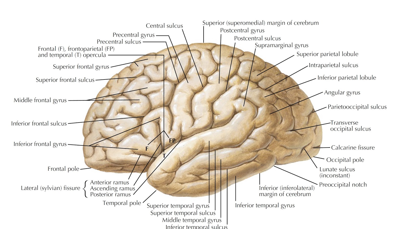
FISSURES OF CEREBRUM
1. Longitudinal Fissure
- runs from posterior to the anterior aspects almost completely dividing it into 2 hemisphere
- The hemispheres are connected in the midline by the corpus callosum (Largest band of crossing or commissural fiber). This is found in the depths of longitudinal fissures.
2. Lateral Sylvian Fissure
- between the frontal and parietal lobes above and temporal lobes below
3. Central of Sulcus (Rolandic Fissure)
- between frontal and parietal lobes
4. Parieto- occipital fissure
- between the occipital and parietal lobes
5. Calcarine Fissure
- found in the occipital lobe perpendicular to parieto occipital fissures around which is the visual center.
LOBES OF THE CEREBRAL CORTEX
1. Frontal lobe
- includes all cortex lying anterior to the central sulcus of Rolando and above the lateral sulcus Sylvius
- center for motor functions and personality
2. Parietal Lobe
- lies posterior to central sulcus of Rolando and above lateral Sylvian Fissure
- center for ordinary sensory functions
3. Temporal Lobe
- lies beneath the lateral sulcus of Sylvius
- center for hearing and olfaction
4. Occipital Lobe
- occupies the posterior extremity of the cerebral hemisphere behind the parieto-occipital fissure
5. Insula (Island of Reil)
- Exposed when the lips of lateral Sylvian Fissure are separated
FUNCTIONS OF THE CEREBRUM
1. Seat of advanced intellectual functions like memory storage, recall, learning and reasoning for comprehension and execution of language.
2. Perceptions of all sensation and sites where one modality of sensation can be integrated with others
3. Initiation of movements
FUNCTIONAL AREAS OF CEREBRAL CORTEX
1. Primary Motor Area or Pre –central gyrus
- lies in the frontal lobe immediately anterior to the sulcus
- controls voluntary movements in the opposite side of the body
2. Pre- frontal Area
- concerned with behavior, character and emotional state of the individual
- concerned with foresight, good judgement, abstract thinking.
3. Primary sensory/Somesthetic Areaor Post-central gyrus
- lies behind the central sulcus of Rolando in Parietal lobe
- ordinary sensation of pain, temperature, pressure and touch, position and movements sensation from the opposite side of the body are received and interpreted here.
4. Motor Speech Area
- Lies in inferior frontal gyrus of dominant hemisphere (usually the left)
5. Sensory Speech Area
- Lies in the temporal lobe posterior to auditory area of dominant hemisphere (usually the left)
- Wernike’s Area
6. Auditory or Hearing Area (transverse gyri of Heschl)
- lies below lateral sulcus within the temporal lobe
- center for hearing
7. Visual Area
- cortex area around the calcarine fissure
- found in occipital lobe
8. Olfactory or smell area
- within the temporal lobe
9. Taste Area
- above lateral sulcus into the deep layers of the sensory area
BASAL GANGLIA
- four paired masses of gray matter embedded in the white matter of the cerebral hemisphere
- include the caudate nucleus (medial portion) and the putamen and globus pallidus (lateral portion) collective called lentiform nucleus
- basal ganglia play an important role in the control of motor function, and injury to them produces unintentional unnecessary movements
B. DIENCEPHALON
- located in the forebrain along with the cerebrum
- made up of:
- Thalamus
- paired mass of gray matter situated below corpus callosum
- highest subcortical sensory integrating center
- all sensory impulses (ordinary and special) should pass the thalamus first before going to cerebral cortex, except for smell which can go directly to cerebral cortex
- Hypothalamus
- involved in the regularization of body temperature, feeding activities, concentration and volume of extracellular fluid, autonomic nervous system responses, endocrine functions
- where pituitary gland is attached to
2. MESENCEPHALON
- Connect the forebrain and hindbrain
3. RHOMBENCEPHALON
- CEREBELLUM (Part of metencephalon)
- The constricted central portion is called vermis (Latin of worm) and the lateral expanded portions the hemispheres.
- resembles the cerebrum in structures, with the gray matter forming a layer of cortex place in the surface
- the cerebrum greatly aids the motor cortex of the cerebral hemispheres in the integration of voluntary movements. Injury does not result in to paralysis of muscles but loss in coordination only of these motor activities.
- PONS (Part of metencephalon)
- lies anterior to the cerebellum and between the midbrain and medulla
- bridge like structure, consisting almost entirely of white matter linking the various parts of the brain.
- MEDULLA OBLONGATA (myelenencephalon)
- continuous with the spinal cord inferiorly and the pons superiorly
- lies ventral to the cerebellum
- has a number of vital regulatory and reflex center s, including those controlling the circulatory system, breathing, swallowing, vomiting, coughing, sneezing
–
VENTRICLES OF THE BRAIN
- spaces inside the brain filled with cerebrospinal fluid
- The Ventricular System includes:
1. Lateral ventricle
- found inside the cerebral hemisphere
- each lateral ventricle communicates within the third ventricle by way of interventricular foramen (foramen of Monroe)
2. Third ventricle
- small slit like cavity in the center of the diencephalon in between the 2 thalami
- continuous with the cerebral aqueduct of Sylvius, a canal which passes through the midbrain
3. fourth ventricle
- lies between the cerebellum on the posteriorly side and the pons and medulla on the anterior side
- communicate within sub arachnoid space through Foramen of Luschka and Magendie.
–
MENINGES
- These are the membranes collectively known as meninges provide protection to the brain and spinal cord.
- From outside in, these are the :
A. Pachymenix
1. Dura mater
B. Leptomininges
1. Arachnoid mater
2. Pia mater
- DURA MATER
- The dura mater (Latin for hard mother) the outer meninx, is made of dense, fibrous tissues.
- these are two portion of the dura mater:
1. cranial
2.spinal
- The cranial dura is arranged in two layers. (outer endosteal and inner meningeal)
- the inner layer becomes continuous with the one layered dura mater in the spinal cord
- The potential space in between dura and bone is epidural space. The one between dura and arachnoid mater is subdural space filled with small amount of serous fluid
- ARACHNOID MATER
- delicate serous membrane located between the dura and pia.
- as the name implies, it has the microscopic appearance of spider web.The subarachnoid space in between the arachnoid and the pia is occupied by the thin, delicate connective tissue trabeculae and intercommunicating channels in which cerebrospinal fluid is contained the cranial portion invest in brain loosely
- PIA MATER
- The pia mater (gentle mother) is a vascular membrane consisting a plexus of fine blood vessels held together by areolar connective tissue
- the cranial portion invest in the surface of the brain and dips down into the sulci
–
CEREBROSPINAL FLUID
- Colorless fluid similar to lymph circulating within the ventricles, the central canal of spinal cord and also within subarachnoid space
- the volume: about 150 ml.
- serves as a water cushion to guard the brain and spinal cord against injury
- Cerebrospinal fluid is continuously formed in all four ventricles by active secretion, principally from capillaries of the choroid plexus (pouch like projections of the pia mater into the ventricles covered with the ependymal lining of the ventricles
CSF PATHWAY
- CSF from choroid plexuses in the lateral ventricles
- interventricular foramen of Monroe
- third ventricle
- iter of aqueduct of Silvius
- 4th ventricle
- Foramen of luschka and Magendie subarachnoid space arachnoid villi
- absorbed in the venous circulation
–
SPINAL CORD
- the elongated almost cylindrical part of CNS
- 45cm long lying within the vertebral canal
- continuous with the medulla oblongata above and extends from the level of foramen magnum to the lower border of 1st lumbar vertebra in adult and level of L3 vertebra in children
- 2 enlargements:
- cervical enlargements (where nerve supply of upper extremities arises from)
- lumbar enlargement (where nerve supply of lower extremities arises from)
- narrows down as conus medullaris, where it gives arises to the thread like filum terminale and terminates in the first coccygeal vertebra .
- lumbar and sacral spinal nerves descend along filum terminale in a bundle known as the cauda equine
–
WEEK 8.2. PERIPHERAL NERVOUS SYSTEM |
|---|
SPINAL AND CRANIAL NERVE
I. SPINAL NERVE
- A bundle of nerve fibers attached to spinal cord
- All are classified as mixed nerves –with sensory and motor fibers
- 31 pairs of spinal nerves arise from spinal cord along almost its entire length and emerge from the vertebral canal through the intervertebral foramina
- spinal segment is the part of spinal cord where a part of spinal nerves is attached
- attached to each spinal segment on the other side are:
1. dorsal root
- containing sensory fiber
2. ventral root
- containing motor fiber
- these two roots join together to the spinal nerve
- 31 pairs of spinal nerves are names from the region of the vertebral column through the which they exit
- there are eight pairs of cervical spinal nerves, 12 thoracic, 5lumbar, 5sacral, 1 coccygeal
PLEXUSES
- Group of nerve fiber from the ventral rami of cervical, lumbar and sacral spinal nerves. posterior rami never from plexuses
1. Cervical Plexus
- formed by the first 4 cervical nerves (c1, c2, c3, c4)
- this supplies the back and sides of the head and front of the neck with ordinary sensory fiber
- most important branch is the phrenic nerve composed of motor fibers supplying the diaphragm
2. Brachial plexus
- lower 4 cervical nerves (C5C6C7C8) and 1st thoracic (T1) supplies the skin and muscles of the upper limb
3. Lumbar Plexus
- from L1-L4 spinal nerves
one of its branches is:
- Femoral Nerve
- Supplies muscle and skin on anterior aspect of the thigh
- Obturator nerve
- supplies muscle and skin of medical aspect of the thigh
4. Sacral Plexus
- from L4L5 S1 S2S3 spinal nerve
the largest branch of which is sciatic nerve (the largest nerve in the body divided into common peroneal nerve and tibial nerve)
* The thoracic spinal nerve does not form plexuses.
T1-T11 spinal nerve –INTERCOSTAL NERVE
T12 spinal nerve- SUBCOSTAL NERVE
Cranial Nerves | Name | Function/s |
|---|---|---|
CNI | olfactory nerve | smell |
CNII | optic nerve | sight |
CNIII | oculomotor | moves the extraocular muscles except lateral rectus and superior oblique |
CNIV | trochlear | motor to superior oblique |
CNV | trigeminal
|
|
CNVI | abducens | motor to lateral rectus muscle |
CNVII | facial |
|
CNVIII | vestibulocochlear | equilibrium and hearing |
CNIX | glossopharyngeal |
|
CNX | vagus | sensory to skin of external auditory meatus, pharynx, larynx, thoracis and abdominal visceral motor to pharynx & larynx |
CNXI | accessory | motor to trapezius, and Sternocleidomastoid |
CNXII | hypoglossal | motor to muscles of the tongue |
WEEK 9. ENDOCRINE SYSTEM |
|---|
Endocrine system
- Made up of widely distributed organs whose secretions (hormones ) reach target tissues or organs through receptors via bloodstream.
Hypothalamus
- Contains neurosecretory neurons that secrete hormones (releasing and inhibiting hormones) which control secretion of hormones from the anterior pituitary.
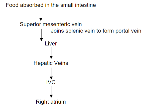
- Hypothalamic releasing and inhibiting hormones are carried directly to the anterior pituitary gland via hypothalamic-hypophyseal portal veins
- Specific hypothalamic hormones bind to receptors on specific anterior pituitary cells, modulating the release of the hormone they produce
- Ex: Thyroid releasing hormone (TRH) from hypothalamus binds to receptors on anterior pituitary cells called thyrotrophs stimulating them to secrete TSH.
Pituitary Gland ( Hypophysis Cerebri)
- Lies in sella turcica of sphenoid bone
- Attaches to hypothalamus by infundibulum
- Two lobes:
- Anterior pituitary (adenohypophysis)
- Posterior pituitary (neurohypophysis)
–
Hormones produced and secreted by the anterior pituitary gland:
- Growth Hormone ( Somatotropin )
- Stimulates liver cells to produce somatomedin C which stimulates growth of all body tissues
- Hyposecretion: dwarfism
- Hypersecretion in children : gigantism
- Hypersection in adults : acromegaly
- Thyroid Stimulating Hormone
- Controls secretion and activities of the thyroid gland
- Hyposecretion: hypothyroidism
- Adrenocorticotropic Hormone (ACTH)
- Stimulates adrenal glands to secrete glucocorticoids, mineralocorticoids and androgens
- Follicle Stimulating Hormone (FSH)
- Causes growth of follicles in the ovaries before ovulation
- Promotes formation of sperms in the testes
- Leutinizing Hormone
- Role in ovulation
- Prolactin
- Helps initiate and maintain milk secretion by mammary gland
–
Hormones stored in the posterior pituitary gland:
1. Oxytocin
- Contracts the uterus during delivery
- Stimulates milk ejection
2. Antidiuretic Hormone ( vasopressin )
- Conserves body water by decreasing urine volume
–
Thyroid Gland
- Found at the lower part of the anterior neck
- Has 2 lobes connected in the midline by isthmus
- Contains follicles (colloid cells) which secrete:
- Thyroxine
- Triiodothyronine
- Actions of thyroid hormones
- Increase basal metabolic rate
- Helps maintain normal body temperature
- Increase rate of glucose, fat and protein metabolism
- Thyrocalcitonin
- Secreted by parafollicular cells
- Regulate calcium homeostasis
- Hypothyroidism
- Hyperthyroidism
Adrenal Glands
- found at the superior pole of kidneys
- Adrenal Cortex
- Mineralocorticoids
- Regulate fluid and electrolyte balance
- Aldosterone
- Glucocorticoids
- Regulate body metabolism and resistance to stress
- Increase blood glucose concentration
- Anti-inflammatory effects
- Cortisol
- Androgens
- Promote libido in females
- Converted to estrogen which stimulates development of secondary sex characteristics
- Cushing’s syndrome
- Hypersecretion of corticosteroids
- Addison’s disease
- Hyposecretion of corticosteroids
2. Adrenal Medulla
- Secretes catecholamines (epinephrine, norephinephrine )
- Enhance effects of sympathetic nervous system
Gland | Hormone | Target tissue |
|---|---|---|
Thyroid gland |
|
|
Parathyroid |
|
|
Adrenal medulla |
|
|
Adrenal cortex |
|
|
Pancreas |
|
|
Testes |
|
|
Ovary |
|
|
WEEK 10. CIRCULATORY SYSTEM: HEART AND BLOOD |
|---|
BLOOD
- A liquid connective tissue that forms part of the cardiovascular system.
- Plays an important role in maintaining homeostasis in a living organism.
–
PHYSICAL PROPERTIES OF BLOOD
1. Average adult = 7-9 % of total body weight
- Male = 5-6 liters of blood
- Female = 4-5 liters of blood
2. Red color of arterial blood is due to oxygenated hemoglobin (Hgb)
FUNCTIONS OF BLOOD
1. Transport
- Oxygen (O2) from lungs to body tissues
2. Protection
- Blood can clot which prevents excessive loss of blood after an injury
- Host defense mechanism thru antibody production
3. Regulation
- Circulating platelets helps maintain hemostasis in all body fluid compartments
- Controls pH acid-base balance thru buffer
- Albumin osmotic pressure helps retain water
- Variable rate of flow of blood thru skin helps dissipate heat to the environment.
–
STRUCTURE OF BLOOD
- PLASMA - Liquid portion of the blood
Made up of the ff:
- Water - 90%, provides the solvent for dissolving and transport of nutrients
- Plasma proteins - Synthesized mostly by hepatocytes (liver cells)
- Albumin - promotes water retention in the blood to maintain blood volume and pressure
- Globulin - acts as a carrier molecule to transport liquid and fat soluble vitamin in the blood used as antibodies – immunoglobulin
- Fibronogen – for blood clotting
- SERUM = plasma minus fibronogen and other protein involved in clotting
- Plasma electrolytes - Inorganic molecules that separate into ions when they are dissolved in water.
- Cations = Possibly charged like sodium Na+, potassium K+, Calcium2+, magnesium Mg2+
- Anion = chloride C1 phospate PO4, iodide
- Nutrients & waste products
- Nutrients – glucose, amino acid, phospholipid, triglyceride, free fatty acid, cholesterol
- Metabolic wastes - lactic acid, nitrogenous waste(urea)
- Gases & buffers
- Oxygen, nitrogen, carbon dioxide – principal gases dissolved in plasma.
2. FORMED ELEMENTS – whole cell, cell fragments
- RBC- red blood cell – erythrocyte
- WBC – white blood cell – leukocyte
- Platelets – thrombocytes
WEEK 10.1 RED BLOOD CELLS |
|---|
RBC – red blood cell - erythrocyte
- Average:
- Male = million/cu.mm.
- Female = 4.8 million/cu.mm
- Shape – biconcave disc – like a doughnut without a hole packed in the middle
- Anucleated – no nucleus
- Absence of cytoplasmic organelles like mitochondria and Endoplasmic reticulum. Thus cannot reproduce itself.
- Contains oxygen carrying protein hemoglobin that gives Blood its red color.
- Plasma membrane - strong and flexible - which allows the red blood cell to be deformed as it is squeezes through small capillaries without rupturing.
- Life span is 120 days
- Old RBC’s are removed by macrophages in spleen
RBC Physiology
- biconcave disc shape provides a larger surface area for gas diffusion
- RBC consists mostly of hemoglobin oxygen carrying globular protein
- As blood flows through capillaries the following occurs:
- In the lungs oxygen binds with heme iron to form oxyhemoglobin. When blood reaches the body tissue capillaries hemoglobin releases oxygen first into interstitial fluid and then to cell for its most cellular metabolism.
- Carbon dioxide, a waste product of cellular metabolism from the tissue, will bind with globin to form carbaminohemoglobin. As blood flows to the lungs carbon dioxide is released by hemoglobin and then exhaled.
Erythropoiesis_ production of red blood cell
- Site of production – red bone marrow of certain bones, Vertebra, ribs sternum, pelvis, upper end of humerus & femur. Hypoxia stimulates the kidney to produce hormone erythropoietin (EPO) which in turn stimulates red bone marrow to produce the red blood cell.
–
Bloodstream
- Erythrocyte – mature red blood cells
- Destruction & removal of RBC – life span 120 days
- As the RBC ages, the protein part of its plasma membrane undergoes a normal degradation process and becomes leaky. Since it has no nucleus it cannot replace the enzyme and protein lost.
- Ultimately, it ruptures. Fragmented particles are engulfed by macrophages in the spleen and liver and the breakdown products are recycled.
–
GENERAL CONSIDERATIONS
SOME PARAMETERS USED IN EXAMINATION FOR RBC
ABNORMALITIES
- Hemoglobin (Hgb) – amount of hemoglobin in blood
- Male = 14-16 gm
- Female = 12-14 gm
* Male has higher Hgb because testosterone found more in male stimulates synthesis of EPO
- Hct – hematocrit (to separate) = number of RBC in whole blood
- Male = 45 – 52 %
- Female = 37 – 48 %
–
RBC DISORDERS
1. Anemia – oxygen carrying capacity of blood is reduced
2. Hemophillia – inherited deficiency of clotting in which bleeding may occur spontaneously or after a minor trauma.
WEEK 10.2 WHITE BLOOD CELLS |
|---|
WBC- white blood cell – leukocyte
- 5,000 – 10, 000 cells/of blood
- has nucleus
- DO NOT have Hgb
- Most live in few days except lymphocyte can live for several months or years
- Combats pathogen by phagocytosis and immune response
- Chemotaxis - process by which neutrophil and other WBC are attached to the chemicals released by microorganism at the site of infection or injury
- Diapedesis - process by which WBC leaves the bloodstream by being able to deform, elongate, squeeze through pores of capillaries to reach injured tissue.
- Once granulocytes and monocyte leaved the bloodstream to fight injury or infection they never returned to the bloodstream.
- Lymphocyte - continuously recirculate when it leaves the bloodstream goes into the interstitial space to interstitial fluid to lymphatic fluid back in blood stream. Very small percent circulate in the bloodstream. Mostly they are found in lymphatic fluid and lymphatic organs.
TYPES OF WBC
A. GRANULAR LEUKOCYTE
- named after their affinity to dye/stain (Wright)
- segmented or multilobulated nuclei
- has granules in cytoplasm
- Neutrophil – 60 – 70% of the WBC PMN – polymorphonuclear
- Most abundant of the WBC nucleus 3-5 lobes
- connected by very thin strand of chromatin
- released in blood in band form – young neutrophil
- BARR Body – inactive X chromosome found only in female important in sex identification
- First line of defense – responds quickly to bacterial infection
- fine granules evenly distributed in cytoplasm contains lysosome. Stains neutral pink – blue in the process of phagocytosis granules are depleted; neutrophil dies together with Microorganism forms pus (yellowish material)
- Eosinophil – 2-4 % of WBC
- 2-3 lobes of nucleus
- larger uniform sized granules than neutrophil
- granules contain lysozymes, peroxidase to destroy intruders.
- Stains pink in acidic dyes.
- Phagocytize Ag – Ab complexes
- Destroy certain parasitic worm
- Combat effects of histamine in allergic reaction by releasing histaminase
- Basophil – 0.5 – 1 %
- coarse large granules which stain dark blue obscuring S-shaped nucleus
- liberates heparin (anticoagulant); slow reacting substance of anaphylaxis SRS-A
B. AGRANULAR/LEUKOCYTE/WBC
- no granules
- have unsegmented nucleus
- Lymphocyte – 20-25%
- 2nd most abundant leukocyte
- round slightly indented nucleus
- cytoplasm forms a rim around the nucleus,
- not phagocytic
- they are produced in the bone marrow and lymphoid tissue
- seen in acute viral infection and chronic bacterial infection
- produces antibodies
- types of lymphocyte:
- B cell
- T cell
- NK cells (natural killer cell)
- Monocyte – 3.8%
- kidney shaped nucleus
- largest of formed elements
- stays in the bloodstream only for 3 days
- capable phagocytosis as it transforms to Macrophages
–
LEUKOPOIESIS - production of WBC
- 150, 000 – 400, 000 cells of blood
- disk-shaped cells fragments
- no nucleus but have many vesicles
- lifespan 7 to 8 days
- not actually blood cell but cellular fragments
- aged platelets are removed by macrophages
- Platelet when it enters the bloodstream pick-up and stores chemical substance that can released later to help seal blood vessel breaks
THROMBOPOIESIS – production of thrombocyte/platelet
HEMOSTASIS – the prevention of blood loss
Mechanism involved:
- Vasoconstrictive Phase - when tissue is damaged and blood vessels break immediately the blood vessel muscular wall constrict to prevent further loss of blood.
- Platelet Phase -
- platelet adhesion - initially when tissue is injured platelets migrate and attach themselves to the collagen fiber of connective tissue underlying the damage area.
- platelet aggregation- As more platelets stick together at the damage area they become more sticky eventually forming a platelet plug. As they interact with one another they release substances from their vesicles like serotonin, which constricts the vascular smooth muscles, which decreases the blood flow and stops the bleeding. Injured blood vessels may continue to constrict only for about 20 mins. If the injury is extensive, the intricate mechanism of clotting continue
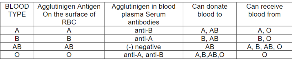
RH System
- so named because it was discovered first in RHESUS MONKEY= RH FACTOR
- D antigen is most important in production of antibodies
- people whose RBC had RH antigen are designated as RH positive and those without are RH negative
- Normal plasma does not contain RH antibodies. However, the immune system starts to make RH+ antibodies that will remain in the blood if an RH- person receives RH+ transfusions. If a second transfusion of RH+ blood is given later, the previously formed anti RH positive antibodies will cause hemolysis(rupture) of the RBC’s in the donated blood.
WEEK 10.3 BLOOD VESSELS |
|---|
BLOOD VESSELS
- Passageway through which blood flows to reach the different tissues in the body.
TYPE OF BLOOD VESSELS
- Arteries
- Veins
- Capillaries
CAPILLARIES
- smallest and most numerous of blood vessels
- prime function - exchange of nutrients and waste materials between the blood and tissue cells.
PULSE
- Traveling pressure was caused by the alternate expansion and recoil after ventricular expansion
- normal pulse rate = 70 - 80 beats per minute
- tachycardia - pulse rate over 100 beats per minute
- bradycardia - pulse rate below 60 beats per minute
- endurance trained athletes exhibit bradycardia
Common Pulse Points
- Superficial temporal artery - infront of ext. acoustic meatus
- Facial artery - anterior to angle of mandible
- Femoral artery - midpoint of inguinal ligament
- Popliteal artery - posterior to the knee
- Radial artery - anterolateral side the wrist
- Dorsalis pedis - dorsum of foot, proximal portion
–
BLOOD PRESSURE
- is the force (energy) in which blood is pushed against the walls of the blood vessels and circulated throughout the body when the heart contracts
- is highest at the arteries near the heart and drops as it reaches the veins
- Systolic Blood Pressure (SBP) is the highest arterial pressure during systole – (ventricular contraction)
- Diastolic Blood Pressure (DBP) is the lowest arterial pressure during diastole – (ventricular relaxation)
SPHYGMOMANOMETER – instrument used to measure blood pressure:
- consist of:
a. inflatable rubber bulb
b. column of mercury of air gauge or electronic display
STEPS:
- Pressure cuff wrapped around the upper arm, usually the left arm above the cubital fossa.
- With the upper bulb, cuff is inflated above the systolic pressure, the artery is compressed. Blood flow stops.
- Stethoscope (instrument used to hear body sound) is applied over the brachial artery at the medial side of the cubital fossa below the cuff.
- Slowly the cuff is inflated and deflated, the turbulent flow of blood produces a tapping sound in the stethoscope and is registered at the column of mercury as systolic pressure.
- As the cuff is deflated further, the sound becomes more faint until it disappears. At this point the pressure is registered as their diastolic blood pressure. The absence of sound indicates that the blood is flowing smoothly in the vessels.
–
MAJOR ARTERIES OF SYSTEMIC CIRCULATION
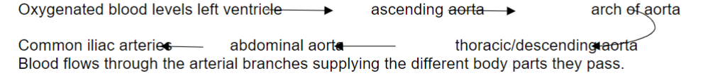
AORTA
- Largest artery of the body
- Arises from the left ventricle of the heart
4 PARTS OF AORTA
- Ascending aorta – arises from the left ventricle
- Branches
- right and left coronary arteries
- Arch of aorta – continuation of the ascending aorta at the back of manubrium sterni
- Branches:
a. brachiocephalic artery
b. left common carotid artery
c. left subclavian artery
- Abdominal aorta - continuation of the thoracic portion that passes through the diaphragmatic opening at the level of T12 into the abdominal cavity
- Branches:
a. direct unpaired visceral branches
1. celiac artery
2. superior mesenteric artery
3. inferior mesenteric artery
b. direct paired visceral branches
1. renal arteries
2. gonadal arteries (ovarian, testicular)
3. middle suprarenal arteries
c. terminal branches
1. 2 common iliac arteries
2. 1 middle sacral artery
–
CIRCLE OF WILLIS
- Common iliac artery at level of the sacroiliac joint divides into
- Internal iliac artery supplies pelvic organs and external iliac artery continues in the thigh as femoral artery
- Femoral artery at the back of knee becomes popliteal artery
- Popliteal artery at lower border of popliteus muscle divides into anterior and posterior tibial arteries
–
MAJOR VEINS OF SYSTEMIC CIRCULATION
- Superior vena cava
- drains oxygenated blood from the head, neck upper limb, thorax into the right atrium
- Inferior vena cava
- drains deoxygenated blood from the lower parts of the body into right atrium
–
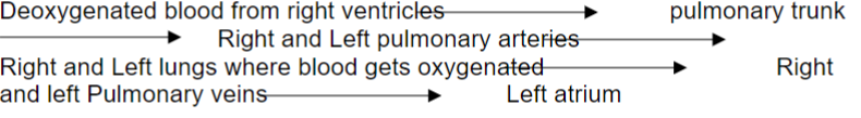
MAJOR VESSELS OF PULMONIC CIRCULATION
- Pulmonary trunk - arises from the right ventricles
- carries deoxygenated blood to the pulmonary arteries on the way to the lungs
- Pulmonary arteries - two in number
- continuation of pulmonary trunk as it enters the lungs
- Pulmonary veins – 4 in number
- carries oxygenated blood from the lungs to the left atrium
CIRCULATORY ROUTES
- SYSTEMIC CIRCULATION
- Arterial division - main vessels is the aorta - largest artery in the body which carries oxygenated blood aortic valve
- Venous division

- Pulmonary Circulation
- Hepatic Portal Circulation
- Fetal Circulation
- circulatory system of fetus differs from that of child and adult for
- 2 main reasons:
1. Fetus gets oxygen and nutrients and eliminates carbon dioxide and waste using the
mother's blood.
2. Fetal lungs, kidneys & digestive system (except liver) are not functional.
WEEK 11. CIRCULATORY SYSTEM: HEART |
|---|
CARDIOVASCULAR SYSTEM
- a closed pump system made up of heart, blood vessels and blood.
HEART
- a cone-shaped structure with apex directed inferiorly and to the left at 5 ICS, MCL (intercostal space, midclavicular line)
- base - opposite to the apex directed posteriorly, superiorly and to the right Anteriorly lies just below the 2nd rib. It is relatively in a fixed position because of its attachment to the great vessels while the apex is free to move.
–
Pericardium – a connective tissue that covers and protects the heart.
- it is made up of:
- Fibrous pericardium
- tough inelastic dense irregular connective tissue
- prevents overstretching of the heart
- provides protection and anchors the heart in the mediastinum
B. Serous pericardium
- thinner more delicate membrane that forms double layer around the heart
- Parietal layer – outer layer fused to fibrous pericardium
- Visceral layer – inner layer also called epicardium (on top of the heart) adheres tightly to surface of the heart
–
HEART PHYSIOLOGY
Properties of Cardiac Muscle Tissue
- Cardiac muscle has a built-in pacemaker that initiates a heartbeat independent of the electro stimulation from the nervous system. Nerve impulse from the autonomic nervous system and blood-borne hormones (such as epinephrine) can only modify the timing and strength of each heartbeat.
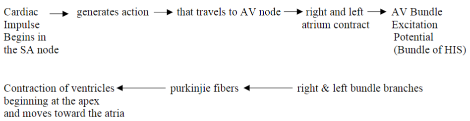
Conduction system of the Heart
- Sinoatrial node or SA node
- located at the superior wall of the right atrium near the entry point of superior vena cava.
- starts the rhythm of electrical excitation (thus the name of PACEMAKER) that causes the heart to contract.
- Atrioventricular node or AV node
- lies at the base of right atrium near the interatial septum
- Atrioventricular bundles (AV bundles) or bundle of HIS
- conducting fibers in interventricular system runs a short distance and then divides into two branches, one branch for each ventricle.
- Purkinje fibers
- cardiac conducting myofibers that course throughout the cardiac muscular wall of ventricle.
–
A. Propagation Of Impulse In The Heart
B. Cardiac Cycle
- all events associated with one heartbeat
- consist of alternating contraction and relaxation of atria and ventricles forcing blood from areas higher pressure to lower pressure
- Systole (contraction) - term refers to the phase of contraction either atrial or ventricular following depolarization
- Diastole (relaxation) - term refers to phase of dilatation following repolarization
- each cardiac has four heart sounds (blood turbulence in a normal heart) only the first and second heart sounds are loud enough to be heard by auscultation (act of listening to sound within the body) with the use of a stethoscope.
- S1 - first heart sound - lubb sound – due to closure of the atrioventricular valve when ventricle contracts in systole
- S2 - second heart sound – dupp sound – due to closure of semilunar valves when ventricle relax in diastole
–
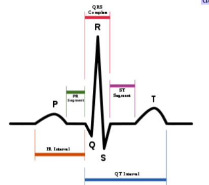
Electrocardiogram (ECG/EKG)
- is a composite record of action potential produces by all the heart muscle fibers during each heartbeat. As action potentials propagate through the heart they generate electrical currents that can be detected at the surface of the body and is recorded by a machine called electrocardiograph.
- P wave - represents atrial depolarization which spreads from the SA node through the contractible fibers in both Atria
- small upward deflection
- QRS complex - represents rapid ventricular depolarization – as action potential spread through ventricular contractile fibers
- Q-downward deflection
- R-large upright, triangular wave
- S-downward wave
- T wave – represents ventricular repolarization occurs just as the ventricles are starting to relax.
- dome-shaped upward deflection