Chapter 1 Bio 201
Section 1.2 Anatomy Terminology & Organ Systems
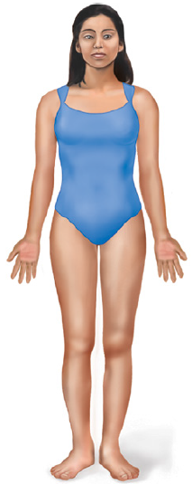
Anatomical Position
Anatomical Position is a position in which:
the body is erect
feet slightly apart (shoulder width)
palms facing forward (anterior/ventral)
thumbs pointing away (laterally) from the body
An understanding of anatomical position is crucial since it provides a universal frame of reference regardless of the body’s actual position.
Keep in mind, the directional terms that follow, will always refer to the body in its anatomical position.
Directional Terms
One of the most daunting tasks for students to overcome in their first anatomy & physiology course is the terminology. Much like learning a new language, the key to success in this course is repetition & being able to integrate the new terms into your everyday vocabulary. Let’s begin our journey into the terminology by becoming familiar with the directional terms.
Anterior: (aka ventral) means toward the front of the body.
Posterior: (aka dorsal) means toward the back/rear of the body.
Superior: means toward the head or upper portion of the body.
Inferior: means toward the tail or lower portion of the body or away from the head or upper portion of the body.
Medial: means towards the midline or center of the body only. Not to be confused with the term median.
Lateral: means away from the midline or center of the body.
Median: means towards the midline or the center of the body or any body part or section that has left & right halves.
Superficial: means toward the surface of the body or skin.
Deep: means away from the surface or toward the body’s interior, as close to the bones, cartilages, or organs.
Intermediate: means its in-between the superficial & deep layers.
The 2 directional terms listed below are used especially in the anatomy of the limbs:
Proximal: means toward the point of origin or something relatively close to the limb's point of attachment, such as the the shoulder, elbow, arm, wrist, hip, leg, knee, or ankle.
Distal: means away from the point of origin or something farther away from the limb's point of attachment.
Lastly, these last 2 directional terms you need to know might seem easy. But it does become challenging when you don't orient yourselves properly. Knowing left from right & vise-versa is an important safety factor to always consider in the medical field. Mislabeling left from right & vise-versa can have severe medical or legal consequences in the medical field, such as injuries or even death.
Note: Every time we are asking you to state whether a structure or body part is in its left or right side, always refer back the cadaver, plastic model, specimen, or image's left or right sides in the anatomical position.
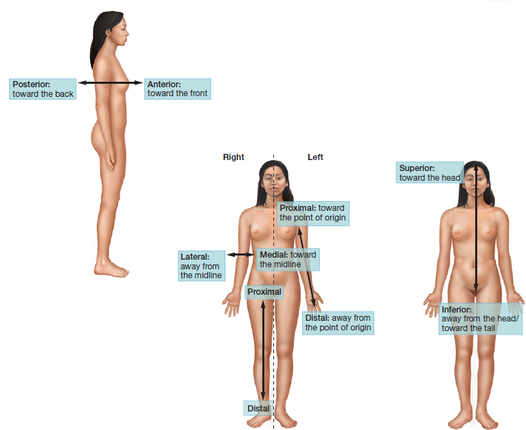
Figure 1.7 Directional Terms
Regional Terms
Regional terms are used in anatomy & physiology in order to make descriptions as specific as possible.
For instance, if someone were to say that they had a deep cut in their chest, it would not be very clear as to where exactly the cut was located. However, if someone were to say they had deep cut in their sternal region, you would have a much better understanding of where the cut was located.
These are new terms that you've never imagine of using or understanding. But remember, in the context of anatomy & physiology, even in the medical field, these terms are utilized & verbalized on a regular basis, so use this opportunity to learn, add, & use them in your vocabulary in a medical setting.
Become familiar with the regional terms shown & listed below.
Note: Moving forward, you must include the word region in your answer for it to be considered a complete answer.
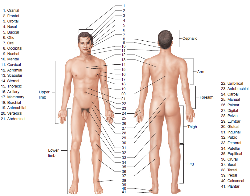
Figure 1.8 Regional Terms
Table 1.1 Regional Terms
Term Definition | |
Adjectives | Location |
Abdominal region | The area over the abdomen that is inferior to the diaphragm & superior to the bony pelvis |
Acromial region | The area over the lateral part of the shoulder that contains the acromion of the scapula |
Antebrachial region | The forearm |
Antecubital region | The anterior upper limb between the forearm & arm, over the elbow joint |
Axillary region | The area in & around the axilla (armpit) |
Brachial region | The anterior & posterior arm (between the elbow and the shoulder) |
Buccal region | The lateral portions of the face corresponding to the cheeks |
Calcaneal region | The heel of the foot |
Carpal region | The wrist |
Cephalic region | The entire head from the chin to the top of the head |
Cervical region | The front & back of the neck (nape) |
Cranial region | The top of the head or the portion of the skull that encases the brain |
Crural region | The anterior leg or the shin |
Digital region | The fingers or the toes |
Femoral region | The thigh |
Frontal region | The forehead |
Gluteal region | The buttock |
Inguinal region | The area along the inguinal ligament that divides the pelvis from the thigh |
Lumbar region | The lower back |
Mammary region | The area around the breast (not to be confused with the thoracic region) |
Manual region | The general area of the hand |
Mental region | The chin |
Nasal region | The nose |
Nuchal region | The ridge that runs along the back of the skull within the occipital region |
Occipital region | The general area of the back of the skull |
Oral region | The mouth |
Orbital region | The area around the eye |
Otic region | The area around the ear |
Palmar region | The anterior hand (the palm of the hand) |
Patellar region | The anterior part of the knee over the patella (kneecap) |
Pedal region | The foot |
Pelvic region | The anterior pelvis |
Plantar region | The bottom of the foot |
Popliteal region | The posterior side of the knee joint |
Pubic region | The area over the pubic bone |
Adjectives | |
Scapular region | The area over the scapula in the superior back |
Sternal region | The area in the middle of the chest over the sternum |
Sural region | The posterior part of the leg (the calf) |
Tarsal region | The ankle region |
Thoracic region | The general chest area (not to be confused with the mammary region) |
Umbilical region | The area around the umbilicus (belly button) |
Vertebral region | The area over the vertebral column (spine) |
Nouns | |
Upper limb | The entire portion of the body from the shoulder to the digits of the hand |
Arm or Upper Arm | The portion of the upper limb from the shoulder to the elbow |
Forearm | The portion of the upper limb from the elbow to the wrist |
Lower limb | The entire portion of the body from the hip to the digits of the foot |
Thigh | The portion of the lower limb from the hip to the knee |
Leg or Lower Leg | The portion of the lower limb from the knee to the ankle |
Serous Membranes & Body Cavities
Internally, the human body is organized into spaces known as body cavities. But before we discuss the body cavities, let's talk about serous membranes first:
Many of these body cavities house specific organs. They are fluid filled & lined with thin layers of tissue known as serous membranes (not serious).
Serous Membranes:
These thin layers of tissue are composed of 2 layers & cells that produce a thin watery substance known as serous fluid.
2 Layers:
An outer parietal layer (some examples: parietal pericardium & parietal pleura).
This layer is attached to the body wall & its structures.
An inner visceral layer (some examples: visceral pericardium & visceral pleura).
This layer is attached to specific internal organs, also known as viscera (singular: viscus not viscous).
Serous fluid:
Serves as a lubricant for the organs allowing them to move freely without friction within the cavity (some examples: pleural fluid & pericardial fluid).
It is found in the space between the parietal & visceral layer (some examples: pleural space & pericardial space).
The body is enclosed between 2 major cavities: anterior & posterior cavities:
Major Cavity 1 - Anterior/Ventral Cavity: Made up of 2 minor cavities: thoracic & abdominopelvic cavities, which contains organs/structures found in the anterior side of the body. The cavities are separated by a thin muscle called the diaphragm.
Minor Cavity 1 - Thoracic Cavity: A cavity located above the diaphragm & encases the organs/structures found in the chest only. Made up of 3 smaller cavities: mediastinum, pleural, & pericardial cavities.
Mediastinum: A space between the lungs, that starts from the base of the neck to the diaphragm.
It contains the heart, esophagus, great blood vessels, trachea, bronchi, lymph nodes, lymph vessels, & some important nerves.
Pleural Cavity: (not plural) Spaces that surround each lung.
It contains the serous membranes called the parietal & visceral pleura.
It also contains the serous fluid called pleural fluid.
Pericardial Cavity: A space between the lungs that contain the heart & other cardiac structures only.
It contains the serous membranes called the parietal & visceral pericardium.
It also contains the serous fluid called pericardial fluid.
"Peri-" means around
Note: The heart is found both in the mediastinum & pericardial cavity. Pay attention to the inclusion of the word "only" in the question.
Minor Cavity 2 - Abdominopelvic Cavity: The space found below the diaphragm, that contains majority of the digestive, urinary, lymphatic, endocrine, & reproductive organs. Made up of 3 smaller cavities: abdominal, peritoneal, & pelvic cavities.
Abdominal Cavity: A space located above the pelvic cavity. It spans from the diaphragm to the the pelvic brim. Contains most of the digestive organs & some urinary, endocrine, & lymphatic organs. Inside this cavity is minor cavity 2, which will hold & suspend the organs in place, otherwise all those organs will pile up at the bottom of the cavity.
Peritoneal Cavity: A space found between the 2-layered serous membrane called the peritoneum.
The 2 layers of the peritoneum are called the:
Outer parietal peritoneum: lines the wall of the cavity. When the posterior midline part of this layer, turns inward it then becomes the visceral peritoneum.
Inner visceral peritoneum: suspends certain abdominal viscera form the body wall, covering their outer surfaces & holding them in place.
It also contains the serous fluid called peritoneal fluid.
Intraperitoneal: Organs that are within the peritoneal cavity,which include the liver, most of the small intestine, much of the colon, the stomach, the spleen, & part of the pancreas.
Retroperitoneal
: Those organs that are posterior to the peritoneal cavity & its other side lie against the posterior body wall, include the kidneys, adrenal glands, the sex organs, the urinary bladder, part of the colon, & part of the pancreas.
Pelvic Cavity: Located below the pelvic brim. Contains some organs of the urinary, digestive, & reproductive systems; such as the urinary bladder, ovaries, rectum, & prostate gland.
Major Cavity 2 - Posterior/Dorsal Cavity: Made up of 2 minor cavities: cranial & vertebral cavities, which pretty much contains the major organs of the nervous system.
Minor Cavity 1 - Cranial Cavity: Contains the brain, enclosed in the skull.
Minor Cavity 2 - Vertebral Cavity: Contains the spinal cord, enclosed in the vertebrae.
Note: Both cavities are lined with membranes called meninges (singular: meninx), instead of serous membranes.
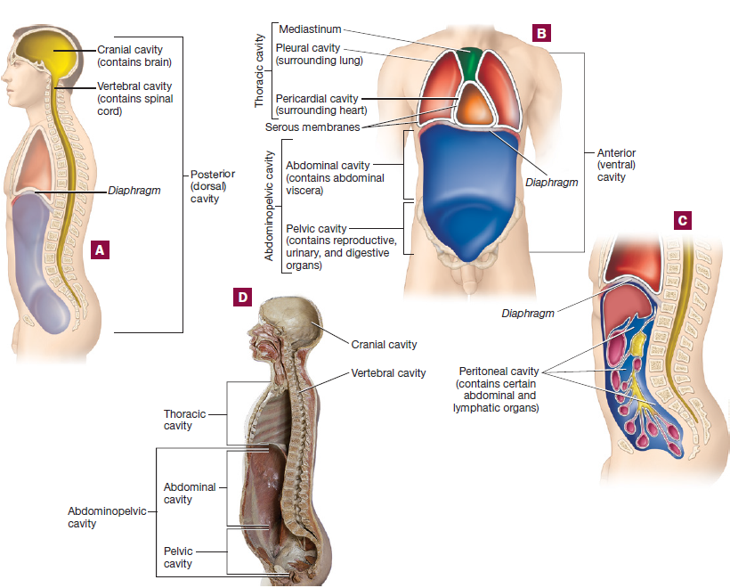
Figure 1.9 Body Cavities
Quadrants
The abdominopelvic cavity can be divided into 4 quadrants or 9 regions. For the purposes of this course, we will focus on the 4 quadrants: right upper, right lower, left upper & left lower. Take note of the organs that can be found within each quadrant. Some organs or parts of an organ may be found in multiple quadrants. Recognize that not all organs can be seen in the image since they may be located deep to the organs in the picture. If you've never heard of these organs before, then this is the time to familiarize yourselves with them.
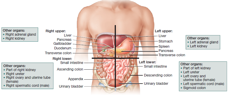
Figure 1.10 Body Quadrants
Planes & Sections
Often times, it is necessary to obtain different views of organs or specific body cavities.
A plane is an imaginary flat surface that passes through the body or its organs.
The sagittal plane divides the body or organs into left & right parts.
A midsagittal plane is when the body or its organs are divided into equal left & right parts.
A parasagittal plane divides the body or its organs into unequal left & right parts.
Note: there is only 1 midsagittal plane, but many parasagittal planes.
Pay close attention to the correct spelling of the term sagittal, which has 2 T's, 1 G, & 1 L.
A frontal/coronal plane divides the body or its organs into anterior/ventral & posterior/dorsal parts.
A transverse/cross section/horizontal plane divides the body or its organs into superior (or proximal) & inferior (or distal) parts.
A section is a cut or slice taken to view the internal anatomy. Shown on figure 1.12
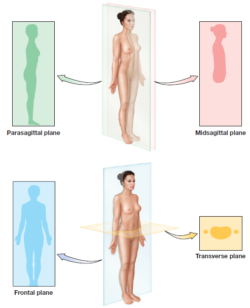
Figure 1.11 Body Planes
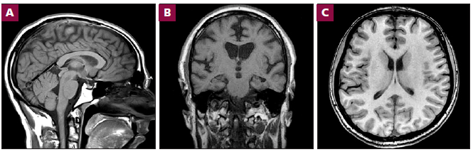
Figure 1.12 Body Sections in an MRI Imaging: A) Sagittal, specifically the Right Midsagittal section; B) Coronal Section; C) Transverse Section
Organ Systems
The human body has 11 organ systems with specific functions & organs associated with them. It is important to recognize the different organ systems, but understand that they all work together to sustain life. The table below provides a brief summary of the principal functions & organs associated with each system.
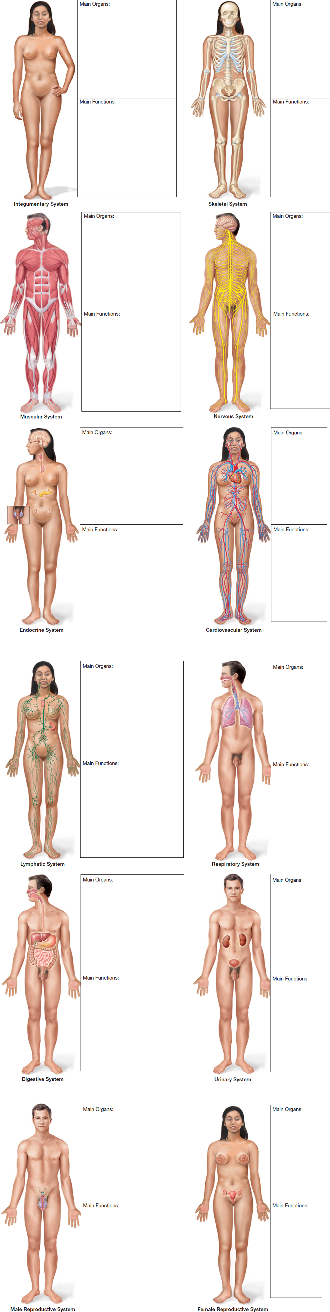
Figure 1.13 Organ Systems
Table 1.2: Summary of the 11 Organ Systems, their function, & the organs found in each system
Organ Systems Summary | ||
Organ System Function Note: Some functions might overlap between multiple organ systems Organs Note: Pay attention & research the ones you are not familiar with. Also, some organs might be found in multiple organ systems | ||
Systems of Protection, Support, & Movement | ||
1) Integumentary System | Protection, thermoregulation, vitamin D synthesis, cutaneous (skin) secretion. | Hair, nails, skin, & cutaneous glands |
2) Skeletal System | Movement, support, protection of internal viscera, electrolyte & acid-base balance, blood formation. | Bones, bone marrow, cartilages & ligaments |
3) Muscular System | Movement, stability, heat production, control of body openings, communication. | Skeletal, smooth, & cardiac muscles & tendons |
Systems of Fluid Transport | ||
4) Cardiovascular System | Fluid, electrolyte & acid-base balance. Distribution of oxygen, wastes, nutrients, hormones, heat, immune cells & antibodies. | Heart & blood vessels (arteries, veins, capillaries, & the great vessels such as the aorta & venae cavae) |
5) Lymphatic System | Detection of pathogens, production of immune cells, defense against disease & recovery of excess tissue fluid. | Lymphatic vessels, trunks, & ducts, lymph nodes, thymus, spleen, Peyer's patches, & tonsils |
Systems of Internal Communication & Control | ||
6) Nervous System | Rapid internal communication, coordination, motor control & sensation. | Brain, spinal cord, nerves, & ganglia |
7) Endocrine System | Slow internal chemical communication & coordination, hormone production. | Pineal gland, pituitary gland, thyroid gland, parathyroid glands, adrenal glands, thymus, pancreas, ovaries & testes |
Systems of Intake & Output | ||
8) Urinary System | Elimination of wastes, stimulation of red blood cell formation, regulation of blood volume & pressure, control of fluid, acid-base & electrolyte balance, detoxification & micturition. | Kidneys, ureters, urinary bladder & urethra |
9) Respiratory System | Acid-base balance, speech, absorption of oxygen & discharge of carbon dioxide. | Nose, pharynx, larynx, trachea, bronchi, alveoli, & lungs |
10) Digestive System | Nutrient breakdown & absorption, & defecation. | Teeth, tongue, salivary glands, pharynx, esophagus, stomach, small & large intestines, liver, gall bladder & pancreas |
Systems of Reproduction | ||
11A) Male Reproductive System | Secretion of sex hormones, production & delivery of sperm. | Testes, vas deferens, epididymides, spermatic ducts, seminal vesicles, bulbourethral glands, prostate gland, scrotum, & penis |
11B) Female Reproductive System | Secretion of sex hormones, production of eggs, site of fertilization & fetal development, fetal nourishment, birth, & lactation. | Ovaries, uterine/fallopian tubes, uterus, vagina & mammary glands |
Identifying Organs
Identify the following organs on your preserved mammal specimen or human torso models. Check off each organ as you identify it, and record the organ system to which it belongs in Table 1.3, using Figure 1.13 for reference. Remember that some organs may function in more than one system.
Adrenal glands
Blood vessels
Bones
Brain
Esophagus
Gallbladder
Heart
Joints
Kidneys
Large intestine
Larynx
Liver
Lungs
Lymph nodes
Pancreas
Skeletal muscles
Skin
Small intestine
Spinal cord
Spleen
Stomach
Testes (male) or ovaries (female)
Thymus
Thyroid gland
Trachea
Urinary bladder
Uterus