Histology section 2
9/16/25
10 pt back thing: pick 5 and find them on UAB and lable them and put them on a PP for 10 points ex. points. — turn in before next week — plus location
3)psudostratified columbnar
4) goblet cells
8) simple columnar
23) fibro cartilage
31) elastic fibers
47) smooth muscle
48) where is 47 found
^ ^ ^ ^ ^
I I I I I
Pick 5
first presentation due two weeks from today
big/gross pasthology can be included, but histology slides prefered
define wher arows are pointed, and explain any medical termonalogy in plain english
show normal tissue and lable it, then show diseased tissue
no more than 10 slides
we will get a list of 5-10 things per thing that you MUST know.
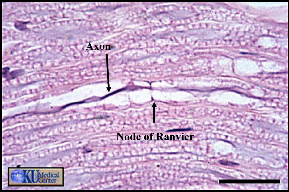
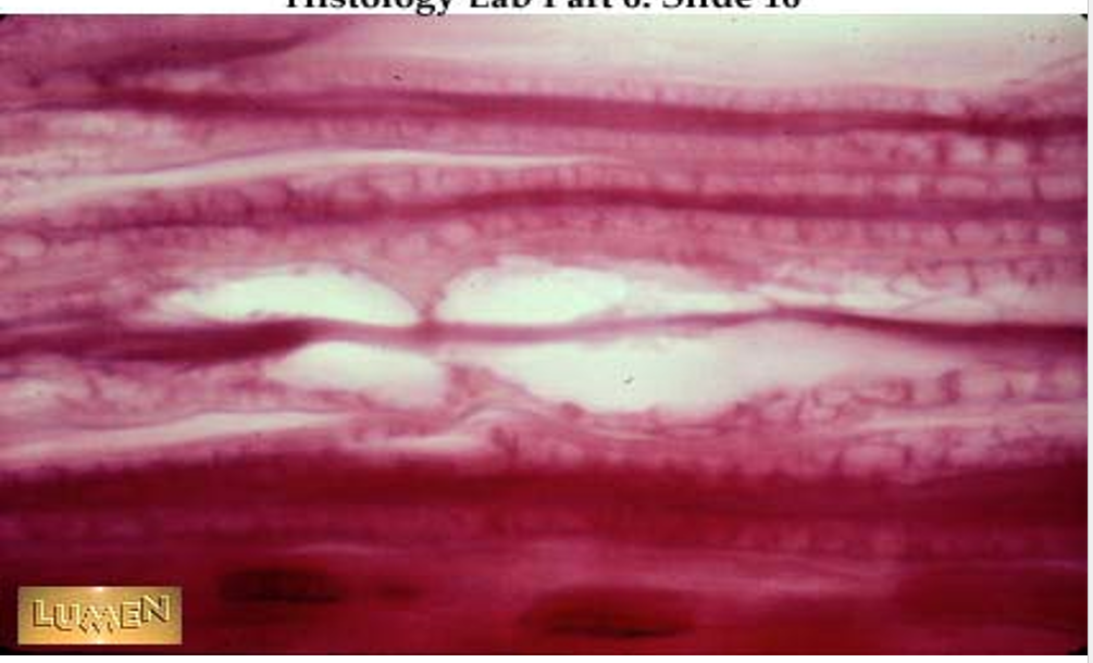
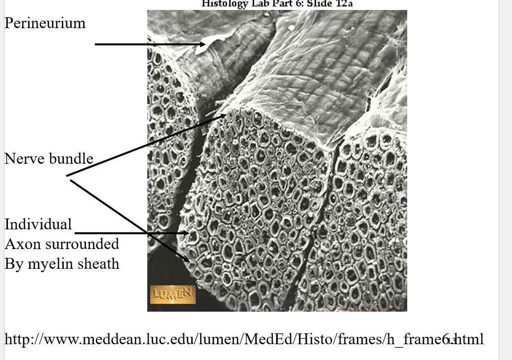
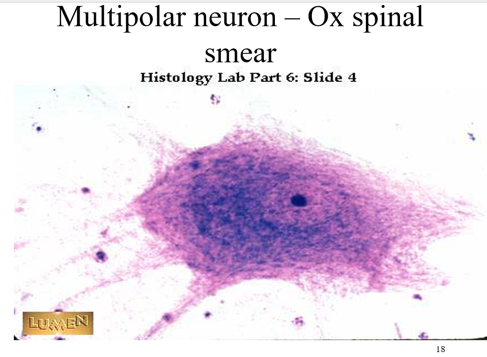
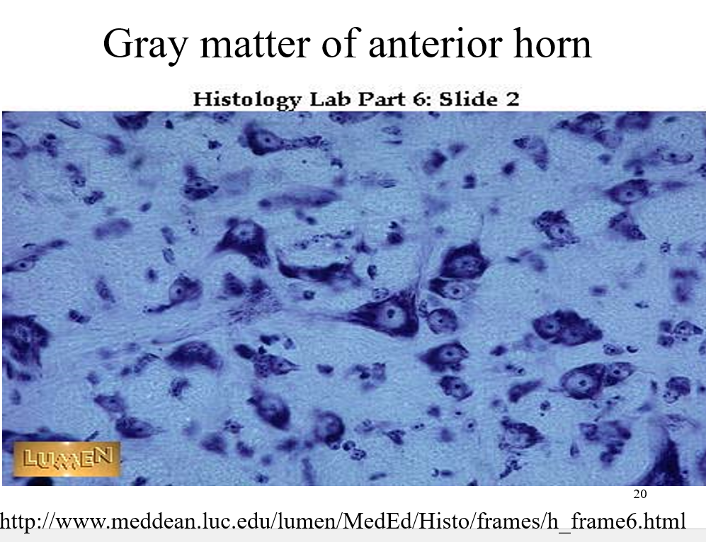
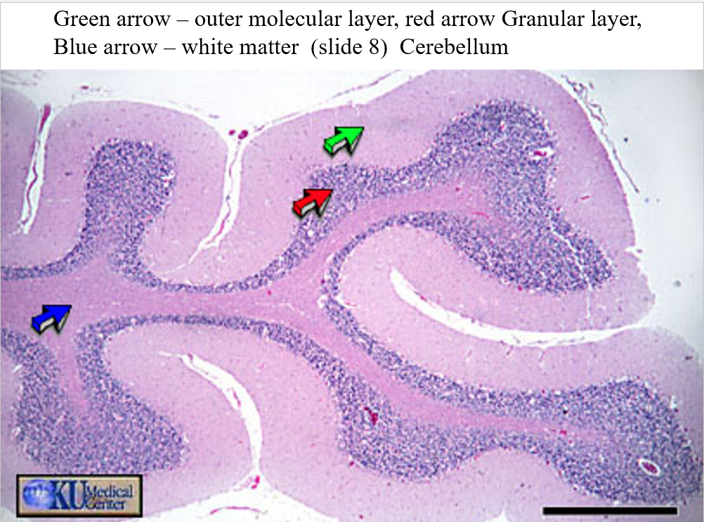
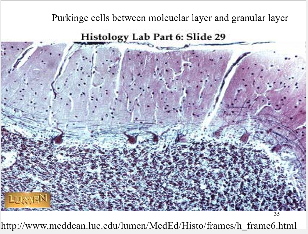
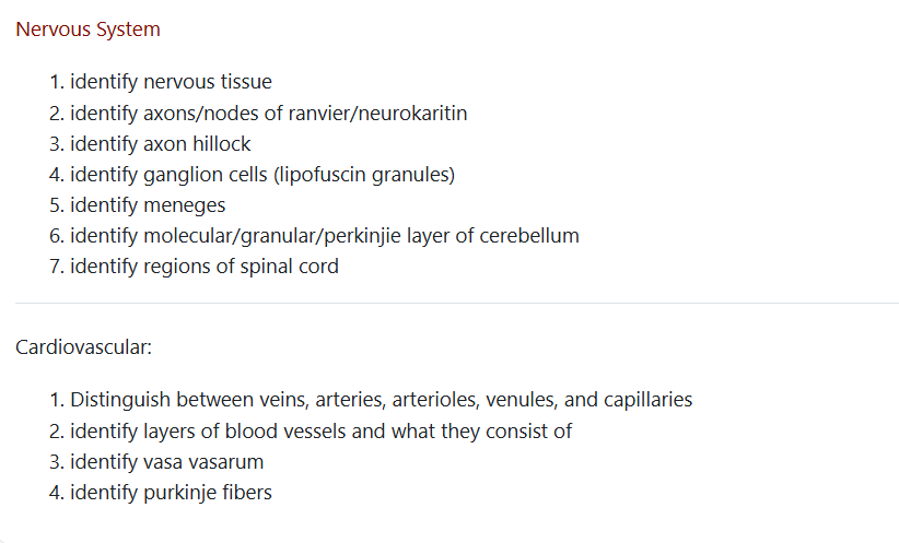
Nervous tissue
•Unique feature of nervous tissue is it ability to transmit impulses and thus communicate with different parts of the body through these impulses.
•Works closely with endocrine system in communication within the body.
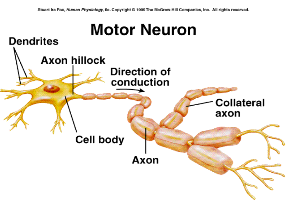
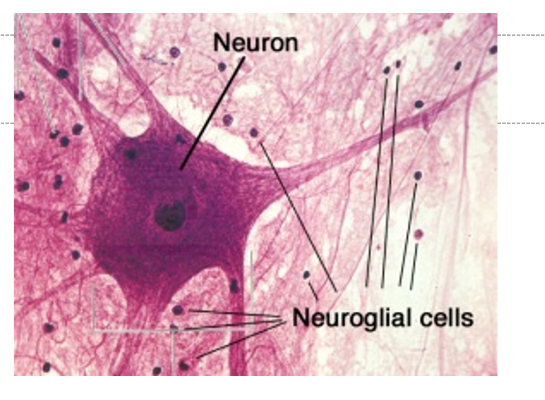
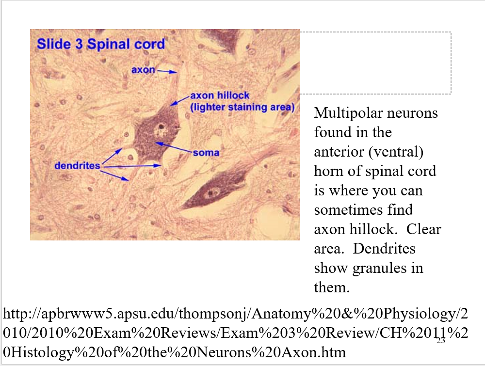
all nurons will have cell body, nuron, and axons
stains will often differentiate b/t white (mylenated) and gray matter (non mylinated)
maylen is like an insulation for the electrical impulses.
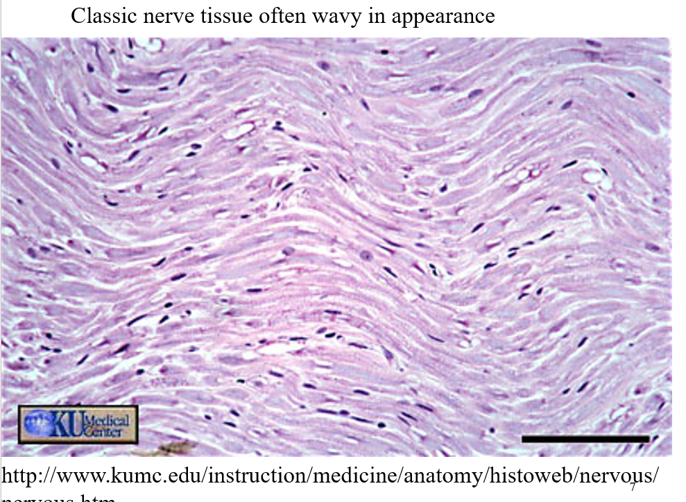
wave like apperence is oftan types nervous tissue
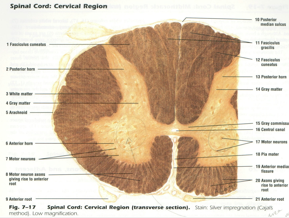
Anterior horn importaint
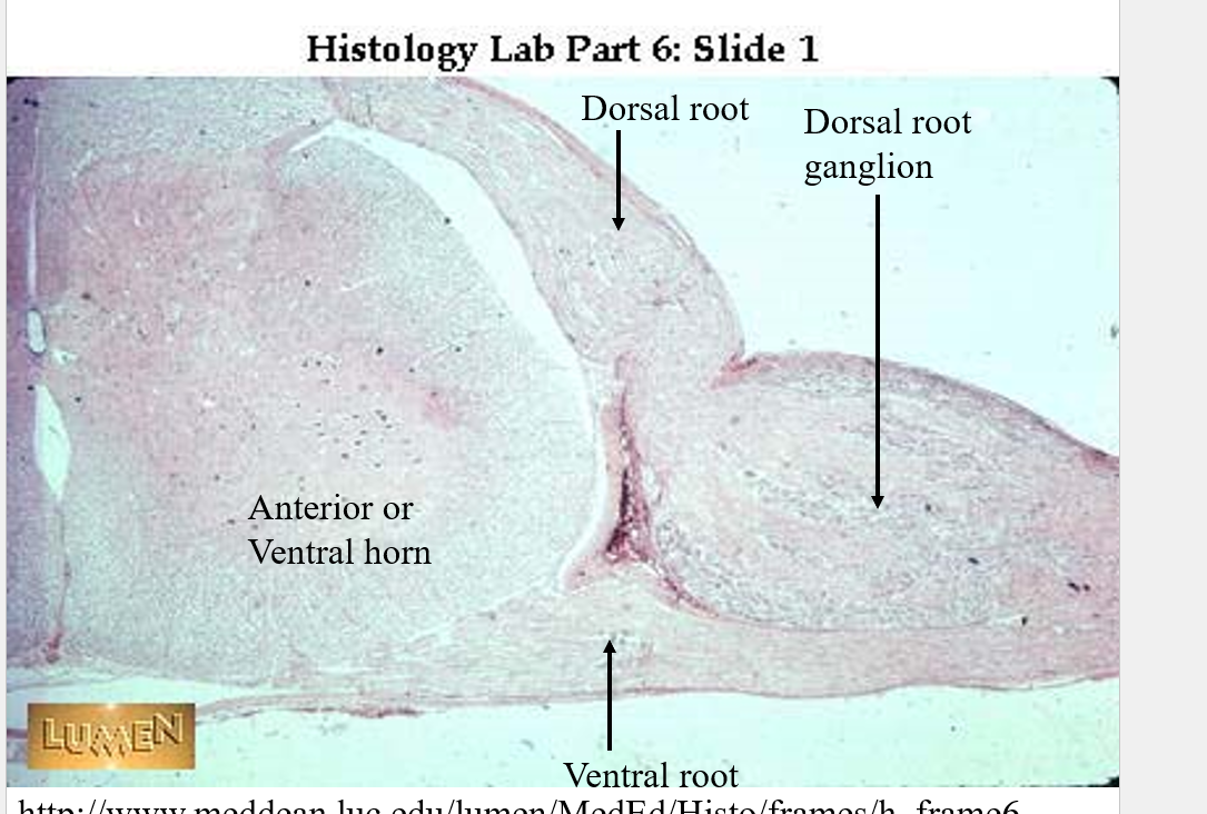
Will need to know the spinalcord anatomy
know front from back
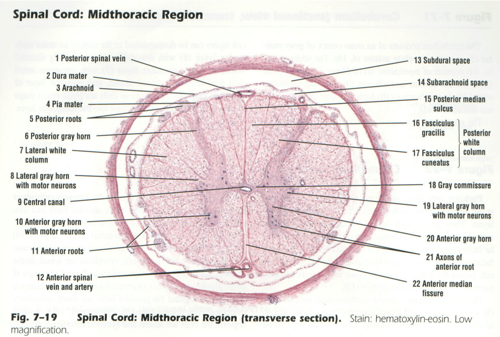
puedes ver los “menengies” que los cosas en “menengititis”
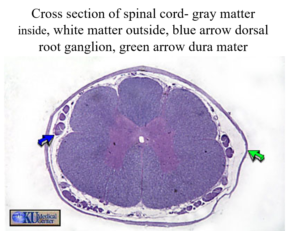
green= dura matter
blue= ganglia = collections of nurons part of the perifrial nurons = sometimes calles minibrains outside of the CNS
neuron = synthsized ribosomes(?)
neuroglial cells the friends of the neuron who keep everything running good and smooth and yay
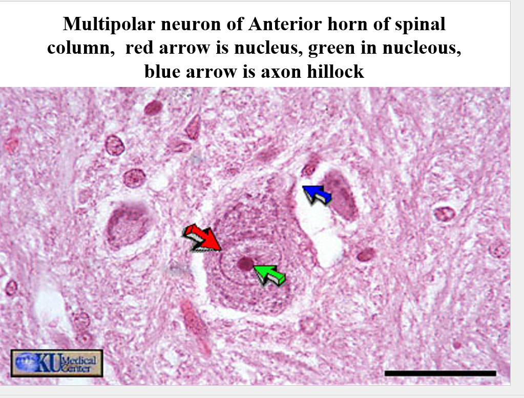
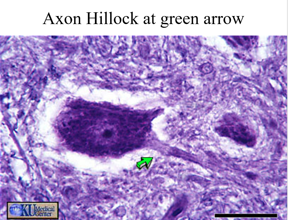
most stains are staining the nissle bodies of the neurons = endoplasmic -______- of neuron
where ever the stain is weaker is the axon not dendrite
spinal cord
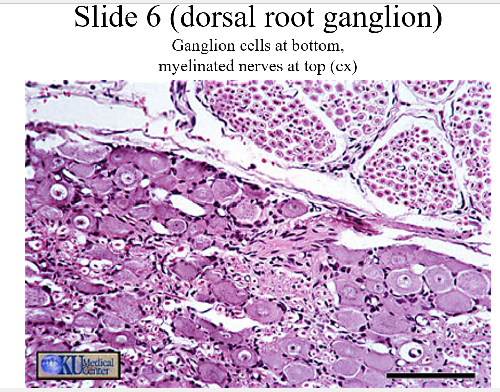
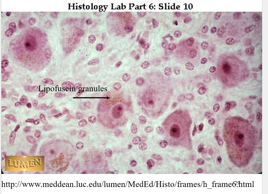
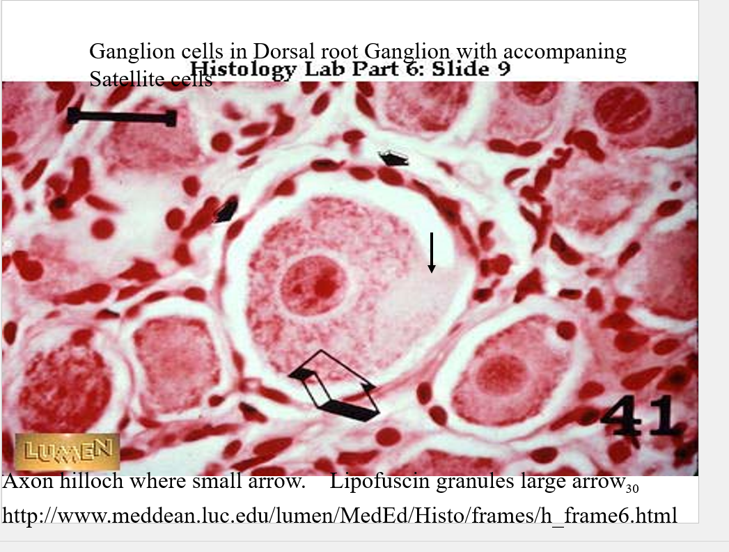
•Sensory neurons have cell bodies located in dorsal root ganglion that is a swelling found on spinal nerve near where it branches close to spinal cord.
•Ganglia are concentration of nerve cell bodies outside the CNS. Contain ganglion cells
•They contain pigment called lipofuscin pigments. Older cells, more pigment.
brain
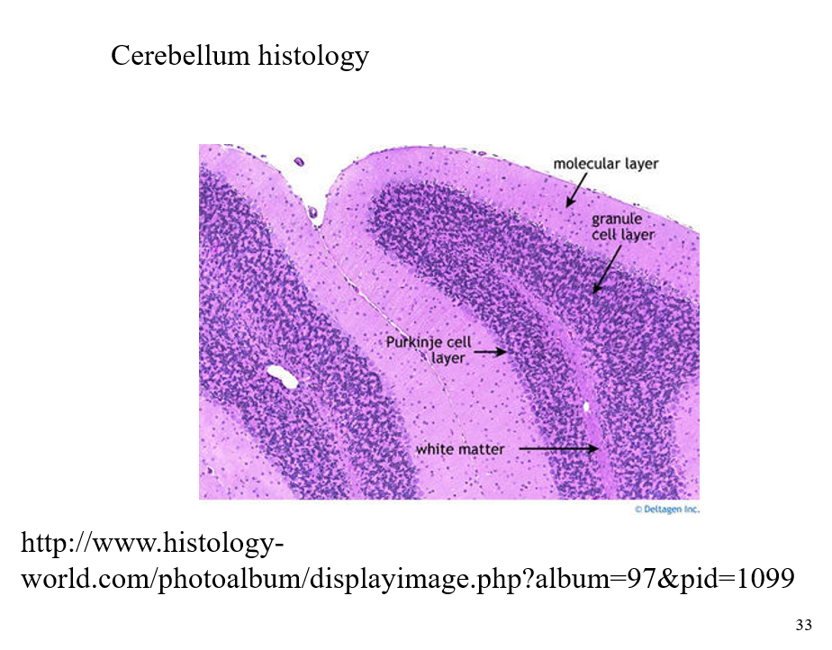
cerebreum and cerebrual cortext are what we’ll mainly deal w/
•Gray matter located on the outside of brain and white matter located on the inside.
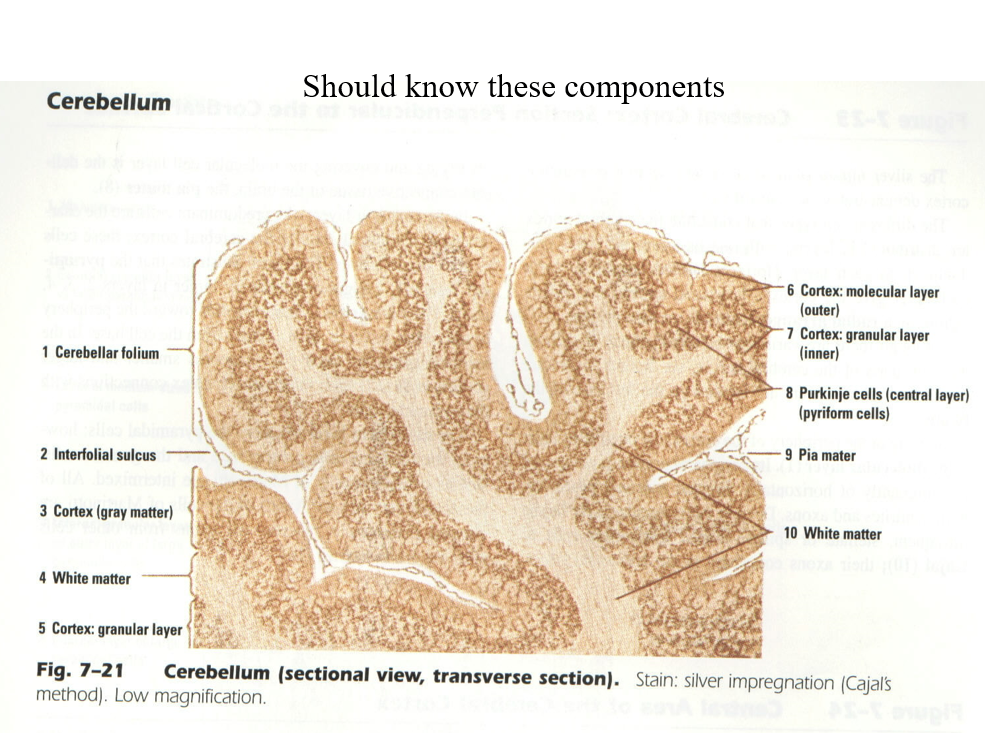
gray matter on outside of the brain and white is on the inside of it
DATE: 9/18/25
Cardiovascular system
•The histological study of the cardiovascular system includes two major components
•Heart – primarily functions as a pump to move blood (and all the things blood carries) through the body.
•Blood vessels – are the tubes that distribute the blood (and its goodies) to the cells and then back to the heart.
•The heart tissue was primarily covered in the unit on muscle.
•The blood vessels include three major divisions.
–Arteries – carry blood from heart to body
–Capillaries – unloading and loading docks
–Veins – carry blood from body to the heart
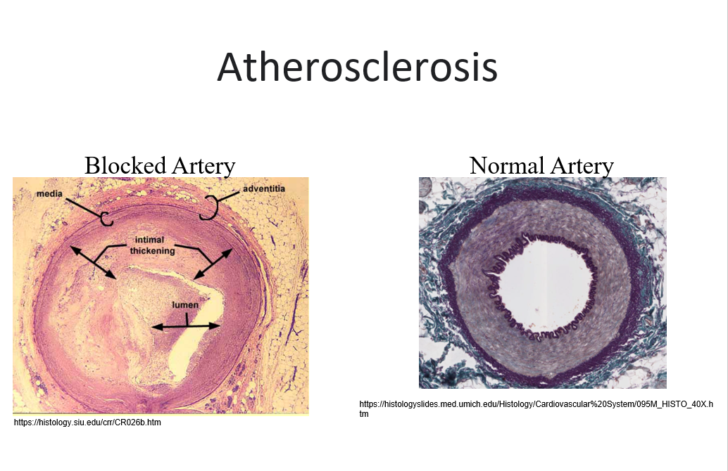
•Arteries have walls that consist of three layers or “coats” often referred to as tunics.
–Tunica intima – is the inner coat
–Tunica media – is the middle layer
–Tunica adventitia or tunica externa is the outer layer of the wall of the blood vessel
Layers differ in different size blood vessels.
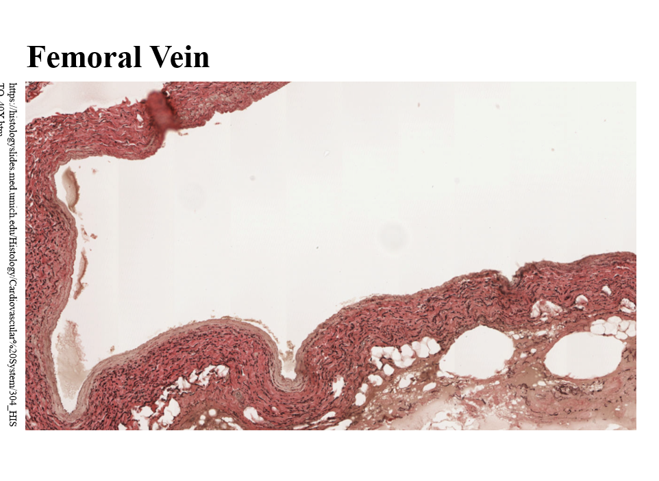
^^veins have these same things but “underdeveloped” tunica media^^
Will have simple squamos and elastic cartlege and stuff
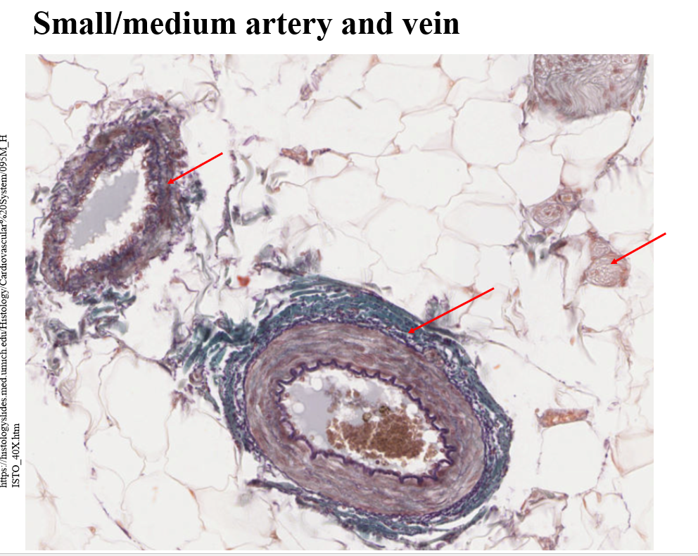
Arteries are consistant, veins arent BC smaller layer of smooth muscle
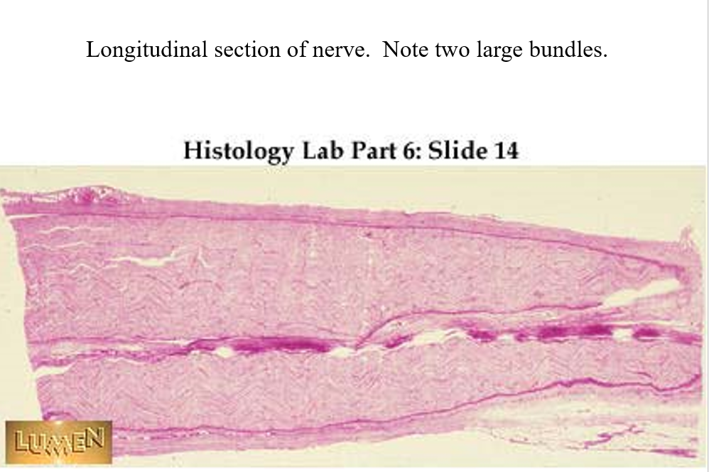
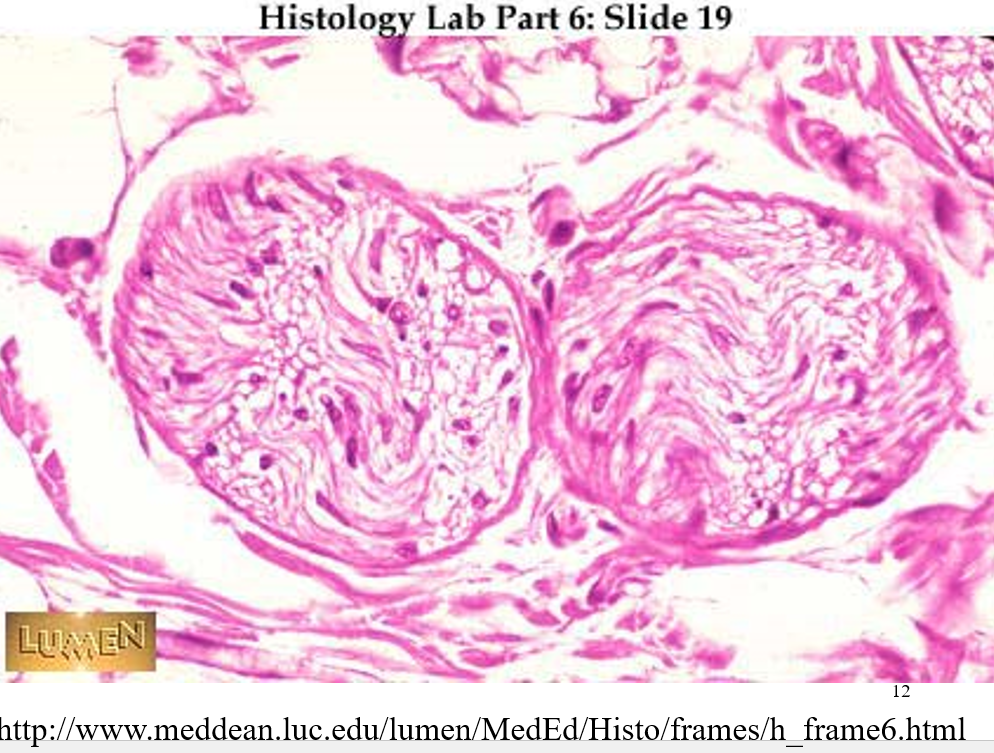
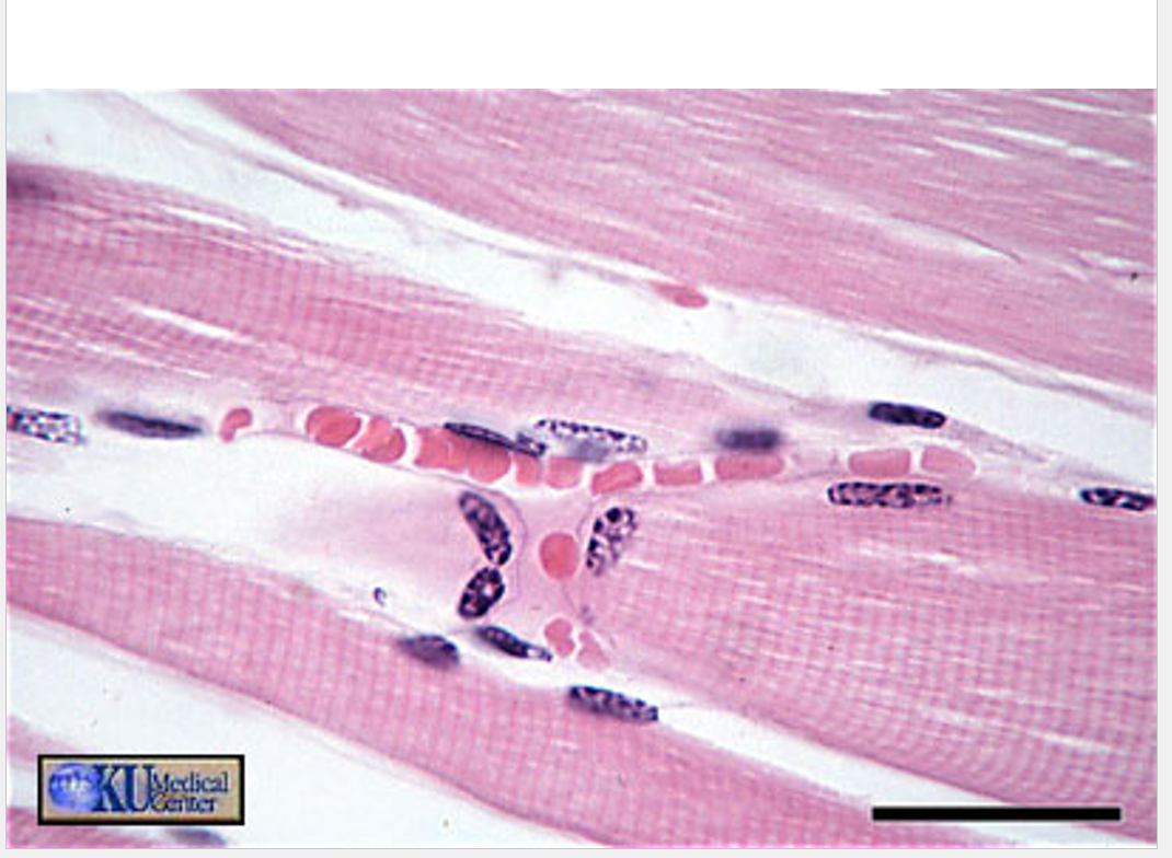
furthests right is a cross section of a nerve
Types of arteries:
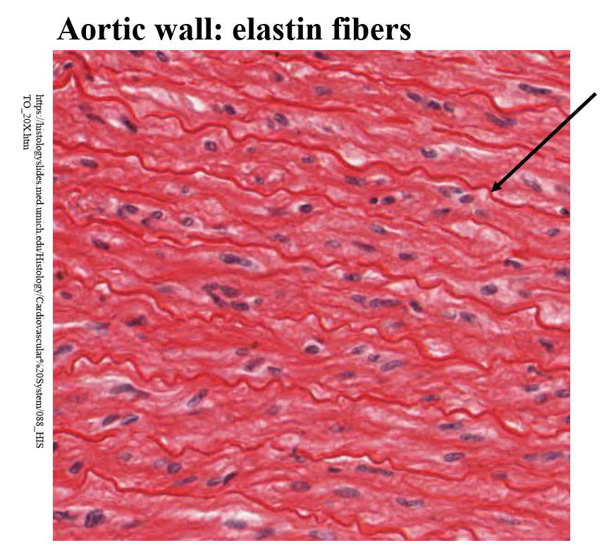
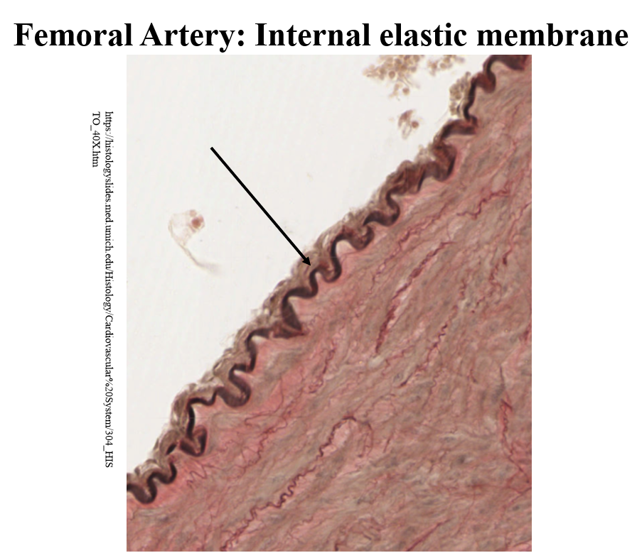
lg or elastic arteries — aorta, pulmonary artery, carotides - tunica media makes up ~ 4/5 of thickness of wall
•Large amounts of smooth muscle with high concentration of elastin fibers.
•Able to withstand high pressures
medium sized arteries often called muscular or distribution arteries
carry blood to most organs
tunica media consists b/t 20-60 layers of smooth muscle w/ some infiltration of elasstic fibers
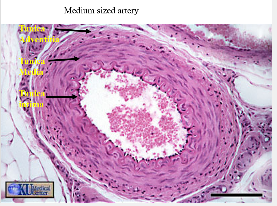
will often see adipose tissue in outer most section
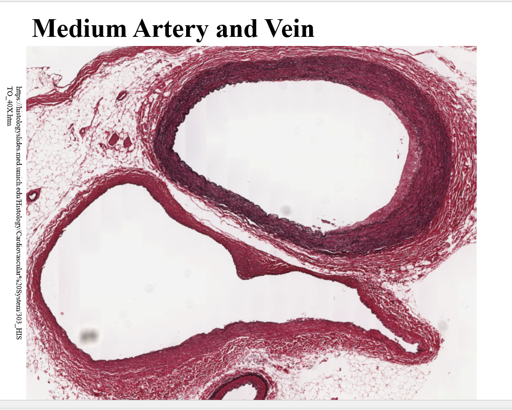
veins have large diametors bc blood resovour and to hold that blood as storage i think
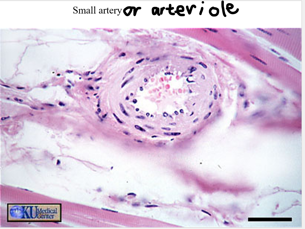
•Small arteries – usually includes vessels with a diameter of 20-100 micrometers.
•Small arteries branch to form many arterioles.
•Small arteries have between 3-20 layers of smooth muscle cells.
•Arterioles have 1-3 layers of smooth muscle cells.
•Arterioles often have pre-capillary sphincters that reduce blood flow into capillary beds.
arterioles feed capilaries
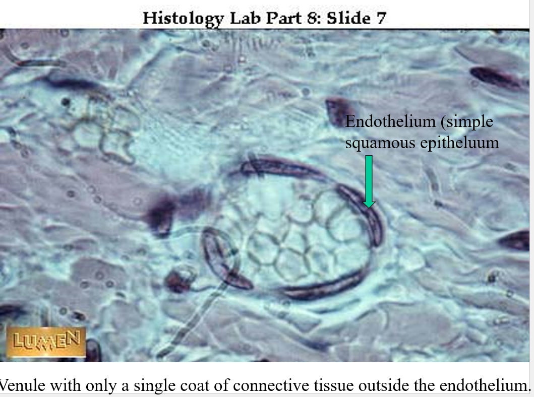
Veins
•Distinction of layers in the walls of veins is not as distinct.
•Contain more connective tissue and less smooth muscle and elastin fibers
•Because of thinner walls, often do not have the round shape of arteries.
•Functionally, arteries serve as reservoirs of pressure. Average pressure in large arteries is close to 120/80 mm Hg.
•Veins serve as reservoir for volume.
•Blood pressure is regulated by shifting blood back and forth from arteries to veins.
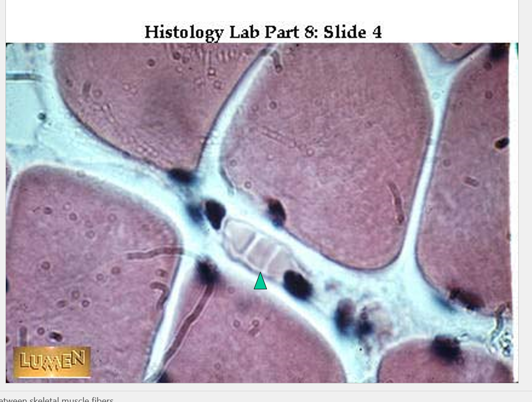
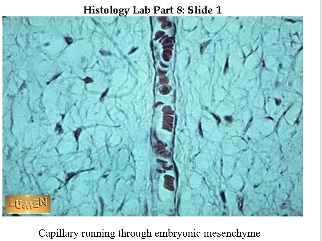
Capillaries:
ONE CELL LAYER THINK,
•Capillaries are the site where materials carried in the blood are unloaded and other materials are loaded into the blood.
•They are known as the loading and unloading docks of the circulatory system.
•Consist of a single layer of simple squamous epithelium.
•Capillaries can vary in size from 7 – 9 micrometers.
•Continuous capillaries have relatively complete walls found in lung, muscle, and skin
•Fenestrated capillaries found in liver, spleen, and lymphatic tissue have holes in walls.
//
DATE: 9/23/25
Neuovascular Bundle
•Arteries, veins, and nerves often are located together especially when they pass through the mesentery.
•This is often referred to as a neurovascular bundle or simply a nerve, artery, vein bundle.
Lymph Vessels
Return fluid from tissues to circulatory system
depend on muscles to move fluid
very thin walled
—resembles veins, best way to tell them apart is that a lymph vessels wont have blood cells/any cells
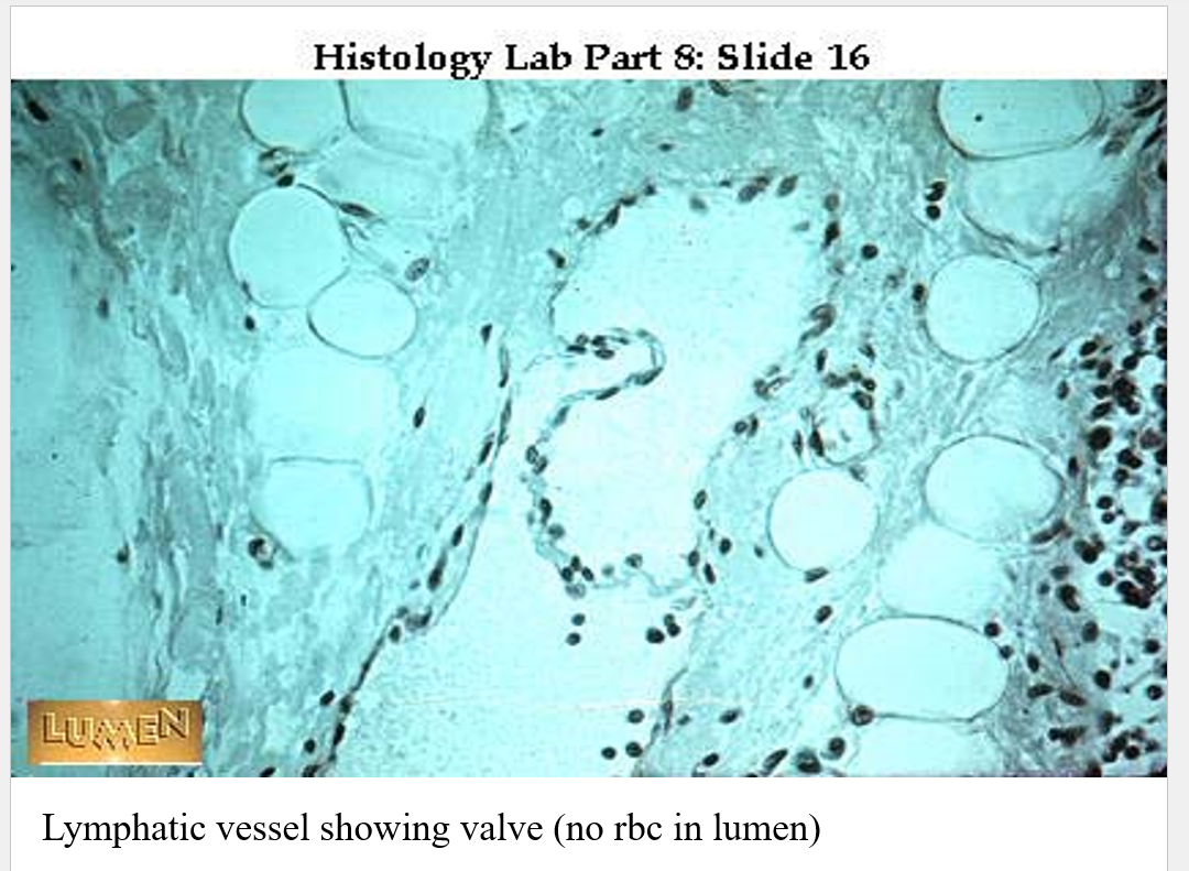
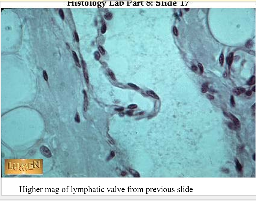
similuner valve in there^^^
Heart structure
can see heart valve
branching pattern characteristic
perkinji fibers - electrical impulses - SA node makes these impulses - heart cells that cave gotten rid of most of their fibers — lookmore empty
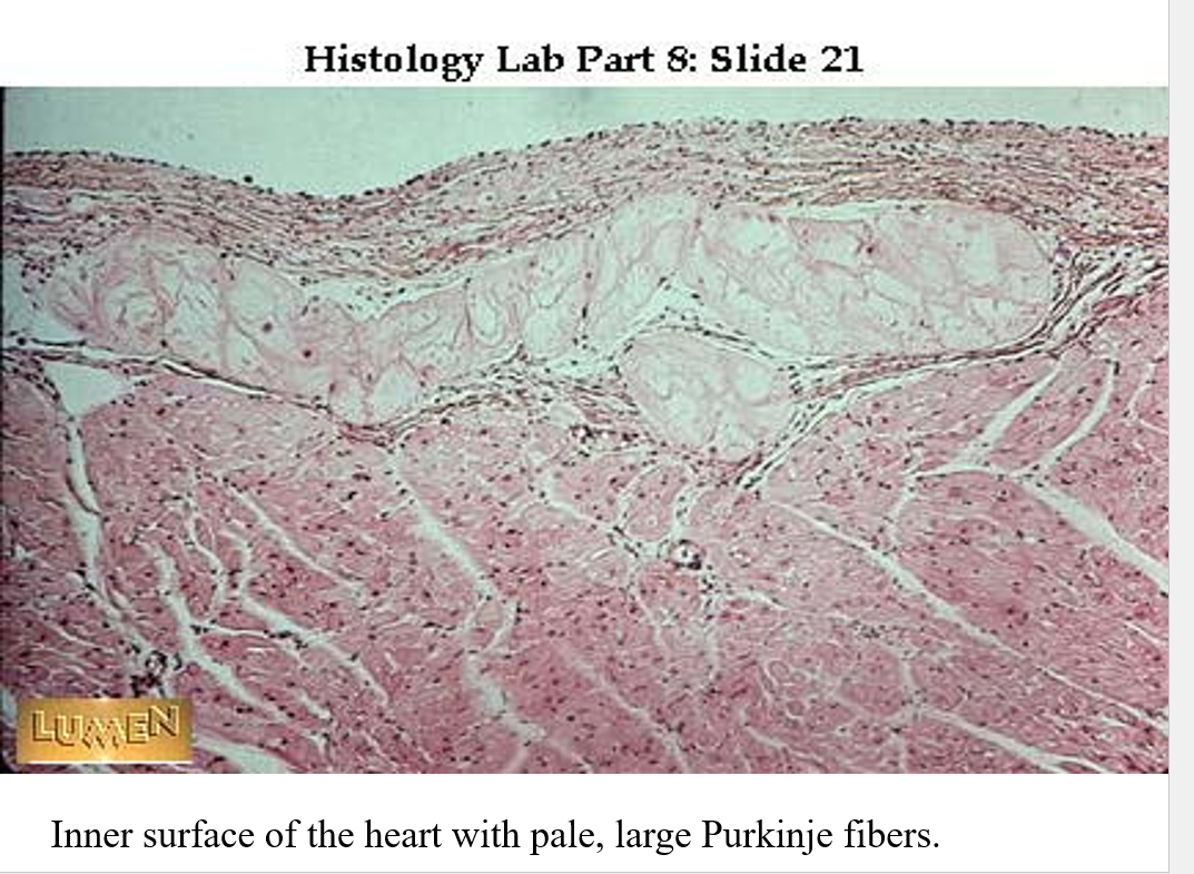
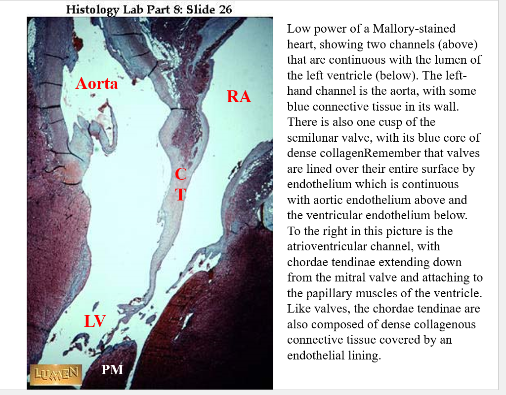
CT corenary tendon — w/out there may be back flow in the heart
PM pulminary muscle
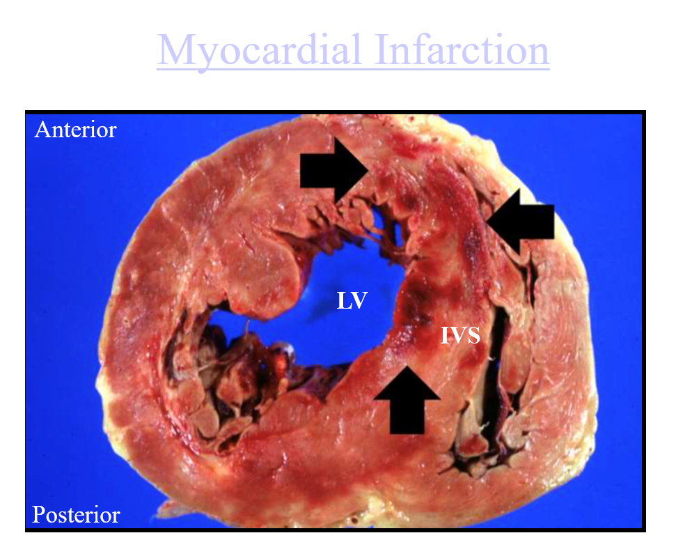
IVS = intraventricular septum — from pt. who had surgery - low BP - 1-2 days later have MI and die.
—most pathology cases Dr. Daft gives us don’t have happy endings
the arrows point to where tissue has died
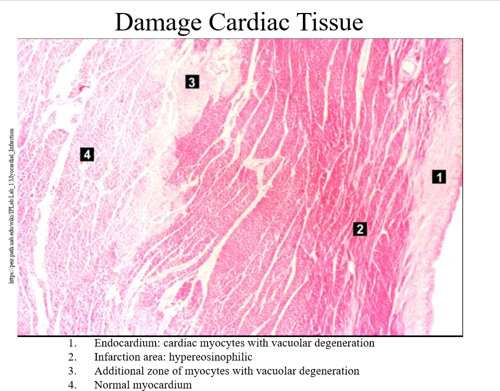
this will be on exam^^^
4 is the normal myocardium - acutual muscle of the heart thats pumping
at 1 vacular degeneration - dead or in the process of dying
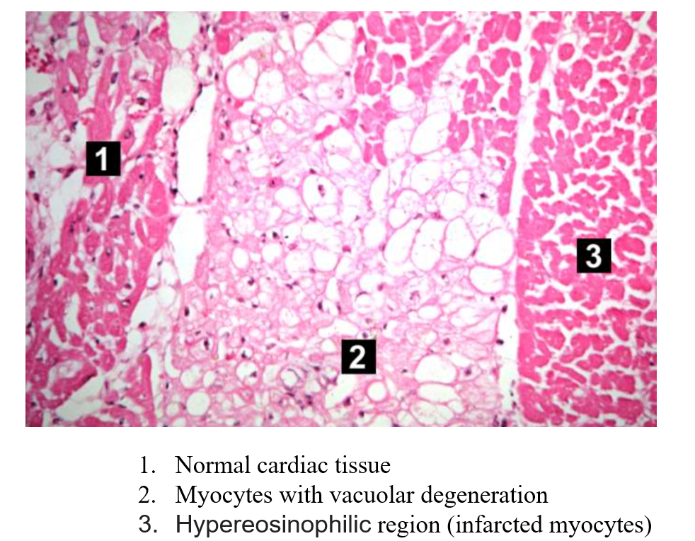
also on test^^^^^
3 lota eosinophils make the stain darker
pathology not hard — know what it looks like normally and be able to see when it’s messed up too
Date: 09/25/2025
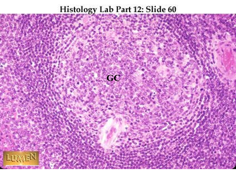
Lymphoid System:
•Made up of a collection of distinct organs
–Thymus, spleen lymph nodes, tonsils
•Made up of diffusely arranged meshwork of lymphatic tissue distributed in other organs (peyer’s patches).
•Nodules consist of reticular fibers that provide a framework for the accumulation of lymphocytes and macrophages.
Classification:
•Primary lymphoid tissue - lymphocytes are produced and undergo development (thymus and equivalent to Bursa of Fabricius in birds.
•Secondary (peripheral) organs – sites where foreign materials are extracted from body fluids and immune responses innitiated Lymph nodes, spleen, peyer’s patches, tonsils
lymphocytes grow and develop in the bone marrow
the secondary organs are sampling whats in the lymph fluis/blood
Primary Lymphatic Organs
•Thymus – very important before birth and shortly after birth
•Produce T cells (t lymphocytes) which cause other lymphatic tissues to become activated.
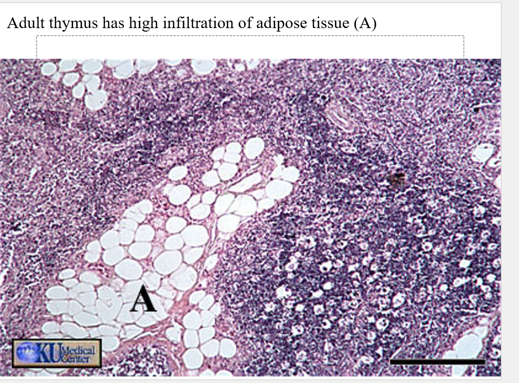
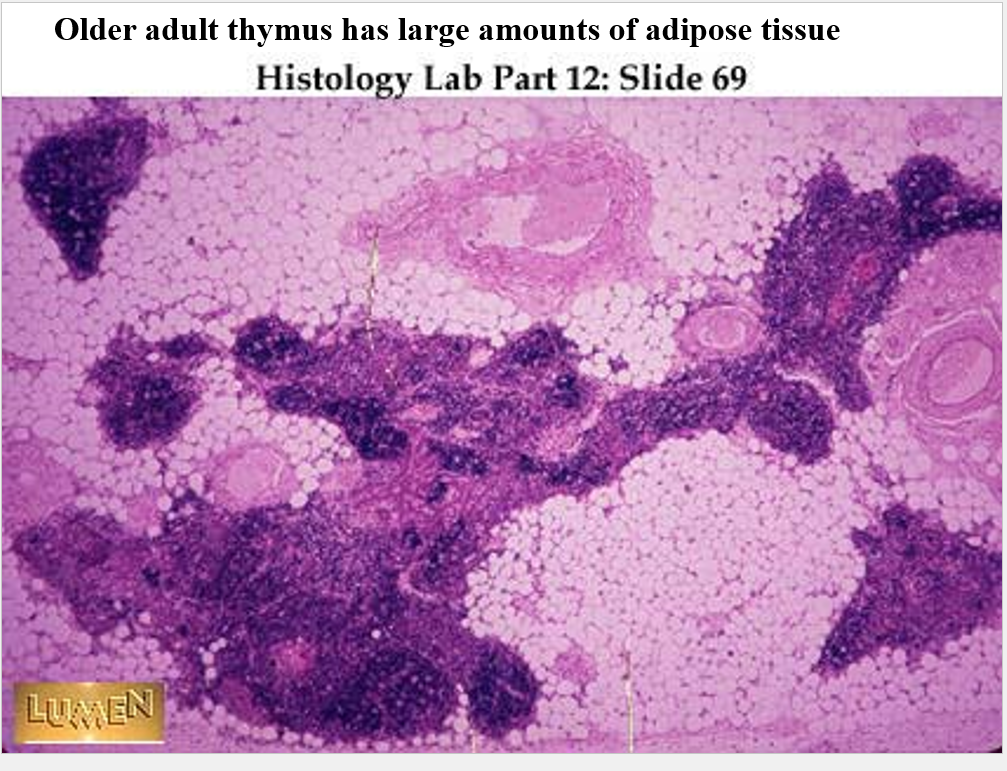
•Thymus begins to be infiltrated by adipose tissue in teen years. Adult mostly fat.
you can tell if younger or older via the adipose tissue content of the thymus (more fat older/less younger)
thymus divided into lobes
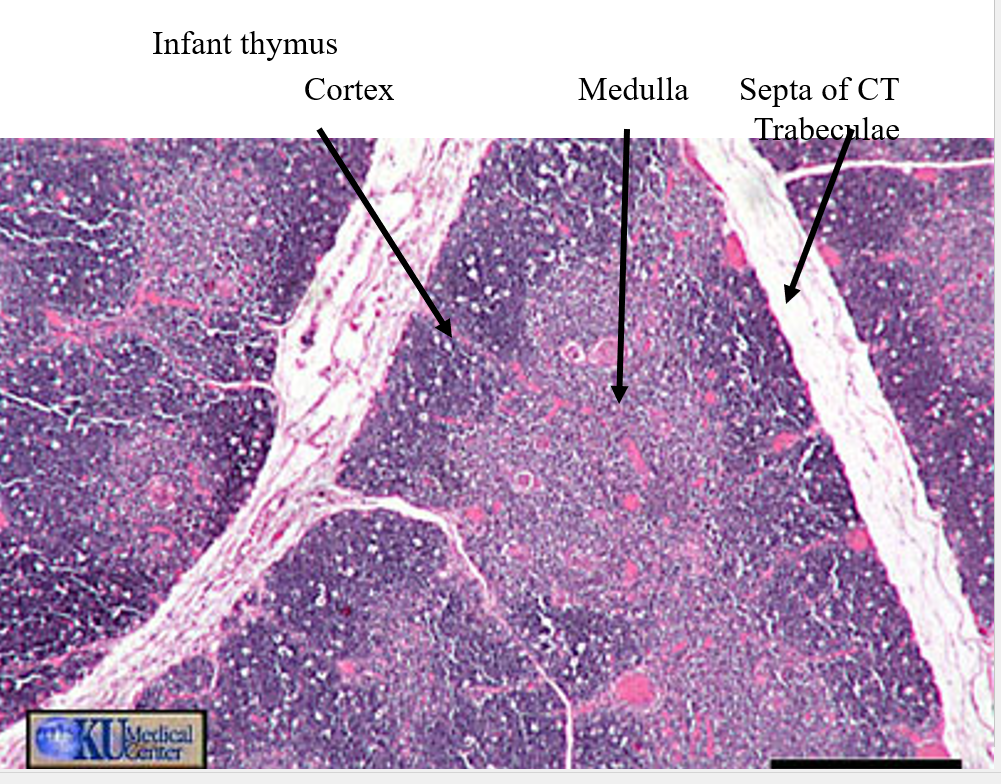
Thymus had T-cells as it’s the T cell training center
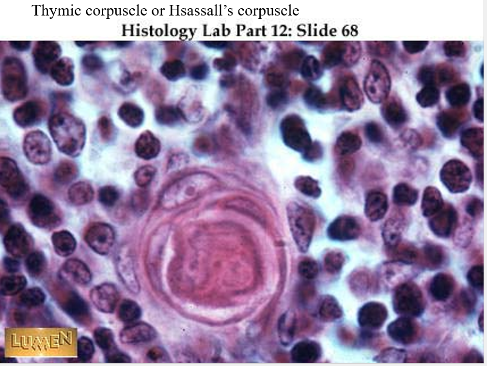
Thymic (Hassall’s) corpuscles
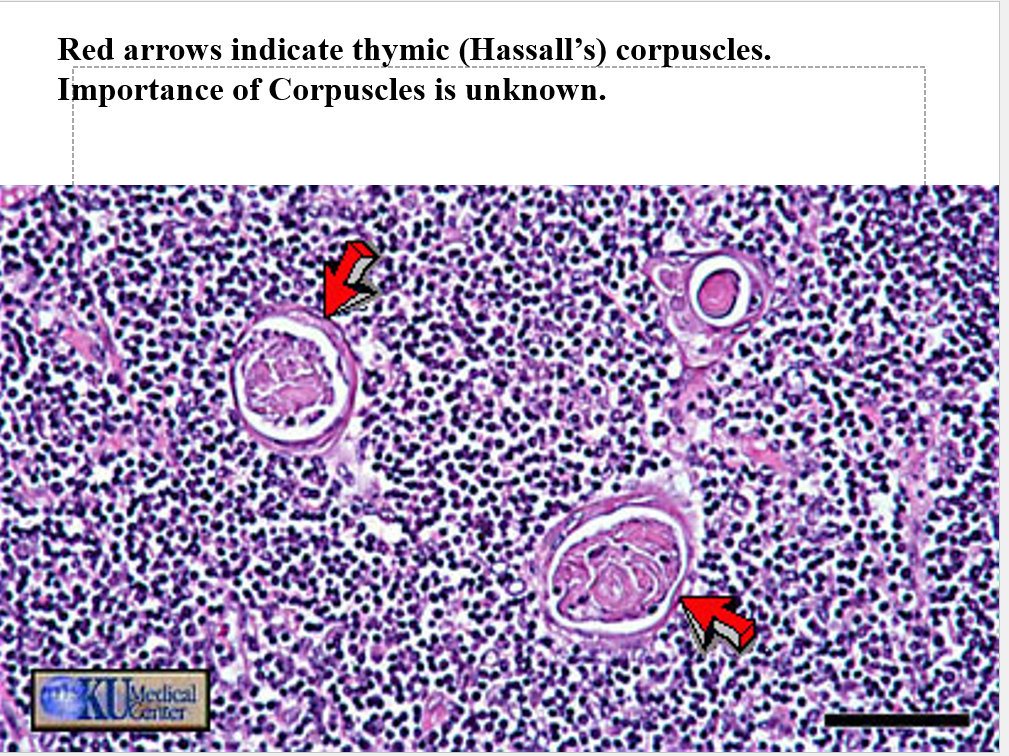
On the power point part of the test there may be a question asking if the thymus is older or younger
blood and lymph fluid are separate in a closed circulatory system
hemolymph is whats in open cerculatory systems as stuff is mixed
Secondary Lymphoid Organs
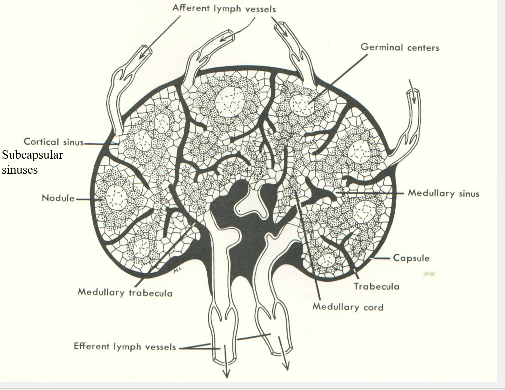
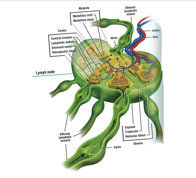
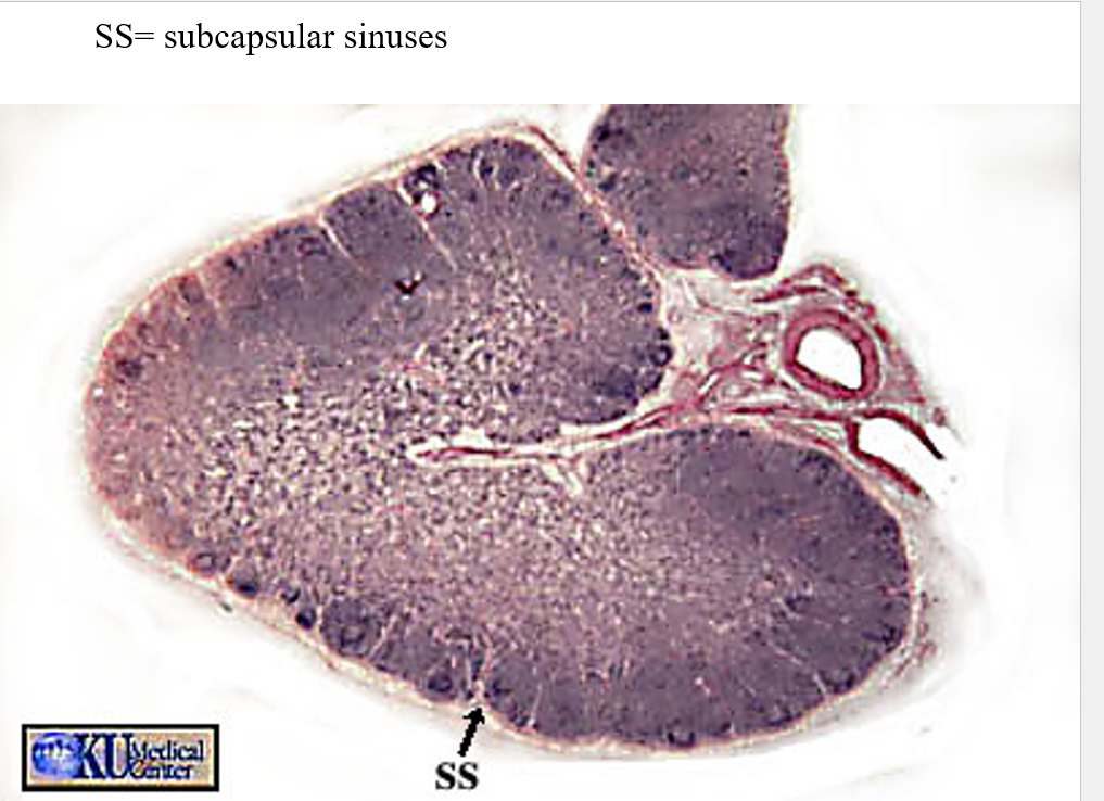
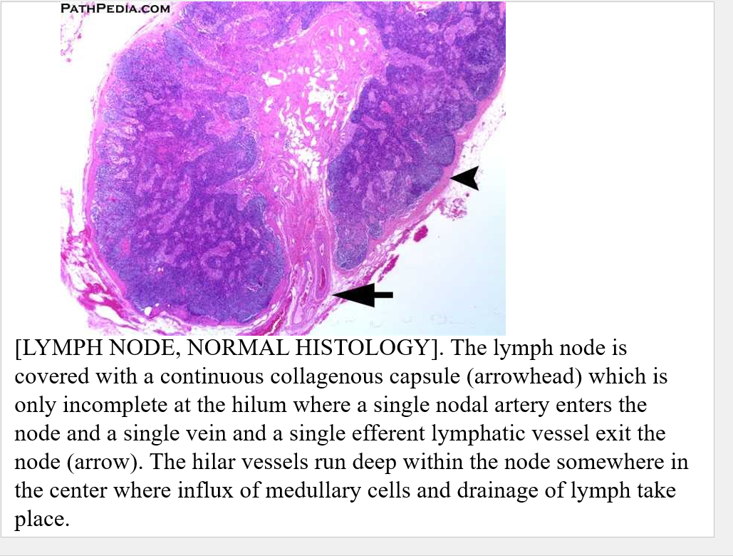
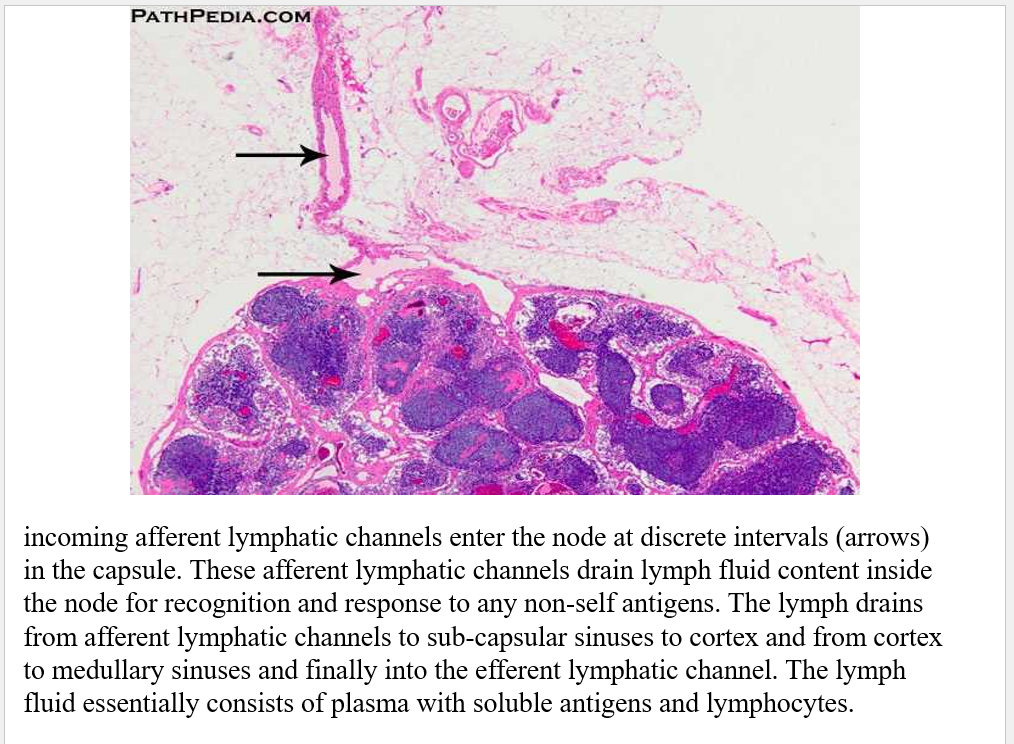
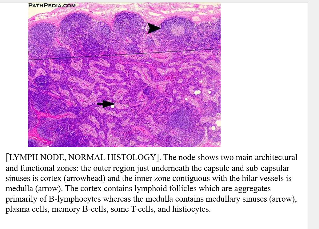
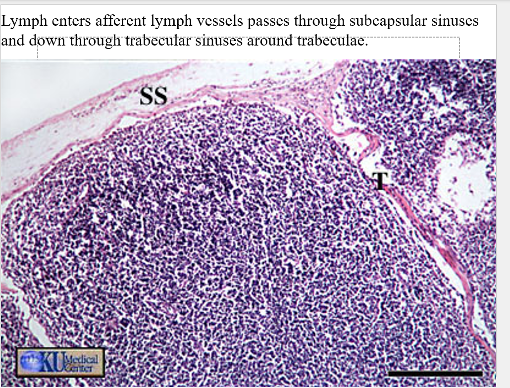
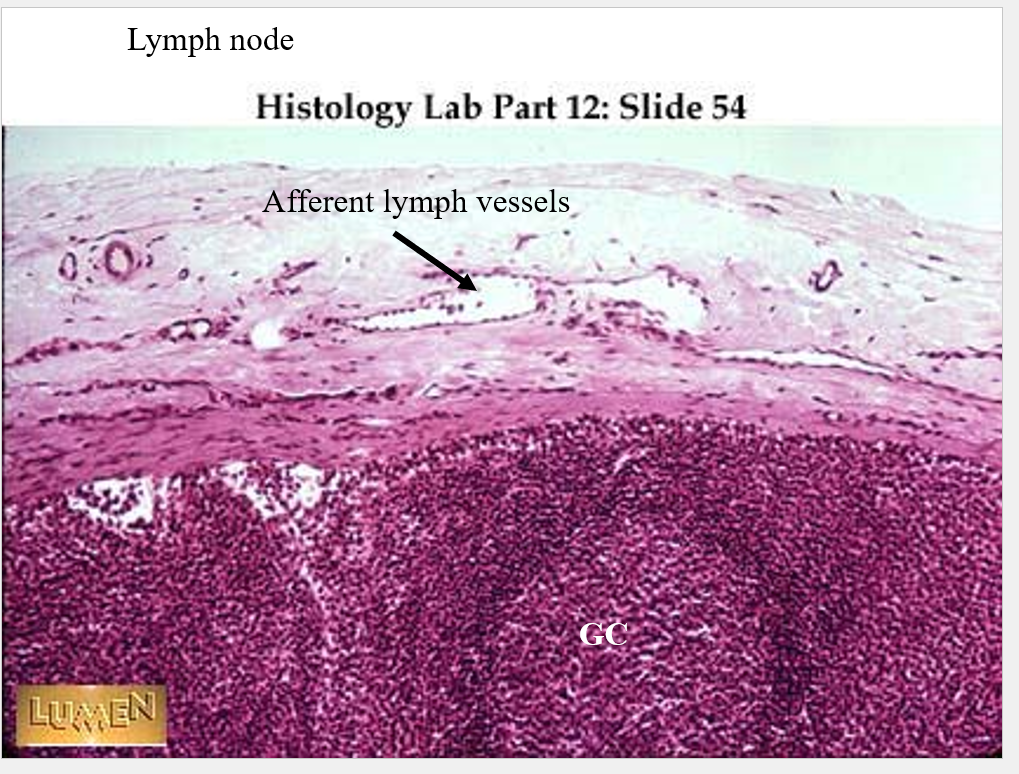
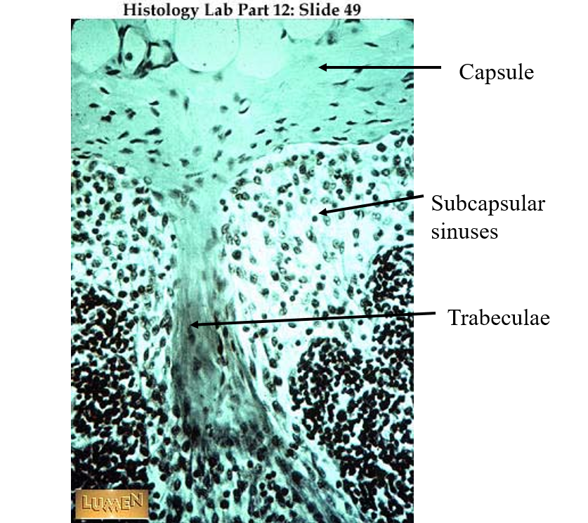
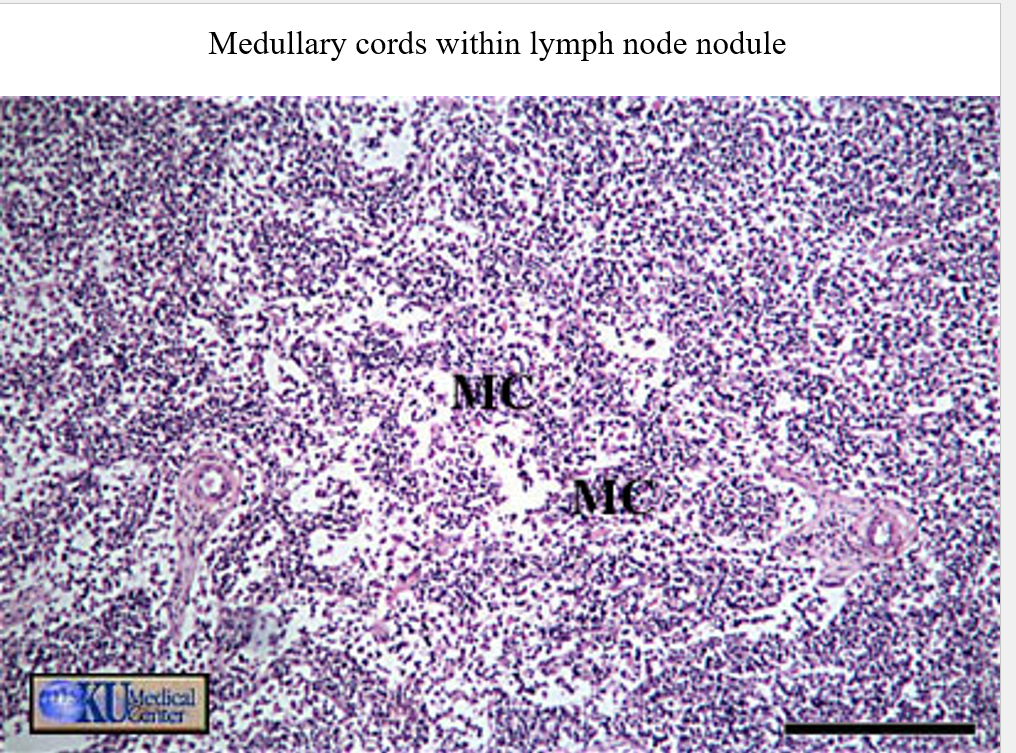
•Lymph node
•A mass of lymphatic tissue enclosed in a capsule of connective tissue
•Scattered along the course of lymph vessels
•Concentrated in area in neck, axilla (arm pits), and groin. Also in breast.
need to know cortex of thymus
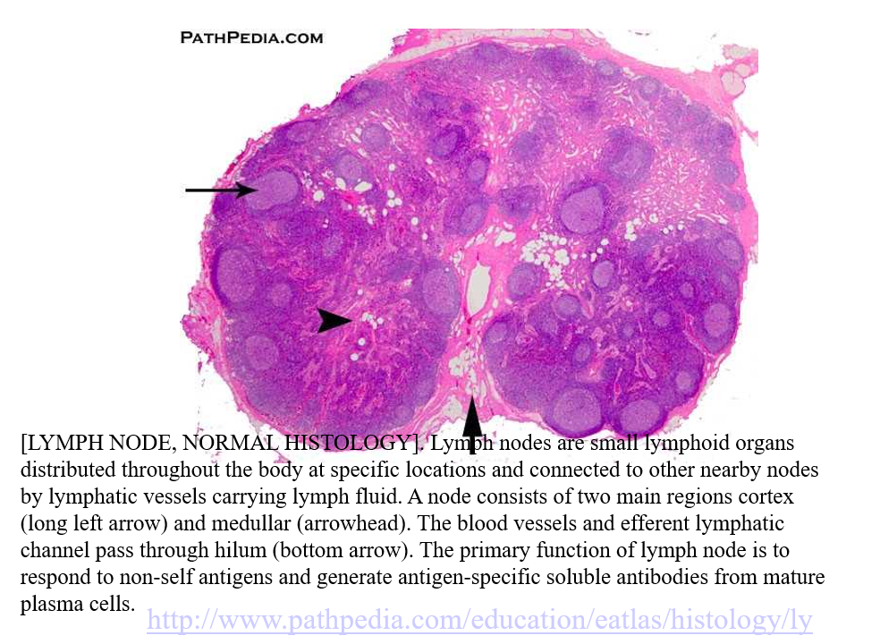
the little arrow is where the blood vessels and efferent lymphatic channel pass through —
DATE: 09/30/25
Presentation day BABY!!!
DATE: 10/02/2025
Secondary lymphatic organs
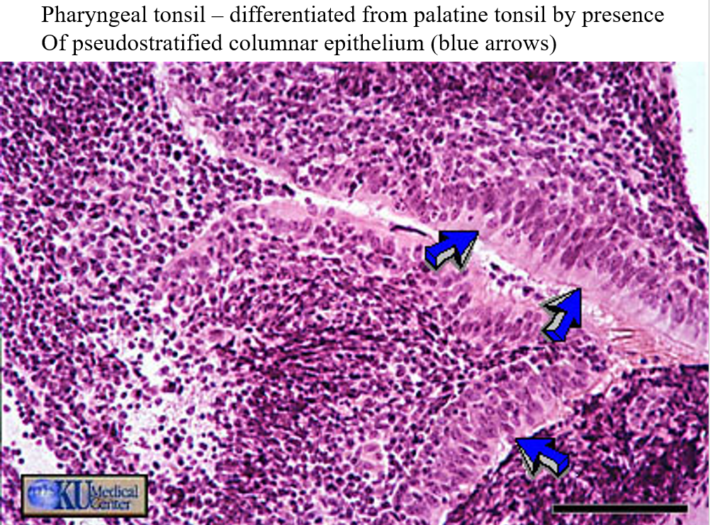
•tonsils-masses of lymphatic tissue embedded in the lining of the throat
•There are three groups, the pharyngeal, palatine, and lingual tonsils. They form a ring of lymphoid tissue around the pharynx.
•Tonsils develop within the layers making up the wall of the pharynx.
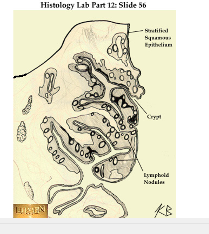
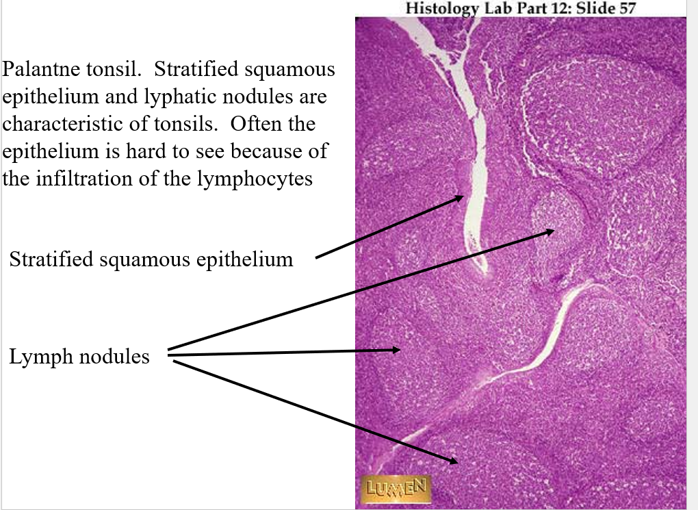
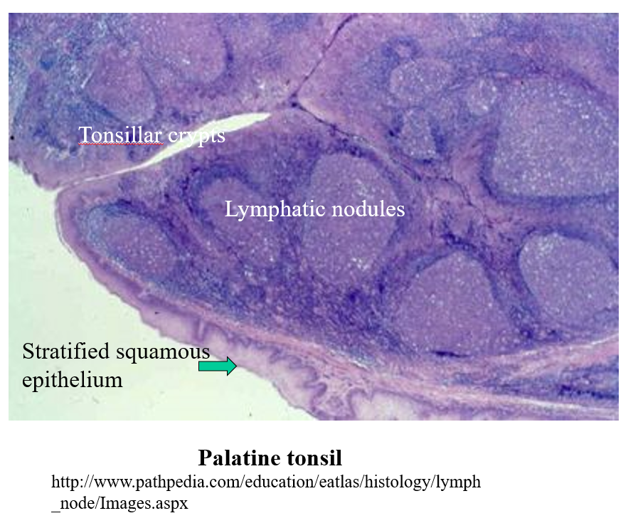
if you see both the tonsilar cripts and lymphatic noduals you are seeing a tonsle, not lymphnode
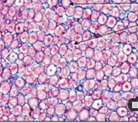
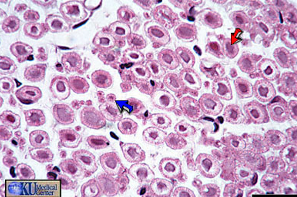
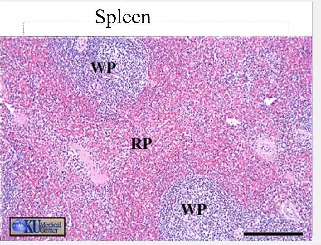
Spleen
spleen serves # of fx to include filtering and storage of blood
has lg area which fx to bring blood into contract w/ lymphocyte where foreign substances can be removed
also removes red and white blood cells
RBCs last only ~120 days
white pulp is a defining characteristic — more purple BC it’s high conc. of WBCs
Red pulp there too, its the reder part

peyer’s patch= individual
typically not in the duaddumu (1st part of intestens) BC they like bacteria, and theres too much acid that kills them
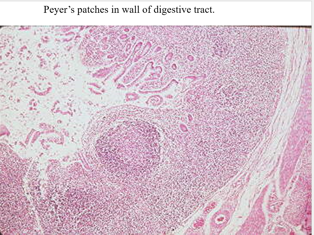
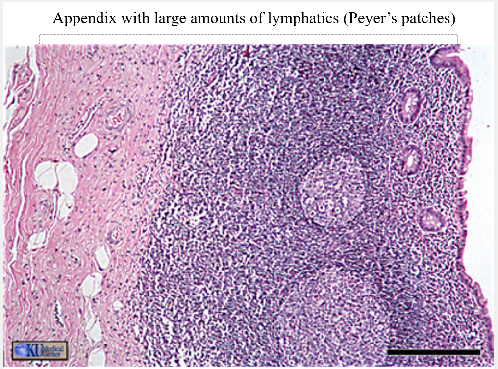
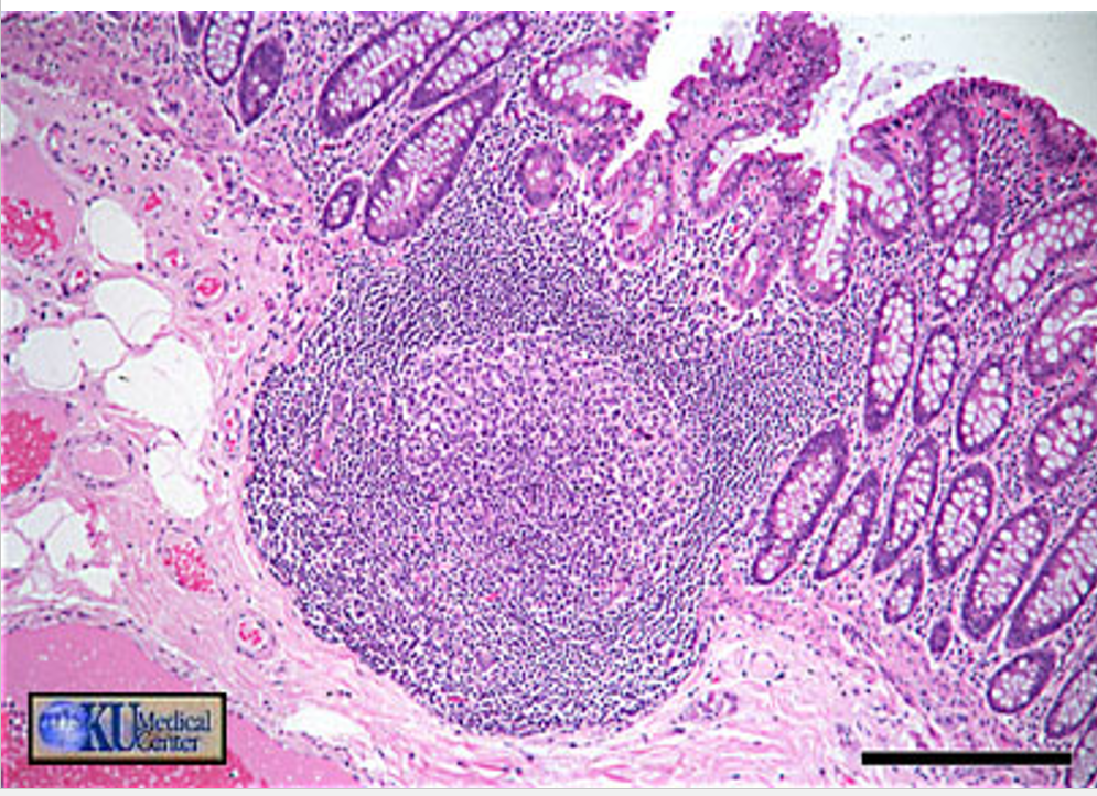
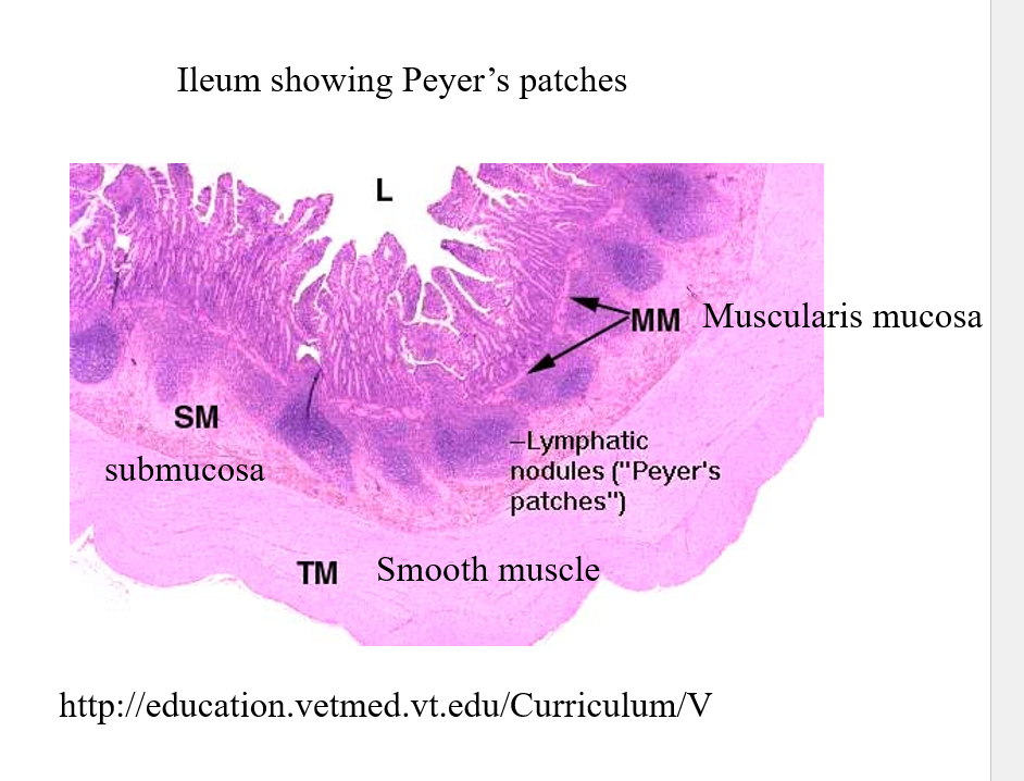
anything in the submucosal layer that’s sectioned off
find and add these to flash cards:
identify meneges - dura, aracnoid, pia
venules
arteriols
layers of blood vessle/artery and what they consist of
vasa vasarum
add the horn stuff - spine