3.2 Nerve Impulses
Nerve impulses
- neurons send messages electrochemically
- chemicals that are ‘electrically charged’ = ions
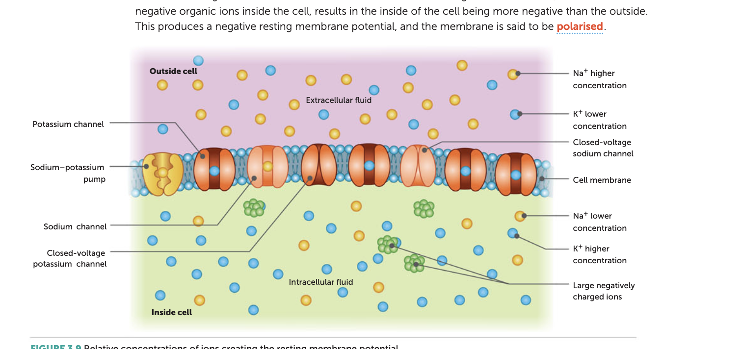
Resting Membrane Potential
- when a neuron is not sending signal - at rest
- inside of a neuron is negative relative to outside (doesn’t mean it is negatively charged, just more negative than outside)
- K+ cross through membrane easily
- Cl- and Na+ have more difficult time crossing
negatively charged protein molecules inside neuron can’t cross membran
Movement of Potassium (K+)
- potassium ions move freely across the membrane
- they will move out of cell into extracellular fluid
- movement of positive ions = potassium leakage → makes it more negative inside cell
- electrical potential difference across the cell membrane builds to a high enough level and causes an electrical resistance
- charge imbalance = leak NOT flood because inside cell is negative - positive attrct to negative
- high concentration → low concentration
- inside (more K+) → outside (less K+)
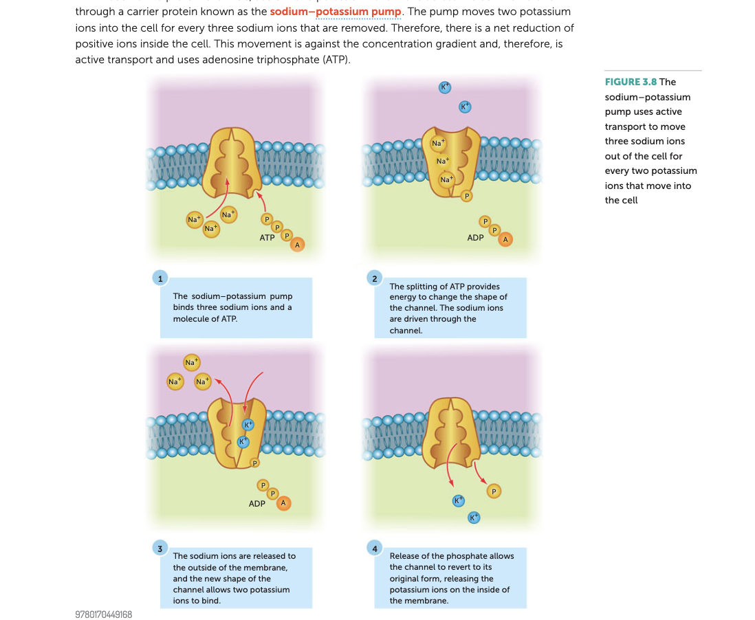
Sodium potassium pumps
- both Na+ and K+ are able to leak through respective channels
- channels change in protein structure (denature) to pump sodium and potassium in and out
- over time, this will decrease potential difference (-70 mV will become closer to 0 mV) = no charged difference - but the neurons will need a charge difference to send messages - need to constantly maintain electrochemicals
- this is because diffusion will never stop - will go from high concentration → low concentration till it reaches equilibrium
- need a way of moving Na+ out of the cell and K+ into the cell - done by the pump
- requires energy as it moves against concentration gradient = ATP = adenosine triphosphate
- ATP transfers a phosphate group = ADP + P - need mitochondria for energy = cellular respiration
- Na+ constantly being pumped in and then out
- K+ constantly being pumped out then in
- more positives out than in = keeps difference, allows negative inside
- moving K+ and Na+ maintains chemical concentration gradient = ⬆️ polarity (more difference in charge) ⬇️ charge
- e.g. diabetes - neurons need glucose → energy to function - always moving or faint
Action Potential (AP)
How do nerve impulses start?
- neurons are stimulated by receptor cells (respond to a stimulus e.g. chemicals, pain, temperature)
- contain special sodium channels that aren’t voltage-gated but gated by appropriate stimulus
- stimulus causes sodium channels to open
- causes sodium ions to flow into channel because of negative inside and concentration gradient
- causes depolarisation of membrane potential → affects voltage-gated sodium channel and starts an action potential
- e.g. chemical-gated in tongue - taste receptor cells open when a certain chemical in food binds to them
- mechanically-gated ion channels in hair cells of inner ear open when they are distorted by sound vibrations
What is action potential?
- resting membrane potential tells us about what happens when a neurons is at rest
- a nerve impulse = whole process, not just action potential - action potential is the letter being sent
- occurs when a neurons sends info down an axon
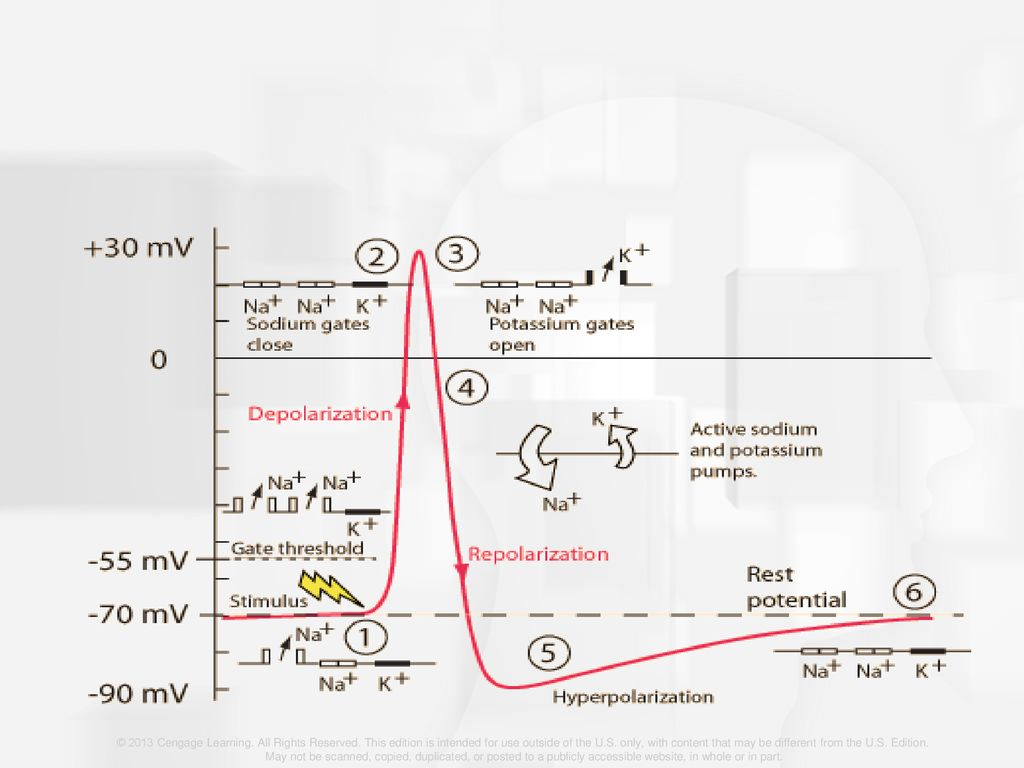
Depolarisation
- flipped polarity
- -55 mV causes more Na+ channels open for 0.5 ms - gate threshold
- causes Na+ to rush in → cell becomes more positive
- membrane potential will reach 30 mV by the time sodium is in the cell
Repolarisation
- depolarisation of membrane causes Na+ channels to close
- K+ channels open
- sodium potassium pump helps return to resting membrane potential
- K+ rush out → make inside more negative - attracted to
- restores original polarity = repolarisation
All-or-none response
- as soon as -55 mV is reached, rest of action potential is triggered - nothing can stop it
- frequency of impulse carries info → strong stimulus = high frequency
- intensity = frequency of action potential
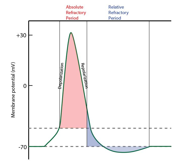
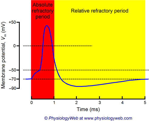
Refractory period
- limit number of AP in a given time
- time after depolarisation where no new AP can start
- impulses travel in 1 direction
- time is needed to restore proteins of voltage sensitive ion channels to original resting conditions
- Na+ channels cannot be opened - can’t be deporalised again
- can last up to 10 milliseconds - limits frequency of impulses
- absolute = second stimulus doesn’t cause new AP
- relative = second AP can be produced ONLY if stimulus is greater than threshold
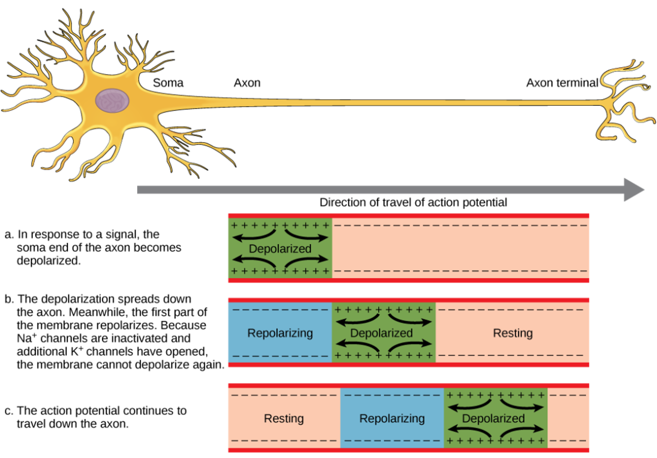
How do impulses travel down the neuron?
- different action potentials along neuron = propogate - triggers down the axon
- depolarises sections along the membrane
How fast are impulses?
- travel 0.1-100 m/s along axons
- allows for fast responses to stimuli
- speed is affected by:
- temperature
- axon diameter - like straw and milkshake - bigger it is, easier it flows
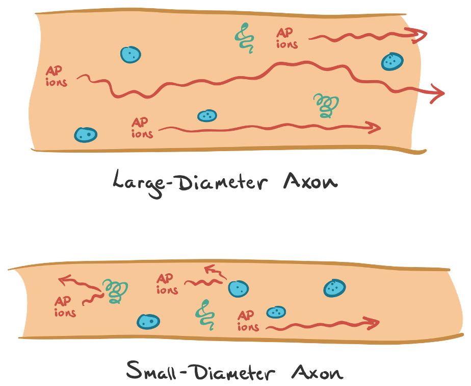
- whether or not it has myelin sheath
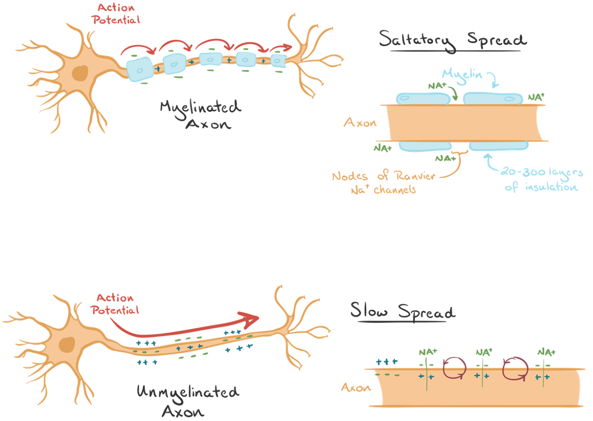
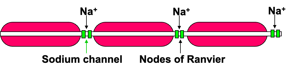
Myelinated Neurons
- axons of many neurons are encased in fatty myelin sheath (Schwann cells)
- where the sheath of one Schwann celll meets the next, the axon is unprotected
- voltage-gated sodium channels of myelinated neurons are confined to spots (nodes of Ranvier)
- in rush of Na+ at one node creates just enough depolarisation to reach threshold of next
- AP jumps from one node to next = saltatory propogation = faster
| Unmyelinated | Myelinated |
|---|---|
| Depolarisation of one area of the cell membrane causes AP to flow onto membrane immediately adjacent to stimulus | Deplorasation of one area of cell membrane causes AP to jump from one node of ranvier to another |
| Nerve impulse/exchange of ions (NOT AP) moves along entire length of neuron/axon | Nerve impulse/exchange of ions (NOT AP) only occurs at nodes of Ranvier or cannot occur where axon is myelinated |
| Lower concentration gradient of ions either side of the membrane | Higher concentration gradient of ions either side of the membrane at nodes of Ranvier |
| Nerve impulse/message (NOT AP) travels along the whole length of the fibre, reducing its speed | AP jumps from one node of Ranvier to the next on the myelinated fibre (saltatory conduction), the impulse can travel faster |
Summary
- nerve impulse conduction is bumping of positive charge down the axon
- AP initiated at one end of axon can only propagate in 1 direction
- AP doesn’t turn back because membrane just behind is in its refractory period (voltage gated Na+ channels are inactivated)
Summarised steps of AP
- A resting neuron has a positive charge on the outside of the membrane and a negative charge on the inside (resting membrane potential -70 mV)
- High concentration of Na+ on the outside and high concentration of K+ on the inside
- greater concentration of negatively charged ions - due to organic anaions than K+ = negative on the inside
- stimulus causes voltage gated sodium channels to open and sodium ions rush in intracellular fluid
- -55 mV is threshold for voltage gated sodium ions
- inward movement of Na+ ions reverses the charge = cell becomes depolarised, polarisation has flipped - inside is positive, outside is negative - membrane potential will reach +30 mV
- After inside of membrane becomes flooded with sodium, gated potassium channels open and allow potassium ions to move to the outside
- as soon as potassium ions are released, sodium ion channels closed (membrane is repolarised)
- hyperpolarisation occurs = too many potassium ions cause the inside of the cell to be more negative than -70 mV
- sodium potassium pump restores concentration of sodium and potassium when membrane is at resting state
Synapse
- junction between branches of adjacent neurons
- neurons don’t join at synapse = small gap (for axon and skeletal muscle = neuromuscular jinction)
- occurs between a branch at the end of an axon and dendrite or the cell body of another neuron
- AP cannot cross synaptic cleft
- impulse is carried by chemicals called neurotransmitters
Neurotransmitters
- made by cell sending impulse (pre-synaptic neuron) and stored in synaptic vesicles at the end of the axon
- cell receiving impulse (post-synaptic neuron) has chemical gated ion channels = neuroreceptors
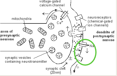
### Synapses explained
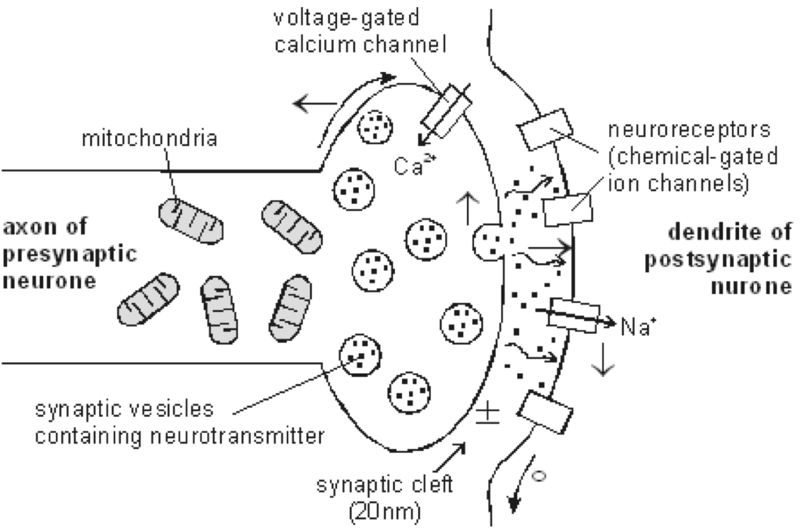
- at the end of the pre-synaptic neuron - voltage gated calcium channels
- when AP reaches synapse, channels open
- calcium ions flow into the cell
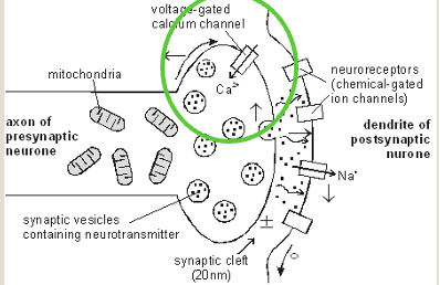
- cause synaptic vesicle to fuse with cell membrane
- neurotransmitters diffuse across synaptic cleft
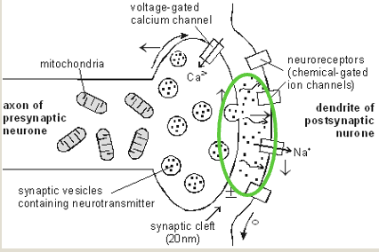
- neurotransmitter binds to neuroreceptors in the post-synaptic membrane
- channels open, Na+ flow in
- causes depolarisation
- AP initiated in post-synaptic neuron
Transmission across a synapse (in steps)
- AP arrives at pre-synaptic axon terminal
- Local depolarisation caused voltage gated calcium ion channels to open
- Calcium ions from extracellular fluid diffuses through presynaptic membrane of axon terminal (AT) and enters cytoplasm of AT
- Calcium causes neurotransmitter vesicles to migrate to pre-synaptic membrane of AT
- Neurotransmitters leave vesicles and enter synaptic cleft through exocytosis
- Neurotransmitter diffuses across synapse to post-synaptic membrane of dendrite of adjacent neuron
- Na+ floods in, causing depolarisation in postsynaptic dendrite
- AP is generated
Function
- prevent impulses travelling in wrong direction
- impulse can pass along an axon in either direction, but can only cross a synapse in one direction because the vesicles are only found in knobs and end plates
- vast number of synaptic connections allow flexiblity
- equivalent to switchboard in an elaborate telephone exchange, enabling messages to be diverted from 1 line to another
What happens to neurotransmitters? + Examples
- broken down by specific enzyme in synaptic cleft
- breakdown products are absorbed by pre-synaptic neuron
- used to re-synthesis more neurotransmitter
- Acetylcholine (ACh)
- released by motor neurons onto skeletal muscle cells
- released by neurons in parasympathetic nervous system
- Noradrenaline
- released by neurons in sympathetic nervous system