EXAM 1
Fundamentals of Anatomy, Physiology, and Homeostasis
Anatomy: The scientific study of body structures and their physical relationships.
Physiology: The scientific study of the functions and mechanisms of the body's structures.
Homeostasis: The dynamic maintenance of a relatively stable internal environment despite continuous external or internal fluctuations.
Feedback Mechanisms in Regulation
Negative Feedback Loops:
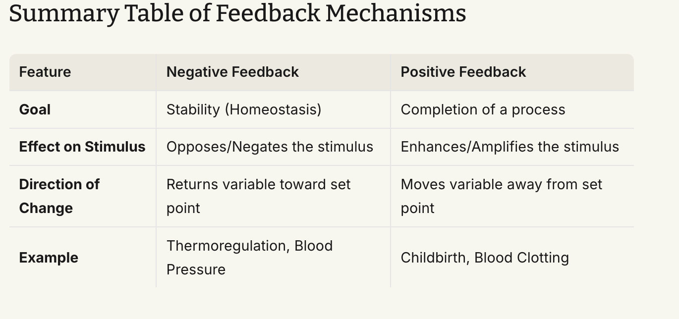
Homeostatic regulation relies primarily on negative feedback systems, which counteract or oppose a detected stimulus to restore physiological parameters to a set target value.
Generic Mechanism:
Stimulus Detection: A change in a physiological variable (stimulus) is detected by sensory receptors.
Communication to Integration Center: Receptors transmit signals to an integration center (typically within the central nervous system or endocrine glands).
Integration & Response Coordination: The integration center analyzes the incoming information, compares it to a set point, answers the question "What is the stimulus?", and coordinates an appropriate response.
Effector Signaling: The integration center communicates signals to target organs (effectors).
Effector Response: Target organs execute a response that directly reverses the direction of the initial stimulus, bringing the variable back toward homeostasis.
Blood Pressure Homeostasis Example:
Stimulus: Blood pressure drops below the physiological baseline of .
Detection: Specialized mechanoreceptors called baroreceptors detect the decreased arterial pressure.
Integration: Baroreceptors communicate the decrease in pressure to the cardiovascular integration center in the brain.
Effector Signal & Response: The integration center sends signals to the vascular system and heart. Blood vessels constrict and heart rate increases ().
Outcome: Systemic vascular resistance and cardiac output increase, causing blood pressure to rise () back to normal.

Body Temperature Homeostasis: Thermoreceptors detect temperature deviations from . A rise in temperature triggers vasodilation and sweating to promote heat loss; a drop in temperature triggers vasoconstriction and shivering to generate and retain heat.
Blood Glucose Homeostasis: Elevated blood glucose triggers pancreatic beta cells to release insulin, promoting cellular glucose uptake and glycogen synthesis in muscle and liver tissues. Low blood glucose triggelevels triggereatic alpha cells to release glucagon, breakiwhich stimulates the breakdown ofd glycogen into free glucose.
System Breakdown: Pathology and disease result when negative feedback mechanisms fail or become overwhelmed, disrupting internal stability.
Positive Feedback Loops:
Mechanisms that amplify or enhance the original stimulus, pushing the physiological system further away from baseline conditions until a definitive endpoint is achieved.
Positive feedback loops are not homeostatic because they do not maintain a stable baseline equilibrium.
Childbirth (Parturition) Example:
Stimulus: The fetal head presses against the uterine cervix, causing cervical stretch ().
Integration: Neural signals from the stretched cervix travel to the hypothalamus and posterior pituitary gland.
Response: The pituitary gland secretes the hormone oxytocin into the bloodstream.
Effector Action: Oxytocin stimulates forceful uterine muscle contractions ().
Positive Loop: Enhanced contractions push the baby further down the birth canal, causing greater cervical stretch, triggering additional oxytocin secretion and even stronger contractions.
Termination: The loop is terminated abruptly by the physical delivery of the baby and placenta, which eliminates cervical stretch.

Blood Clotting Example: Vessel damage exposes extracellular matrix proteins, adhering platelets, and initiating chemical mediator release. Released factors attract and activate more platelets, forming a localized cascade that rapidly seals the vascular defect.
Anatomical Terminology, Landmarks, Planes, and Cavities
Universal Surface Terminology
Standardized anatomical terms describe surface locations unambiguously. Standard terminology ensures that healthcare professionals worldwide communicate accurate locations regardless of language or setting (e.g., "right posterior carpal region" universally designates the back side of a patient's right wrist).
Core Orientation Rule: When observing an individual, THEIR RIGHT IS YOUR LEFT.
Anatomical Position: The universal baseline posture where the individual stands upright, facing forward, feet parallel and flat on the floor, with upper limbs at the sides and palms facing anteriorly (forward). All anatomical positional references assume the body is in this standard position.
Anatomical Surface Landmarks
Anterior Body Landmarks:
Abdominal: Abdomen region
Antebrachial: Forearm
Antecubital: Front of the elbow
Axillary: Armpit
Brachial: Upper arm
Buccal: Cheek
Carpal: Wrist
Cervical: Neck
Coxal: Hip
Digital: Fingers or toes
Femoral: Thigh
Fibular/Peroneal: Lateral side of the lower leg
Frontal: Forehead
Inguinal: Groin
Mammary: Breast
Mental: Chin
Nasal: Nose
Oral: Mouth
Orbital: Eye socket
Palmar: Pald
Patellar: Front of the knee (kneecap)
Pedal: Foot
Pelvic: Pelvis
Pubic: Genital region
Sternal: Breastbone
Tarsal: Ankle
Thoracic: Chest
Umbilical: Navel
Posterior Body Landmarks:
Acromial: Point of the shoulder
Calcaneal: Heel of the foot
Cephalic: Head
Dorsum: Back surface
Gluteal: Buttock
Lumbar: Lower back between ribs and hips
Manus: Hand
Occipital: Base of the skull
Olecranal: Back of the elbow
Otic: Ear
Plantar: Sole
Popliteal: Posterior knee area
Sacral: Bone area between the hips
Scapular: Shoulder blade
Vertebral: Spinal column
Directional Terminology
Superior / Inferior: Superior refers to a structure closer to the head or higher up; Inferior refers to a structure closer to the feet or lower down.
Cranial / Caudal: Anatomical direction terms used primarily in quadruped animals and aquatic species. Cranial indicates toward the head; Caudal indicates toward the tail.
Anterior / Posterior: Anterior (ventral in quadrupeds) indicates the front of the body; Posterior (dorsal in quadrupeds) indicates the back of the body.
Medial / Lateral: Medial indicates position closer to the longitudinal midline of the body; Lateral indicates position farther from the midline.
Proximal / Distal: Used exclusively for appendages (arms and legs) relative to their trunk attachment point. Proximal indicates closer to the attachment point; Distal indicates farther from the attachment point.
Superficial / Deep: Superficial indicates proximity to the exterior body surface; Deep indicates location further internal from the body surface.
Body Planes and Sections
Sagittal Plane: Vertical plane running anterior to posterior, dividing the body into right and left portions.
Midsagittal (Median) Plane: Passes directly through the midline, dividing the body into equal left and right halves.
Parasagittal Plane: Off-center vertical plane dividing the body into unequal left and right portions.
Frontal (Coronal) Plane: Vertical plane dividing the body into anterior (front) and posterior (back) portions.
Transverse (Horizontal) Plane: Cross-sectional horizontal plane dividing the body into superior (upper) and inferior (lower) portions.
Oblique Plane: Section cut diagonally at an angle other than .
Clinical Application: Body planes guide diagnostic imaging modalities (such as CT scans and MRIs) to isolate sections and accurately locate anatomical lesions, tumors, or internal injuries.
Abdominopelvic Quadrants and Regions
Four Abdominopelvic Quadrants:
Right Upper Quadrant (RUQ): Contains the liver, gallbladder, right kidney, portions of the small and large intestines.
Right Lower Quadrant (RLQ): Contains the cecum, appendix, right ovary/uterine tube or right spermatic cord, right ureter.
Left Upper Quadrant (LUQ): Contains the stomach, spleen, left lobe of liver, body of pancreas, left kidney, portions of small and large intestines.
Left Lower Quadrant (LLQ): Contains the left ovary/uterine tube or left spermatic cord, sigmoid colon, left ureter.
Nine Abdominopelvic Regions:
Right Hypochondriac Region: Superior right region under the cartilage of the ribs.
Epigastric Region: Superior middle region located above the stomach.
Left Hypochondriac Region: Superior left region under the cartilage of the ribs.
Right Lumbar Region: Middle right region near the waist.
Umbilical Region: Centermost region surrounding the navel.
Left Lumbar Region: Middle left region near the waist.
Right Iliac (Inguinal) Region: Inferior right region near the hip bone.
Hypogastric (Pubic) Region: Inferior middle region located below the navel.
Left Iliac (Inguinal) Region: Inferior left region near the hip bone.

Body Cavities
Dorsal Body Cavity: Enclosed by bone and fluid-filled membranes to protect central nervous tissue.
Cranial Cavity: Formed by the cranium; houses the brain.
Vertebral (Spinal) Cavity: Formed by the vertebral column; houses the spinal cord.
Ventral Body Cavity: Divided by the diaphragm into two main compartments containing visceral organs.
Thoracic Cavity: Superior cavity enclosed by the chest wall.
Mediastinum: Central compartment housing the esophagus, trachea, thymus, and major vessels.
Pericardial Cavity: Fluid-filled space surrounding the heart.
Pleural Cavities: Left and right fluid-filled spaces surrounding each lung.
Abdominopelvic Cavity: Inferior cavity extending from the diaphragm to the pelvic floor.
Abdominal Cavity: Superior portion housing the digestive organs (stomach, intestines, liver, spleen), kidneys, and ureters.
Pelvic Cavity: Inferior portion enclosed by pelvic bones; houses the urinary bladder, internal reproductive organs, and rectum.
Peritoneal Cavity: Serous membrane-lined space containing peritoneal organs.
Chemical Foundations, Cellular Transport, and Bioenergetics
Chemical Foundations of Physiology
Ions: Atoms or molecules carrying a net electrical charge resulting from the gain or loss of electrons.
Cations: Positively charged ions (,, , ).
Anions: Negatively charged ions ().
Essential Physiological Ions:
Sodium (): Principal extracellular cation; essential for osmotic balance, maintaining extracellular fluid volume, generating resting membrane potential (RMP), and initiating action potentials.
Potassium (): Principal intracellular cation; crucial for establishing resting membrane potential, repolarization phase of action potentials, and maintaining cell volume.
Calcium (): Vital extracellular and sarcoplasmic ion; required for muscle contraction, neurotransmitter release, blood clotting, and bone mineralization.
Magnesium (): Key intracellular cation; functions as an enzyme cofactor, stabilizes nucleic acid structures, and binds ATP.
Chloride (): Major extracellular anion; maintains electrical neutrality, balances cation distributions, and aids in fluid balance.
Biological Macromolecules
Carbohydrates:
Monomers: Monosaccharides (e.g., glucose).
Polymers: Polysaccharides (e.g., glycogen stored in skeletal muscle and liver cells).
Function: Primary energy substrate for cellular respiration and ATP synthesis. Monosaccharides cross membranes via transporter proteins.
Lipids:
Building Blocks: Fatty acids and glycerol.
Types & Polymers:
Triglycerides: Three fatty acids bound to glycerol; long-term energy storage in adipose tissue.
Steroids: Non-polar four-ring carbon structures (e.g., cholesterol, steroid hormones).
Phospholipids: Structural lipids forming plasma membranes.
Proteins:
Monomers: Amino acids.
Polymers: Polypeptides and functional proteins.
Function: Function as enzymes, membrane channels, transporters, structural fibers, and signaling molecules. Large protein structures cannot pass freely through lipid bilayers.
Nucleic Acids:
Monomers: Nucleotides.
Polymers: Deoxyribonucleic acid (DNA), Ribonucleic acid (RNA), and Adenosine Triphosphate (ATP).
Function: Genetic coding, protein synthesis, and direct cellular energy transfer.
Bioenergetics & Cellular ATP Production
Glucose enters the cytosol and undergoes glycolysis to yield pyruvate and a net gain of ATP.
Pyruvate enters the mitochondria in the presence of oxygen to undergo aerobic respiration, yielding large amounts of ATP ().
Stored glycogen in liver and muscle tissue is converted back into glucose when cellular energy demand increases.

Plasma Membrane Architecture and Fluid Compartments
Fluid Compartments:
Intracellular Fluid (ICF): Fluid contained within cells. Characterized by high concentrations of , proteins and phosphate ions.
Extracellular Fluid (ECF): Fluid outside cells. Characterized by high concentrations of, , and. ECF compartments include Interstitial Fluid (IF), blood plasma, lymph, and cerebrospinal fluid.
Phospholipid Bilayer:
Amphipathic structure with hydrophilic phosphate heads exposed to aqueous ICF and ECF, and hydrophobic fatty acid tails forming an inner non-polar core.
Selective Permeability: Small, non-polar hydrophobic molecules (,, fatty acids, steroid hormones) diffuse freely across the bilayer. Ions (,, , ), polar molecules (water, glucose), and large macromolecules cannot cross without specific membrane transporter proteins.
Cellular Transport Mechanisms
Passive Transport: Movement of substances down their concentration gradient (from high concentration to low concentration) without expending cellular metabolic energy (ATP).
Simple Diffusion: Passive net movement of solute particles down a concentration gradient across a permeable lipid bilayer.
Osmosis: Diffusion of water molecules across a selectively permeable membrane toward a region of higher solute concentration.
Channel-Mediated Diffusion: Passage of specific ions through transmembrane protein channels.
Leak Channels: Continuously open channels allowing passive ion movement down electrochemical gradients.
Gated Channels: Protein channels that open or close in response to chemical (ligand), electrical (voltage), or physical (mechanical) stimuli.
Carrier-Mediated Passive Transport (Facilitated Diffusion): Transmembrane carrier proteins bind specific molecules (e.g., glucose) on one side of the membrane, undergo a conformational change, and release the molecule on the opposite side down its concentration gradient.
Active Transport: Movement of solutes against their concentration gradient (from low concentration to high concentration), requiring cellular energy expenditure in the form of ATP.
Primary Active Transport (Pumps):
Sodium-Potassium Pump (): Membrane-bound active carrier protein that hydrolyzes ATP to move ions out of the cell and ions into the cell per cycle. This process maintains the resting membrane potential (RMP) and low intracellular concentration.

Epithelial Tissue Architecture, Characteristics, and Specialized Functions
Essential Functions of Epithelial Tissue
Epithelial tissue covers exposed body surfaces, lines internal passageways, hollow organs, and body cavity tracts, and forms secretory glands.
Four Essential Functions:
Physical Protection: Protects underlying tissues from mechanical abrasion, physical trauma, dehydration, chemical destruction, and biological pathogens.
Control Permeability and Regulate Absorption/Secretion: Controls the passage of all substances entering or exiting the internal environment across cell membranes.
Provide Specialized Secretions: Specialized epithelial cells (glandular cells) produce secretions discharged onto physical surfaces for protection/lubrication or into blood/interstitial fluid (hormones).
Sensory Perception: Innervated by sensory nerves sensitive to local environmental stimuli, transmitting sensory signals to the central nervous system.
Essential Characteristics of Epithelial Tissues
1. Polarity:
Epithelial cells exhibit structurally and functionally distinct surfaces:
Apical Surface: Free, unattached surface exposed to the external environment, internal lumen, or body fluid cavity. May possess specialized apical structures:
Microvilli: Non-motile plasma membrane folds that increase surface area to maximize absorption and secretion.
Cilia: Motile, hair-like protein projections that beat rhythmically to propel fluids, mucus, or materials across the cell surface.
Lateral Surface: Side surfaces bound to neighboring epithelial cells via specialized intercellular junctions.
Basal / Basolateral Surface: Bottom surface anchored to the underlying pasement membrane.

2. Intercellular Attachment & Cell Junctions:
Gap Junctions: Composed of transmembrane protein complexes called connexons (each formed by six connexin proteins) aligning between adjacent cells to form aqueous channels. Promote direct diffusion of ions and small molecules between cells, enabling rapid intercellular communication.

Tight Junctions (Zonula Occludens): Formed by interlocking junctional proteins that fuse adjacent plasma membranes into continuous bands near the apical surface. Prevent fluid and solute movement between adjacent cells (paracellular pathway). Below tight junctions lies an adhesion belt attached to microfilaments of the terminal web.

Desmosomes (Spot Desmosomes): Strong intercellular connections composed of dense protein plaques and cell adhesion molecules linked to intermediate filaments. Provide high mechanical strength to withstand stretching and twisting forces.
Hemidesmosomes: Specialized partial desmosomes located on the basal membrane surface. Anchor the epithelial cell to the extracellular protein fibers of the underlying basement membrane.

3. Avascularity:
Epithelial tissues contain no direct blood vessels. Epithelial cells receive nutrients, oxygen, and metabolic exchange exclusively via diffusion from blood capillaries located in underlying connective tissue (areolar tissue). Shallow surface cuts confined to the epithelium do not bleed due to this avascular nature.
4. Regeneration:
Epithelial cells experience high rates of wear, tear, and damage, with average lifespans ranging from 2 to 6 days. The tissue continuously regenerates through stem cell division and differentiation in basal layers to replenish lost apical cells. (e.g., following smoking cessation, pulmonary stem cells replace damaged simple squamous alveolar structures over time).
Structural Classification of Epithelia
Epithelia are classified by cell shape and layer number:
Cell Shape Classification:
Squamous: Thin, flat, scale-like cells with flattened disk-like nuclei.
Cuboidal: Cube-shaped cells with spherical, central nuclei.
Columnar: Tall, rectangular cells with elongated nuclei situated near the basal region.
Transitional: Variable-shaped cells capable of stretching and changing shape (from cuboidal to flattened squamous appearance) in response to organ distension.
Cell Layer Classification:
Simple: A single layer of cells attached to the basement membrane. Specialized for diffusion, absorption, filtration, and secretion, offering minimal mechanical protection.
Stratified: Two or more layers of cells stacked upon the basement membrane. Specialized for structural protection against physical wear and abrasion.
Pseudostratified: A single layer of column-shaped cells of varying heights; appears multi-layered because nuclei are positioned at different levels, though all cells contact the basement membrane.

Specific Epithelial Types: Locations and Functions
Simple Squamous Epithelium:
Locations: Lines blood and lymphatic vessels (endothelium), lines ventral body cavities (mesothelium of serosa), forms pulmonary alveoli in lungs, forms glomeruli in kidneys.
Functions: Enables rapid gas exchange, nutrient/waste diffusion, filtration, and fluid secretion.
Simple Cuboidal Epithelium:
Locations: Kidney tubules (proximal and distal convoluted tubules), small secretory ducts of glands (e.g., thyroid gland).
Functions: Active secretion and reabsorption of solutes, ions, and water.
Simple Columnar Epithelium:
Locations: Lines the stomach, small intestine, and large intestine (gastrointestinal tract).
Functions: Nutrient absorption and secretion of digestive enzymes and protective mucus (possesses apical microvilli and intercalated goblet cells).
Pseudostratified Ciliated Columnar Epithelium (PSCC):
Locations: Lines the nasal cavity, trachea, and primary bronchi of the respiratory tract.
Functions: Secretes protective mucus (via goblet cells) to trap inhaled particles and pathogens; cilia beat rhythmically to propel mucus up and out of the respiratory tract.
Stratified Squamous Epithelium:
Locations: Epidermis of skin (keratinized); oral cavity, pharynx, esophagus, anus, and vagina (non-keratinized).
Functions: Provides a thick barrier protecting underlying tissues against severe mechanical abrasion, physical trauma, dehydration, and chemical attack.
Stratified Cuboidal Epithelium:
Locations: Ducts of sweat glands and mammary glands.
Functions: Protection, secretion, and conduit transport.
Stratified Columnar Epithelium:
Locations: Portions of the pharynx, epiglottis, anus, and male urethra.
Functions: Protection and specialized secretion.
Transitional Epithelium:
Locations: Urinary bladder, ureters, and renal pelvis.
Functions: Recoils and stretches repeatedly to permit distension without tissue tearing.
Physiological Case Study: Renal Epithelial Glucose Reabsorption
Millions of nephrons serve as functional filtering units within each kidney. Blood is filtered through a capillary bed called the glomerulus (composed of simple squamous epithelium) into the renal tubule (lined by simple cuboidal epithelium).
Mechanism of Tubular Reabsorption:
Filtration: Water, ions, and small solute molecules (such as glucose) pass out of blood capillaries into the renal tubule lumen.
Basolateral Primary Active Transport: Pumps on the basolateral membrane hydrolyze ATP to pump out of the cell into interstitial fluid, maintaining a low intracellular concentration.
Apical Secondary Active Transport (Cotransport): Sodium-glucose cotransporter proteins on the apical surface use the downhill lectrochemical gradient to pull glucose into the cell against its concentration gradient.
Basolateral Facilitated Diffusion: Glucose exits the basolateral membrane down its concentration gradient via carrier proteins into the interstitial space and re-enters blood capillaries.
Structural Adaptations: Proximal convoluted tubule (PCT) simple cuboidal cells feature extensive apical microvilli (brush border) for increased surface area, abundant mitochondria to fuel mps, and leaky tight junctions that allow paracellular water and solute reabsorption.

Connective Tissues, Tissue Membranes, and Organ Systems Framework
Connective Tissue Architecture and Components
Connective tissue (CT) connects, supports, cushions, and protects body structures. CT consists of three basic components:
Specialized Cells: Synthesize, maintain, or defend the tissue.
Fibroblasts: Active cells that produce and secrete protein fibers and ground substance.
Fibrocytes: Mature, less active fibroblasts that maintain matrix fibers.
Adipocytes: Fat cells storing energy in triglyceride droplets.
Chondrocytes: Cartilage cells residing within protective cavities called lacunae.
Osteocytes: Bone cells residing within lacunae.
Macrophages & Leukocytes: Phagocytic and immune defense cells.
Extracellular Protein Fibers:
Collagen Fibers: Thick, straight, unbranched collagen protein bundles; highly flexible with exceptional tensile strength (resists pulling forces).
Elastic Fibers: Thin, branching elastin protein strands; stretchable and elastic, returning to original length after tension release.
Reticular Fibers: Thin, delicate, branching collagen networks; form a supportive framework (stroma) in soft organs.
Ground Substance: Clear, colorless extracellular fluid filling spaces between cells and fibers. Ranges in consistency from liquid (blood plasma) to viscous gel (loose CT/cartilage) to calcified solid (bone).
Extracellular Matrix (ECM): The combined non-cellular material surrounding cells, composed of Ground Substance + Extracellular Fibers.
Major Functions of Connective Tissues
Structural Support & Framework: Establishes body architecture (bones, cartilage, dense CT).
Fluid & Solute Transport: Circulates nutrients, gases, wastes, and cells (blood and lymph).
Protection of Delicate Organs: Encases and cushions vital organs (cranium, ribs, adipose padding around kidneys).
Surrounding & Interconnecting Tissues: Connects muscle to bone (tendons) and bone to bone (ligaments).
Energy Storage: Stores metabolic energy reserves as triglycerides in adipose tissue.
Immune Defense: Protects against biological pathogens via circulating white blood cells and tissue macrophages.
Detailed Classification of Connective Tissues
Loose Connective Tissues: Fibers are loosely organized with abundant ground substance, offering flexible support and movement.
Areolar Connective Tissue: Contains fibroblasts, collagen, and elastic fibers in a viscous ground substance. Highly vascularized; cushions organs, supports epithelia (forms lamina propria), and facilitates nutrient diffusion.
Adipose Tissue: Composed primarily of closely packed adipocytes with minimal matrix. Functions in shock absorption, thermal insulation, and energy storage. Located deep to the skin (buttocks, breasts) and surrounding organs (eyes, kidneys).
Dense Connective Tissues: Densely packed collagen fibers engineered for high mechanical strength.
Dense Regular Connective Tissue: Parallel, tightly packed collagen bundles interspersed with rows of fibroblasts. Provides strong directional attachment and resists unidirectional tensile stress. Located in tendons (connecting muscle to bone) and ligaments (connecting bone to bone).
Dense Irregular Connective Tissue: Interwoven, non-uniform meshwork of collagen bundles. Resists multi-directional physical stresses and prevents organ overexpansion. Located in the dermis of skin, organ capsules, and submucosa of the digestive tract.
Elastic Connective Tissue: Dominated by elastic fibers. Allows expansion and elastic recoil. Located in blood vessel walls (aorta) and respiratory airways.
Supporting Connective Tissues:
Cartilage: Firm gel matrix containing chondrocytes housed within small spaces called lacunae. Avascular (lacks blood vessels; receives nutrients via diffusion through matrix).
Hyaline Cartilage: Matrix contains fine collagen fibers. Provides stiff, flexible support and reduces friction between bony surfaces. Located in tracheal rings, costal cartilages, and articular joint surfaces.
Elastic Cartilage: Matrix rich in dense elastic fibers. Provides flexible structural support, maintaining original shape after distortion. Located in the external ear (pinna) and epiglottis.
Fibrocartilage: Matrix packed with thick collagen fiber bundles. Resists strong compression and prevents bone-to-bone contact. Located in intervertebral discs, pubic symphysis, and knee menisci.
Osseous Tissue (Bone): Rigid, hard matrix composed of calcified calcium salts and collagen fibers, containing osteocytes housed in lacunae. Provides structural support, organ protection, mineral storage, and mechanical leverage for movement.
Fluid Connective Tissues:
Blood: Liquid matrix (plasma) carrying formed elements:
Red Blood Cells (Erythrocytes): Anucleated biconcave discs containing hemoglobin; transport and
White Blood Cells (Leukocytes): Nucleated immune cells containing prominent purple-staining nuclei under light microscopy; defend against infection.
Platelets: Membrane-bound cell fragments essential for blood clotting.
Tissue Membranes
Tissue membranes combine an epithelial layer bound to an underlying connective tissue layer by an acellular basement membrane. They line body cavities and cover internal organs.
Serous Membranes (Serosa):
Structure: Composed of simple squamous epithelium (mesothelium) supported by a layer of loose areolar connective tissue.
Function: Line closed ventral body cavities with no environmental exposure. Secretes watery serous fluid into the cavity to lubricate organ surfaces and minimize friction.
Layers: Composed of a parietal layer (lining the cavity wall) and a visceral layer (covering the organ surface).
Types:
Pleura: Lines pleural cavities and covers the lungs.
Pericardium: Lines the pericardial cavity and covers the heart.
Peritoneum: Lines the peritoneal cavity and covers abdominal organs.
Mucous Membranes (Mucosa):
Structure: Composed of an epithelial layer (varies by location) anchored to an underlying loose areolar connective tissue layer called the lamina propria.
Function: Line internal hollow organ passageways (lumens) open to the external environment (respiratory, digestive, urinary, and reproductive tracts). Secretes protective mucus (via goblet cells or multicellular glands) to lubricate surfaces, trap foreign debris, and shield underlying cells from acidic or enzymatic damage.


Integrative Histology of Major Organs
Trachea (Windpipe):
Mucosa Layer: Pseudostratified ciliated columnar epithelium with goblet cells (traps and clears particles) + dense irregular connective tissue/lamina propria (anchors epithelium and provides blood supply).
Structural Layer: C-shaped Hyaline Cartilage rings (provide rigid support to maintain airway patency and prevent collapse) connected posteriorly by the Trachealis Muscle (smooth muscle that contracts or relaxes to alter airway diameter).
Outer Layer: Adventitia (areolar CT anchoring trachea to surrounding neck structures).
Epiglottis:
Surface Layer: Stratified squamous epithelium (protects against mechanical abrasion from food during swallowing) + dense irregular CT (lamina propria).
Internal Core: Elastic Cartilage (provides flexible structural support, bending over the larynx glottis during swallowing and recoiling post-swallowing).
Esophagus:
Mucosa Layer: Non-keratinized stratified squamous epithelium (protects against abrasion from swallowed food) + areolar CT (lamina propria).
Submucosa Layer: Dense irregular CT with mucous glands.
Muscular Layer: Smooth muscle (muscularis externa) contracting to propel food boluses.
Outer Layer: Adventitia (areolar CT).
Ileum (Distal Small Intestine):
Mucosa Layer: Simple columnar epithelium with apical microvilli and goblet cells (maximizes nutrient absorption) + areolar CT (lamina propria).
Submucosa Layer: Dense irregular CT.
Muscular Layer: Smooth muscle (muscularis externa).
Serosa Layer: Visceral Peritoneum (simple squamous mesothelium + areolar CT secreting serous fluid).
Musculoskeletal Connection (Muscle-Tendon-Bone Interface):
Skeletal Muscle: Multinucleated striated myocytes generating physical force.
Tendon: Dense Regular CT composed of parallel collagen fibers transmitting muscle pull to bone.
Bone: Calcified osseous tissue (collagen + calcium salts) providing rigid skeletal support.
Hyaline Cartilage: Covers articular surfaces and forms growth plates.
Aorta (Great Arterial Vessel):
Tunica Intima: Endothelium (simple squamous epithelium) + areolar CT.
Tunica Media: Concentric smooth muscle layers interspersed with abundant elastic fibers (allows arterial expansion during cardiac systole and elastic recoil during diastole to maintain blood pressure).
Tunica Externa: Dense irregular CT.
Lumen: Transports blood (fluid CT containing red blood cells for oxygen transport).
Integumentary System (Skin):
Epidermis: Keratinized stratified squamous epithelium (keratin provides a tough, waterproof barrier against abrasion and pathogen entry). Avascular.
Dermis: Dense Irregular CT containing stratified cuboidal/simple cuboidal sweat and sebaceous glands, smooth muscle (arrector pili muscle controlling piloerection), and sensory receptors (nervous tissue detecting touch, temperature, and pain).
Hypodermis: Subcutaneous layer composed of areolar CT and adipose CT (insulates, cushions, and stores energy).
Muscle and Nervous Tissue Overview
Muscle Tissue Architecture and Types
Function: Muscle tissue contracts to generate force, move the body, propel fluids through internal tracts, and maintain posture.
Terminology: Muscle cells are termed myocytes. The cell membrane is the sarcolemma; the contractile unit is the sarcomere. The root words myo- and sarco- specifically designate muscle tissue.
Three Types of Muscle Tissue:
Skeletal Muscle: Composed of long, cylindrical, striated, multinucleated myocytes under voluntary control. Anchored to bone by tendons composed of dense regular connective tissue.
Cardiac Muscle: Composed of branching, striated myocytes (cardiomyocytes) with single central nuclei under involuntary control. Cells interconnect at specialized junctions called intercalated discs (containing gap junctions and desmosomes). Located exclusively in the heart wall to pump blood.
Smooth Muscle: Composed of non-striated, spindle-shaped myocytes with single central nuclei under involuntary control. Located in walls of hollow organs, blood vessels, digestive tract, and airways to propel fluids and substances.
Nervous Tissue Architecture
Function: Nervous tissue conducts electrical nerve impulses and releases chemical neurotransmitters (e.g., acetylcholine) to communicate rapidly across body organs, maintaining homeostasis.
Neuron: The primary functional cell of nervous tissue; specialized to generate and transmit electrical action potentials.
Neuroglia (Neuroglial Cells): Specialized supporting cells that protect, nourish, insulate, and support neurons.
Organ Systems Summary Table
Organ System | Major Organs | Major Functions of the System |
|---|---|---|
Nervous | Brain, spinal cord, peripheral nerves, sensory organs | Directs immediate responses to stimuli, coordinates activity of other organ systems, provides electrical/chemical communication. |
Cardiovascular | Heart, blood, blood vessels (arteries, veins, capillaries) | Transport blood cells, water, nutrients, metabolic wastes, oxygen, and carbon dioxide regulates temperature. |
Integumentary | Skin, hair, nails, sweat glands, sebaceous glands | Protects against environmental hazards, regulates body temperature, provides sensory information. |
Skeletal | Bones, cartilages, associated ligaments, bone marrow | Provides structural support and protection for tissues, stores calcium and minerals, forms blood cells. |
Muscle | Skeletal muscles, tendons | Provides movement, provides protection and support for other tissues, generates heat to maintain body temperature. |
Endocrine | Pituitary gland, thyroid gland, pancreas, adrenal glands, gonads | Directs long-term changes in activities of other organ systems, adjusts metabolic activity and energy use. |
Urinary | Kidneys, ureters, urinary bladder, urethra | Excretes waste products from blood, controls water balance and urine volume, regulates blood ion concentrations and pH. |
Respiratory | Nasal cavities, pharynx, larynx, trachea, bronchi, lungs, alveoli | Delivers air to alveoli for gas exchange, provides oxygen to the bloodstream, removes carbon dioxide, produces sound. |
Lymphatic | Spleen, thymus, lymphatic vessels, lymph nodes, tonsils | Defends against infection and disease, returns tissue fluid (lymph) to the bloodstream. |
Reproductive | Male: Testes, epididymis, ductus deferens, prostate, penis | Produces sex cells (sperm/oocytes) and hormones, supports embryonic development from fertilization to birth (female). |