MICROBIO (Intro)
Introduction to Clinical Microbiology and Specimen Analysis: Bacteriology
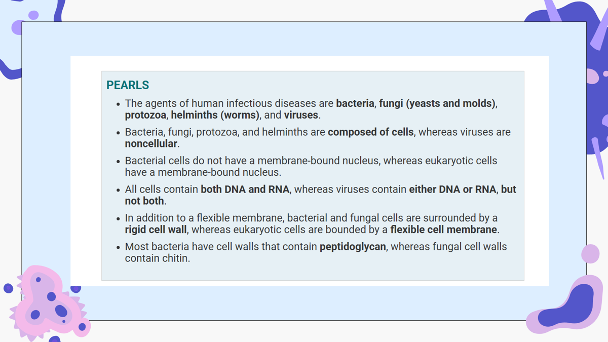
Compare and contrast the structural components of viruses, bacteria, fungi, protozoa, and helminths
Feature | Viruses | Bacteria | Fungi | Protozoa | Helminths |
|---|---|---|---|---|---|
Cell Type | Noncellular | Prokaryote | Eukaryote | Eukaryote | Eukaryote |
Nucleus | None | No nucleus (DNA in cytoplasm) | True nucleus | True nucleus | True nucleus |
Genetic Material | DNA or RNA | DNA in cytoplasm (single circular DNA) | DNA in multiple chromosomes surrounded by a nuclear membrane | DNA in multiple chromosomes surrounded by a nuclear membrane | DNA in multiple chromosomes surrounded by a nuclear membrane |
Cytoplasm | None | Present | Present | Present | Present |
Organelles | None | No membrane-bound organelles; contains 70S ribosomes | Mitochondria and lysosomes | Mitochondria and lysosomes | Mitochondria and lysosomes |
Cell Membrane / Cell Wall | Not described | Rigid peptidoglycan cell wall | Flexible cell membrane containing sterols | Flexible cell membrane containing sterols | Flexible cell membrane containing sterols |
Replication | Replicate only within host cells | Binary fission | Mitosis | Mitosis | Mitosis |
Energy Production | Uses host cell | Glycolysis and proton gradient across cell membrane | Has its own organelles | Has its own organelles | Has its own organelles |
Key Structural Differences
Viruses | Bacteria | Fungi, Protozoa & Helminths |
|---|---|---|
Noncellular | Prokaryotic cells | Eukaryotic cells |
DNA or RNA | DNA only | DNA only |
No cytoplasm | Cytoplasm present | Cytoplasm present |
No organelles | 70S ribosomes only | Mitochondria and lysosomes present |
Depend on host cells for protein synthesis, energy generation, and replication | Produce their own energy and reproduce by binary fission | Have a true nucleus, organelles, and reproduce by mitosis |
Inner core of genetic material (DNA or RNA) | Rigid peptidoglycan cell wall | Flexible cell membrane containing sterols |
Compare and contrast the structural components of eukaryotes
Feature | Fungi | Protozoa | Helminths |
|---|---|---|---|
True nucleus | ✔ | ✔ | ✔ |
DNA | ✔ | ✔ | ✔ |
Multiple chromosomes | ✔ | ✔ | ✔ |
Nuclear membrane | ✔ | ✔ | ✔ |
Cytoplasm | ✔ | ✔ | ✔ |
Organelles (mitochondria, lysosomes) | ✔ | ✔ | ✔ |
Flexible cell membrane with sterols | ✔ | ✔ | ✔ |
Divide by mitosis | ✔ | ✔ | ✔ |
Fungi – includes yeasts and molds.
Protozoa – a separate group of eukaryotic microorganisms.
Helminths – parasitic worms.
Describe the structural components of bacterial cells, including:
Cell wall-
Gram-Positive vs. Gram-Negative Cell Wall
Feature | Gram-Positive | Gram-Negative |
|---|---|---|
Peptidoglycan | Thick | Thin |
Outer membrane | No | Yes |
Lipopolysaccharide (LPS) | No | Yes |
Endotoxin | No | Yes |
Teichoic acids | Present | Absent |
Lipoteichoic acids | Present | Absent |
Gram stain color | Purple | Pink |
Gram Staining:
Crystal violet – primary stain
Iodine – acts as a mordant to help fix the dye
Acetone – removes the dye from Gram-negative bacteria
Safranin – pink counterstain
Cell wall = support + shape + protection
Made of peptidoglycan (unique to bacteria)
Gram-positive: Thick peptidoglycan, teichoic acids, stains purple,
■Alternating layers of N-acetylglucosamine (NAG) and N-acetylmuramic acids (NAM)cross linked by peptide bridges.
Gram-negative: Thin peptidoglycan + outer membrane containing LPS (endotoxin), stains pink
Endotoxin (LPS) is associated with fever and shock.
Cytoplasmic membrane-
The cytoplasmic membrane is the inner membrane of a bacterial cell. It is located just inside the peptidoglycan cell wall and is made of a phospholipid bilayer.
Functions:
Active transport of molecules into the cell
Energy generation by oxidative phosphorylation
Synthesis of cell wall precursors
Secretion of enzymes and toxins
Cytoplasm:
The cytoplasm is an amorphous matrix containing the bacterial cell's internal components.
Contains: Ribosomes, plasmids, Nucleoid
Function: Protein synthesis
70S ribosomes
50S subunit
30S subunit
Clinical importance: Many antibiotics work by targeting bacterial ribosomes.
Plasmids are:
Small circular, double-stranded DNA molecules that replicate independently.
Nucleoid:
Contains: DNA molecule
Flagella and pili:
Flagella
Structure:
Long, whip-like structures
Made of flagellin
Function:
Propel bacteria through chemotaxis (movement toward or away from chemical signals)
Pilli
Structure:
Hair-like structures
Made of pilin
Found on Gram-negative bacteria
Shorter than flagella
Functions:
Mediate bacterial attachment to human cell receptors
Help bacteria adhere to epithelial cells
Sex pilus facilitates bacterial attachment
Bacterial spores:
Bacterial spores are produced when nutrients become depleted.
Structure:
Form inside the vegetative bacterial cell
Thick, keratin-like protective coat
Contain:
DNA
Minimal cytoplasm
Cell membrane
Peptidoglycan
Small amount of water
■Medical importance:
●Resistant to heat, dehydration, radiation and chemicals
●Metabolically inactive
○Produced by bacillus (antrax) and clostridium (tetanus, botulism)
Structure | Key Features | Function | Clinical Importance |
|---|---|---|---|
Cell wall | Peptidoglycan; Gram (+) thick, Gram (−) thin + outer membrane | Support, protection, shape | Determines Gram stain; Gram (−) LPS endotoxin causes fever and shock |
Cytoplasmic membrane | Phospholipid bilayer | Transport, energy production, cell wall synthesis, secretion | Usually lacks sterols (except Mycoplasma) |
Cytoplasm | Ribosomes, plasmids, nucleoid, nutrients | Protein synthesis and DNA storage | Ribosomes are antibiotic targets; plasmids carry resistance genes |
Flagella | Long, flagellin protein | Movement (chemotaxis) | Allows spread within the body |
Pili (Fimbriae) | Short, pilin protein | Attachment to host cells | Important for colonization and infection |
Bacterial spores | Thick protective coat; metabolically inactive | Survival in harsh environments | Resistant to heat, dehydration, radiation, and chemicals; produced by Bacillus and Clostridium |
Discuss the concept of normal flora, and identify common members of the normal flora in anatomic locations including the skin, respiratory, gastrointestinal, and genitourinary systems.
Normal flora (also called the human microbiota or microbiome) refers to the microorganisms that normally live on or inside the human body.
These microorganisms include:
Bacteria
Fungi
Viruses
Protozoa
They form complex communities in different body sites. Normally, they live in balance with the host, but imbalances can lead to infection and disease.
Most normal flora are commensals, meaning they:
Live symbiotically on or within the human body
Rarely cause disease under normal conditions
Common Members of Normal Flora:
Anatomic Location | Common Normal Flora |
|---|---|
Skin | Staphylococcus species and diphtheroids |
Respiratory (Oropharynx) | Streptococci and anaerobes |
Gastrointestinal (Large intestine) | Enterococci and enteric bacilli |
Genitourinary (Vagina) | Lactobacilli |
Term | Definition |
|---|---|
Normal flora (Microbiota) | Microorganisms that normally live on or in the body |
Commensals | Organisms that live symbiotically with the host and rarely cause disease |
Colonization | Presence of microorganisms without tissue invasion or symptoms |
Infection | Presence of bacteria in the body with or without disease |
Colonization ≠ infection because colonization has no tissue invasion and no symptoms.
Describe the concepts of colonization, pathogenicity, virulence, infection, disease, and carrier states.
Term | Definition (Based on the slides) | Key Point |
|---|---|---|
Colonization | The presence of bacteria on a surface without causing disease. There is no tissue invasion, the person is asymptomatic, and the bacteria may be part of the normal flora. | Colonization does NOT equal infection. |
Pathogenicity | The ability or potential of a microorganism to cause disease. | Determines whether an organism is capable of causing illness. |
Virulence | The characteristics of bacteria that enhance their ability to cause disease. It reflects how sick the organism can make a patient. | Higher virulence = greater severity of disease. |
Infection | The presence of bacteria in the body, with or without disease. | An infection may or may not produce symptoms. |
Disease | A condition in which the infection becomes symptomatic and contagious. | The patient develops recognizable signs and symptoms of illness. |
Carrier State | A person who harbors a microorganism but remains asymptomatic. The slides use Typhoid Mary as the classic example. | The individual has the organism but does not show symptoms. |
Discuss the determinants of bacterial pathogenesis, such as transmission, adherence to cell surfaces, invasion, inflammation, and intracellular survival, toxin production, and immunopathogenesis.
Bacterial pathogenesis is the process by which bacteria cause disease. According to the slides, infectious disease occurs when microorganisms overpower the host's defenses. Two important factors that determine whether disease develops are the:
Infectious dose (the number of organisms needed to cause infection) and the organism's virulence (its ability to produce harmful factors).
Determinant | Description (from the slides) | Examples from the slides |
|---|---|---|
Transmission | The way bacteria spread from one host or source to another. Different bacteria use different routes of transmission. | Respiratory droplets (Group A Streptococcus, Neisseria meningitidis, Corynebacterium diphtheriae), direct contact (S. aureus), fecal-oral (Salmonella, Shigella, Vibrio cholerae, Campylobacter jejuni), sexual transmission (Neisseria gonorrhoeae, Chlamydia trachomatis, Treponema pallidum), vertical transmission during childbirth (Group B Streptococcus, N. gonorrhoeae, Chlamydia trachomatis), soil (Clostridium tetani, Bacillus anthracis), tick bite (Rickettsia rickettsii, Borrelia burgdorferi), cat scratch (Bartonella henselae). |
Adherence to cell surfaces | Many bacteria must first attach to host cells before causing disease. The slides identify pili (fimbriae) as structures that mediate bacterial attachment to human cell receptors. | Pili of gram-negative bacteria; sex pilus facilitates attachment; Neisseria gonorrhoeae uses pili for adherence to mucosal surfaces. |
Invasion and inflammation | Some bacteria invade tissues, grow locally, and trigger an inflammatory response. The slides state that invasion causes redness, edema, warmth, and pain. | Shigella invades the distal ileum and colon; Salmonellainvades intestinal tissues; Campylobacter jejuni causes intestinal inflammation with bloody stool. |
Intracellular survival | Certain bacteria survive and multiply inside host cells, allowing them to avoid immune defenses. | Legionella pneumophila prevents phagolysosome fusion and replicates inside cells; Mycobacterium tuberculosis survives in macrophages; Chlamydia species replicate intracellularly after conversion from elementary bodies to reticulate bodies. |
Toxin production | Bacteria produce toxins that damage tissues or cause disease symptoms. The slides describe two major types: exotoxins(secreted proteins) and endotoxins (lipopolysaccharide [LPS] in gram-negative bacteria). Both can produce symptoms such as fever even when bacteria are absent. | Exotoxins: S. aureus, Clostridium tetani, Clostridium botulinum, Bacillus anthracis, C. difficile. Endotoxin: LPS of gram-negative bacteria, responsible for fever and shock. |
Immunopathogenesis | Disease caused by the body's immune response rather than direct bacterial damage. The slides explain that antibodies against bacterial components can cross-react with normal tissuesor form immune complexes that damage tissues. | Streptococcus pyogenes can cause rheumatic fever (heart and joints) and glomerulonephritis (kidneys). Campylobacter jejuni is associated with Guillain-Barré syndrome because antibodies cross-react with neurons. |
Infectious dose = Number of bacteria needed to cause infection.
Virulence = How capable the organism is of causing disease.
Pili (fimbriae) = Attachment to host cells.
Exotoxins = Secreted proteins.
Endotoxins = LPS in gram-negative bacteria; can cause fever and shock.
Immunopathogenesis = Immune response damages the host (e.g., rheumatic fever, glomerulonephritis, Guillain-Barré syndrome).
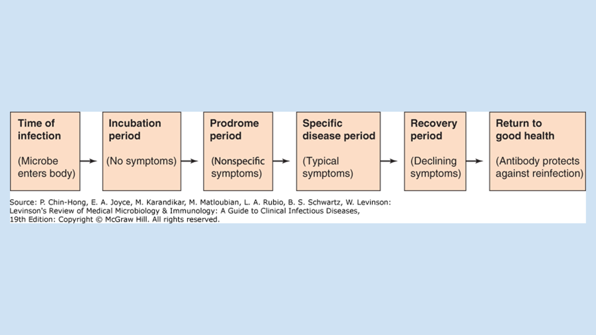
Describe the typical stages of an infectious disease (incubation, prodrome, specific illness, and recovery periods).
An infectious disease typically progresses through four stages. Each stage represents a different point in the infection, from the time the organism enters the body until recovery. According to the lecture, the stages are incubation, prodrome, specific illness, and recovery (convalescence).
Stage | Description | Key Features |
|---|---|---|
1. Incubation Period | The time between acquiring the organism or toxin and the onset of symptoms. | • No symptoms yet • Length is variable depending on the organism |
2. Prodrome Period | The stage when nonspecific symptoms begin to appear. | • Fever • Malaise (feeling unwell) • Loss of appetite |
3. Specific Illness Period | The stage when the patient develops the overt, characteristic signs and symptoms of the disease. | • Disease-specific symptoms become apparent |
4. Recovery (Convalescence) Period | The illness begins to resolve, and the patient returns to baseline health (most of the time). | • Symptoms improve • Recovery occurs • IgG and IgA antibodies help protect against future infection |
Explain proper methods of specimen collection, transport, and processing to facilitate laboratory diagnosis of bacterial infections.
Bacterium | Specimen Collected | Laboratory Diagnosis |
|---|---|---|
Staphylococcus aureus | Smear | Gram stain showing gram-positive cocci in grape-like clusters; coagulase test. |
Neisseria meningitidis | Blood and spinal fluid (CSF) | Smear and culture; gram stain shows gram-negative cocci in CSF. |
Neisseria gonorrhoeae | Urethral or cervical discharge | Gram stain and nucleic acid amplification test (NAAT). |
Bacillus anthracis | Smear | Large gram-positive rod chains. |
Clostridium perfringens | Tissue or wound exudate | Smear showing large gram-positive rods. |
Clostridioides difficile | Stool | ELISA (detects toxin) and NAAT. |
Corynebacterium diphtheriae | Throat specimen | Throat culture. |
Listeria monocytogenes | Clinical specimen | Gram stain and culture. |
Gardnerella vaginalis | Vaginal specimen | Microscopy showing clue cells and vaginal pH > 4.5. |
Escherichia coli | Blood agar culture | Lactose-fermenting pink colonies; O157:H7 does not ferment sorbitol. |
Salmonella | Stool sample | Non-lactose fermenting (colorless) colonies. |
Shigella | Stool sample | Stool culture; methylene blue stain can detect neutrophils. |
Vibrio cholerae | Stool sample | Stool culture; slow lactose fermentation. |
Campylobacter jejuni | Stool specimen | Culture on blood agar containing antibiotics. |
Helicobacter pylori | Gastric biopsy | Gram stain of gastric mucosa; urease positive; urea breath test may also be used. |
Proteus mirabilis | Urine | Urine culture; swarming motility, positive urease, alkaline urine. |
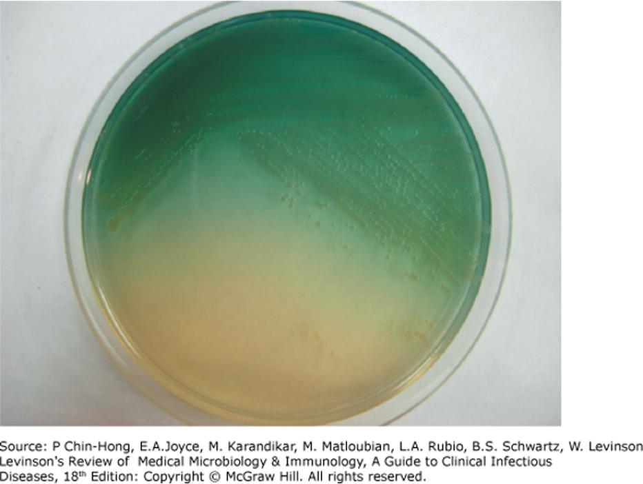
Pseudomonas aeruginosa | Clinical specimen | Gram stain; production of blue-green (pyocyanin) and yellow-green (pyoverdine) pigments. |
Providencia spp. | Urine | Urine culture. |
Morganella morganii | Urine | Urine culture. |
Bordetella pertussis | Nasopharyngeal swab | Culture from nasopharyngeal specimen. |
Bartonella henselae | Blood/clinical specimen | Serology (difficult to culture). |
Legionella pneumophila | Urine | Urinary antigen test; gram stain has poor visualization. |
Mycoplasma pneumoniae | Respiratory specimen | PCR; culture is not practical because it takes too long and the organism lacks a cell wall. |
Chlamydia pneumoniae | Respiratory specimen | PCR; culture is difficult because living cells are required. |
Chlamydia trachomatis | Clinical specimen | NAAT. |
Chlamydia psittaci | Clinical specimen | NAAT. |
Treponema pallidum | Blood/clinical specimen | RPR test. |
Rickettsia rickettsii | Clinical specimen | Clinical diagnosis; PCR. |
Mycobacterium tuberculosis | Clinical specimen | Acid-fast stain. |
Mycoplasma pneumoniae lacks a cell wall, so Gram stain is poor and beta-lactam antibiotics are ineffective. PCR is used for diagnosis.
Legionella pneumophila stains poorly on Gram stain; the preferred test is the urinary antigen test.
Chlamydia pneumoniae cannot be cultured easily because it requires living cells; PCR is the preferred test.
Mycobacterium tuberculosis is identified with an acid-fast stain because its mycolic acid-rich cell wall retains the stain after alcohol washing.
Neisseria gonorrhoeae diagnosis includes NAAT, an important test repeatedly emphasized in the slides.
Acid-fast stain → Mycobacterium tuberculosis
NAAT → Neisseria gonorrhoeae, Chlamydia trachomatis
PCR → Mycoplasma pneumoniae, Chlamydia pneumoniae, Rickettsia rickettsii
Urinary antigen test → Legionella pneumophila
Urease test → Helicobacter pylori
Describe the staining techniques and morphologic characteristics utilized in the identification and classification of bacteria.
Bacteria are identified and classified based on:
Morphologic characteristics (shape and arrangement)
Staining techniques, which distinguish bacteria according to differences in their cell wall structure.
Shape + Arrangement = Identification
Staphylococcus → Gram (+) cocci in grape-like clusters
Streptococcus → Gram (+) cocci in chains or pairs
Streptococcus pneumoniae → Lancet-shaped diplococci
Neisseria → Gram (-) kidney bean-shaped diplococci
Shape | Description |
|---|---|
Cocci | Round (spherical) bacteria |
Bacilli | Rod-shaped bacteria |
Spirochetes | Spiral-shaped bacteria |
Pleomorphic | Variable shapes |
Shape is determined by cell wall.
Arrangement also helps identify bacteria.
Arrangement | Description |
|---|---|
Diplococci | Pairs of bacteria |
Chains | Long chains |
Clusters | Grape-like clusters |
Examples:
Bacteria | Morphology |
|---|---|
Staphylococcus spp. | Gram (+) cocci in grape-like clusters |
Streptococcus spp. | Gram (+) cocci in chains or pairs |
Streptococcus pneumoniae | Lancet-shaped diplococci |
Neisseria spp. | Gram (-) kidney bean-shaped diplococci |
Bacillus anthracis | Large gram (+) rods with square ends |
Listeria monocytogenes | Small gram (+) rods in V- or L-shaped formations |
Clostridium tetani | Terminal spore gives a "tennis racket" appearance |
Bordetella pertussis | Small gram (-) coccobacillus |
Treponema pallidum | Thin spiral-shaped spirochete |
Borrelia burgdorferi | Large spiral-shaped spirochete |
Gram Staining:
Step | Function |
|---|---|
1. Crystal violet | Primary purple stain |
2. Iodine | Acts as a mordant to help retain the dye |
3. Acetone (organic solvent) | Removes the dye from Gram-negative bacteria |
4. Safranin | Pink counterstain |
Acid-Fast Staining:
Some bacteria cannot be adequately identified using a Gram stain.
Mycobacterium tuberculosis:
The slides describe Mycobacterium tuberculosis as:
Acid-fast bacillus (AFB)
Slender rod
Gram staining does not work well in Mycoplasma because it lacks sterols in its cytoplasmic membrane
Acid-fast stain is used for Mycobacterium tuberculosis because its mycolic acid-rich cell wall prevents decolorization with alcohol.
Identify media commonly used to promote, inhibit, and differentiate bacteria
Hemolysis | Appearance on Blood Agar | Example |
|---|---|---|
α (Alpha) hemolysis | Incomplete lysis of red blood cells; green discoloration | Streptococcus pneumoniae and other α-hemolytic streptococci |
β (Beta) hemolysis | Complete lysis of red blood cells; clear zonearound colonies | Streptococcus pyogenes, Streptococcus agalactiae |
γ (Gamma) hemolysis | No hemolysis | Gamma-hemolytic streptococci |

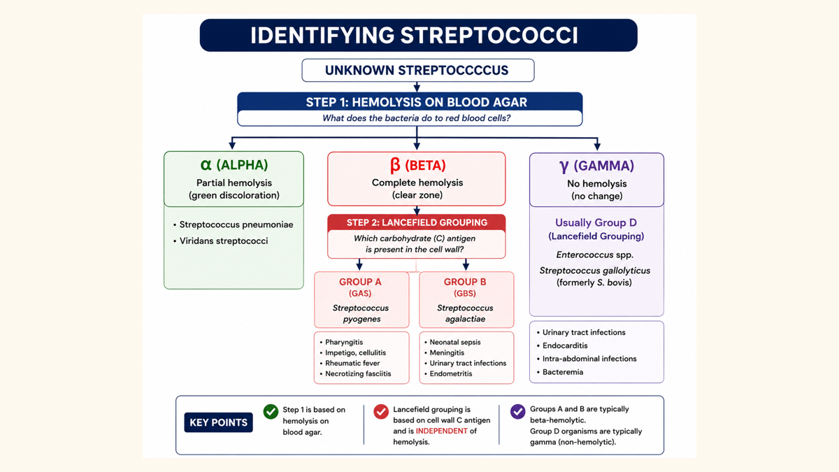
Once it is determined that the organism is β-hemolytic, it moves to Lancefield grouping based on the carbohydrate antigens found in the cell wall.
Discuss important properties of anaerobic bacteria and discuss their clinical significance.
●Escherichia Coli
Straight gram (-) rod
Most abundant facultative anaerobe in colon and feces
●Ferment lactose
Escherichia Coli
Straight gram-negative rod
Most abundant facultative anaerobe in the colon and feces
Ferments lactose
Organism / Property | Important Properties | Clinical Significance | ⭐ Professor's High-Yield Notes |
|---|---|---|---|
Normal anaerobes | Part of the normal flora of the oropharynx; most are commensals and rarely cause disease. | Can contribute to infection if the normal microbiota becomes imbalanced. | Know that anaerobes are normal flora of the oropharynx. |
Escherichia coli | Most abundant facultative anaerobe in the colon and feces; ferments lactose. | Common cause of UTIs, neonatal meningitis, and diarrhea. | ⭐ Memorize: Most abundant facultative anaerobe. |
Campylobacter jejuni | Microaerophilic; grows best in 5% oxygen rather than atmospheric (20%) oxygen. | Causes watery then bloody diarrhea; associated with Guillain-Barré syndrome. | ⭐ Professor emphasized 5% oxygen and that it is microaerophilic. |
Clostridium tetani | Spore-forming bacterium found in soil; enters through wounds; produces a potent exotoxin. | Causes tetanus with muscle spasms, lockjaw (trismus), risus sardonicus, opisthotonos, and respiratory failure. | ⭐ Terminal spore = "tennis racket" appearance. |
Clostridium botulinum | Spore-forming; spores found in soil and contaminated canned foods; exotoxin blocks acetylcholine (ACh) release. | Causes descending paralysis, diplopia, dysphagia, ptosis, and respiratory failure. | ⭐ Infants <1 year should NOT eat honey. Organism is not typically cultured. |
Clostridium perfringens | Spore-forming; grows in traumatized muscle; produces toxins and enzymes that damage tissue and generate gas. | Causes gas gangrene (necrotizing fasciitis) with pain, edema, necrosis, and crepitus. | ⭐ Large "boxcar" gram-positive rods; gas in tissues is a key finding. |
Clostridioides difficile | Colonizes the large intestine; overgrows after antibiotics suppress normal flora; produces exotoxins. | Causes pseudomembranous colitis with non-bloody diarrhea, fever, and abdominal pain. | ⭐ Clindamycin is the antibiotic specifically highlighted as being associated with C. difficile infection. |
Discuss the clinical significance, transmission, pathogenesis, laboratory diagnosis, and clinical findings associated with the following bacteria:
a. Gram-Positive Cocci
i. Staphylococcus spp.
S. aureus: Grape-like clusters, Coagulase (+), Catalase (+), MRSA (treat with Vancomycin). Pyogenic (pus/abscess), Enterotoxin (undercooked food), Toxic shock syndrome (tampons), Exfoliatin (scalded skin syndrome).
S. epidermidis: Coagulase (-), Hospital-acquired, Artificial materials (prosthetic valves/joints, central lines).
S. saprophyticus: Coagulase (-), Genital tract mucosa, UTIs.
ii. Streptococcus spp.
S. pyogenes (Group A): Chains/pairs, Beta-hemolytic, Bacitracin sensitive. Pharyngitis, Impetigo, Scarlet fever, Necrotizing fasciitis, Rheumatic fever, Glomerulonephritis.
S. agalactiae (Group B): Beta-hemolytic, Bacitracin resistant. Prolonged rupture of membranes (>18 hours), Neonatal sepsis, Meningitis.
S. pneumoniae: Alpha-hemolytic, Lancet-shaped diplococci. Pneumonia, Otitis media, Meningitis. Risk factors: Splenectomy, Alcoholism.
b. Gram-Negative Cocci (Diplococci)
i. Neisseria spp.
N. meningitidis: Paired kidney bean shape. Airborne droplets, Military recruits. Meningitis, Meningococcemia.
N. gonorrhoeae: Sexually transmitted. Pili (adherence). Purulent vaginal/penile discharge, PID, Infertility. NAAT test.
c. Gram-Positive Rods
i. Bacillus spp. (Spore-forming)
B. anthracis: Square ends. Edema factor, Lethal factor. Black eschar, Wool sorters disease (hemorrhagic mediastinitis).
B. cereus: Reheated rice. Short incubation (vomiting), Long incubation (watery diarrhea).
ii. Clostridium spp. (Spore-forming)
C. tetani: Tennis racket terminal spore. Spastic paralysis, Lock jaw (trismus), Risus sardonicus (grimace), Opisthotonos (arched back).
C. botulinum: Canned/vacuum-packed foods. Infant botulism (honey). Descending weakness/paralysis (diplopia, dysphagia, ptosis).
C. perfringens: Gas gangrene, Necrotizing fasciitis, Crepitus, Hemolysis.
C. difficile: Pseudomembranous colitis, Post-antibiotics. ELISA for endotoxin.
iii. Corynebacterium diphtheriae (Non-spore forming)
Club-shaped. Thick, gray adherent pseudomembrane, Airway obstruction.
iv. Listeria monocytogenes (Non-spore forming)
V- or L-shaped. Unpasteurized milk, Undercooked meats. Immunocompromised (solid organ transplants), Pregnant (abortion, neonatal meningitis).
v. Gardnerella vaginalis
Gram-variable (thin cell wall). Clue cells, pH > 4.5. Bacterial vaginosis (malodorous white/gray discharge).
d. Gram-Negative Rods
i. Enterobacterales
1. Escherichia coli: Lactose fermenter. UTIs, Neonatal meningitis, Traveler's diarrhea. O157:H7 Strain: Undercooked beef, Non-sorbitol fermenting, Bloody diarrhea, Hemolytic Uremic Syndrome (HUS).
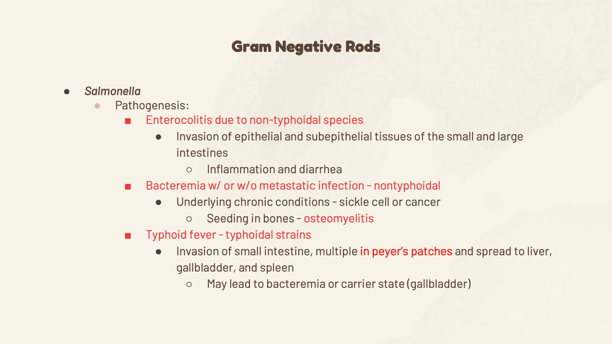
2. Salmonella spp.: Non-lactose fermenter. Nontyphoidal: Poultry, eggs, reptiles. Gastroenteritis, Osteomyelitis (sickle cell). Typhoidal (S. typhi): Humans only, Peyer's patches, Enteric (typhoid) fever, Rose spots, Chronic gallbladder carrier.
3. Shigella spp.: Non-lactose fermenter, Humans only. 4 F's (Fingers, Flies, Food, Feces). Bloody diarrhea with mucus.
4. Klebsiella spp.: Lactose fermenter, Large polysaccharide capsule, Mucoid appearance. Currant jelly sputum. Alcoholics, Diabetics.
5. Enterobacter spp.: (Not included in slide sources).
6. Serratia spp.: Red pigment (prodigiosin). Healthcare-associated (medical devices/catheters).
7. Proteus spp.: Non-lactose fermenter, Urease positive, Highly motile (swarming motility). Alkaline urine, Struvite kidney stones.
8. Providencia spp.: Non-lactose fermenter. Complicated UTIs, Catheters.
9. Morganella spp.: Non-lactose fermenter, Urease positive. Complicated UTIs.
ii. Non-Enterobacterales
1. Vibrio spp.: Comma-shaped. Cholera toxin. Rice-water stools.
2. Campylobacter spp.: Comma/S-shaped, Microaerophilic (5% oxygen). Poultry/dogs/cattle. Bloody diarrhea, Guillain-Barre syndrome.
3. Pseudomonas spp.: Strict aerobe, Non-lactose fermenter. Blue-green pigment (pyocyanin), Yellow-green pigment (pyoverdine). Cystic fibrosis pneumonia, Burn infections (green pus), Swimmer's ear.
4. Helicobacter spp.: Strongly Urease positive. Urea breath test. Gastritis, Peptic ulcers.
5. Haemophilus spp.: (Not included in slide sources).
6. Bordetella spp.: Coccobacillus, Encapsulated. Damages cilia. Whooping cough.
e. Atypical/Miscellaneous Bacteria
i. Bartonella spp.
Cat scratch/bite. Regional lymph node enlargement, Angiomas (in immunocompromised).
ii. Legionella spp.
Intracellular. Environmental water (AC units, hot tubs). Urinary antigen test. Legionnaire disease (Atypical pneumonia + Hyponatremia), Pontiac fever.
iii. Mycoplasma spp.
No cell wall (Gram stains poorly, beta-lactam resistant). Atypical/walking pneumonia. Most common in ages 5-15. PCR test.
iv. Chlamydia spp.
Obligate intracellular. Life cycle: EB Enters (Infectious), RB Reproduces (Replicating).
C. pneumoniae / C. psittaci (birds): Atypical walking pneumonia.
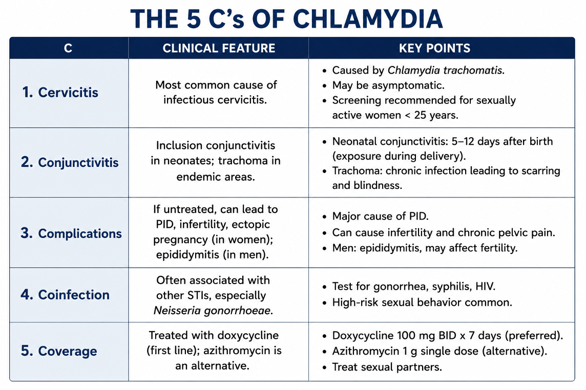
C. trachomatis: 5 C's (Cervicitis, Conjunctivitis-neonatal, Chlamydia STI, Chronic blindness/Trachoma, Choice test = NAAT).
v. Treponema pallidum
Spirochete, Corkscrew motility. Syphilis (4 P's: Primary-Painless chancre, Secondary-Palms & soles rash, Persistent/Latent-no symptoms, Progressive/Tertiary-Gummas, aortitis, neurosyphilis). Test: RPR/VDRL (Screen) -> FTA-ABS/TP-PA (Confirm).
vi. Rickettsia spp.
Obligate intracellular. Tick bite (Dermacentor). RMSF Memory Ticks: Recent tick bite, Rash (wrists/ankles first, spreads to palms/soles), Rickettsia (vasculitis), Rapid Doxycycline.
vii. Borrelia spp.
Large spirochete. Deer tick (Ixodes). Two-tier testing (ELISA then Western blot). Lyme disease: Bull's-eye rash (Erythema migrans), Facial nerve palsy, AV block (Lyme carditis), Arthritis of the knee.
f. Mycobacterium spp.
Acid-Fast Bacillus (AFB). Granuloma formation in macrophages. Weight loss, Night sweats, Hemoptysis, Chronic cough
a. Gram-Positive Cocci
i. Staphylococcus species (spp.)
Clinical Significance: Gram (+) cocci arranged in grape-like clusters.
S. aureus is the most common cause of serious illness, and 90% in the US are MRSA (resistant to penicillins, treated with Vancomycin).
S. epidermidis (skin) and
S. saprophyticus (genital tract mucosa) are coagulase-negative and do not produce toxins.
Transmission: Humans are reservoirs (nose is the main site). Spread via hand contact and shedding from lesions. S. epidermidis is almost always hospital-acquired (via artificial materials).
Pathogenesis: S. aureus produces coagulase, which walls off bacteria and prevents neutrophil access, and produces powerful toxins.
Laboratory Diagnosis: Gram stain reveals Gram (+) cocci in grape-like clusters. S. aureus is coagulase (+) and catalase (+).
Clinical Findings:
S. aureus: Pyogenic infections (abscess, cellulitis, folliculitis, impetigo, sepsis, endocarditis), and Toxigenic infections (gastroenteritis from undercooked food, toxic shock syndrome from tampons, scalded skin syndrome from exfoliatin toxin).
S. epidermidis: Infections of artificial materials (prosthetic valves/joints, central lines).
S. saprophyticus: Urinary tract infections.
ii. Streptococcus spp.
Clinical Significance: Gram (+) cocci in chains or pairs.
Classified by hemolysis on blood agar: Alpha (green, incomplete lysis), Beta (clear, complete lysis), or Gamma (no hemolysis).
Transmission:
Group A: Respiratory droplets/direct contact.
Group B: Vertical transmission during childbirth. S. pneumoniae: Humans are natural hosts (not communicable).
Pathogenesis: Group A: Causes pyogenic, toxigenic, and immune-mediated damage. Group B: Risk increases with prolonged rupture of membranes (>18 hours) or premature birth (<37 weeks). S. pneumoniae: Preys on lowered host resistance (alcoholism, splenectomy).
Laboratory Diagnosis: Categorized by hemolysis. Group A is bacitracin sensitive; Group B is bacitracin resistant. S. pneumoniae appears as lancet-shaped diplococci or short chains.
Clinical Findings:
Group A (S. pyogenes): Pharyngitis, impetigo, cellulitis, scarlet fever, necrotizing fasciitis, rheumatic fever, glomerulonephritis.
Group B (S. agalactiae): Neonatal sepsis, meningitis, pneumonia.
S. pneumoniae: Pneumonia, otitis media, sinusitis, meningitis.
b. Gram-Negative Cocci (Diplococci)
i. Neisseria spp.
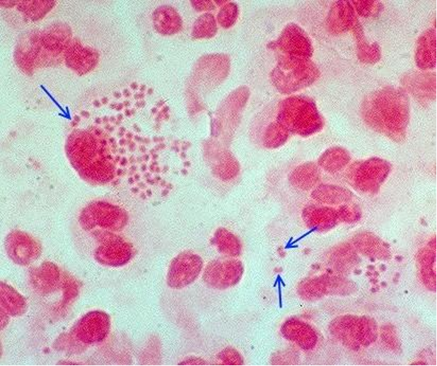
Clinical Significance: Gram (-) cocci that resemble paired kidney beans.
Transmission: N. meningitidis: Airborne droplets, highly contagious (military recruits). N. gonorrhoeae: Sexually transmitted or vertical transmission.
Pathogenesis: N. meningitidis: Asymptomatic carriers harbor it in the upper respiratory tract; disseminates via bloodstream. N. gonorrhoeae: Uses pili for adherence to mucosa; resistant to antibodies and complements.
Laboratory Diagnosis: N. meningitidis: Smear/culture of blood and spinal fluid. N. gonorrhoeae: Nucleic acid amplification test (NAAT) or Gram stain of discharge.
Clinical Findings: N. meningitidis: Meningitis, meningococcemia. N. gonorrhoeae: Urethritis, purulent vaginal/penile discharge, PID, infertility.
c. Gram-Positive Rods
i. Bacillus spp.
Clinical Significance: Spore-forming Gram (+) rods. B. anthracis has square ends.
Transmission: B. anthracis: Cutaneous (soil), respiratory, or GI (meat). B. cereus: Spores survive steaming/drying on grains (reheated rice).
Pathogenesis: B. anthracis: Produces edema factor (fluid outpouring) and lethal factor (cell death/apoptosis). B. cereus: Produces enterotoxins.
Laboratory Diagnosis: B. anthracis: Smear shows large Gram (+) rod chains. B. cereus: Not typically tested.
Clinical Findings: B. anthracis: Black eschar (necrotic skin lesion), "wool sorters disease" (hemorrhagic mediastinitis, shock). B. cereus: Rapid vomiting (4 hours) or watery diarrhea (18 hours).
ii. Clostridium spp.
Clinical Significance: Spore-forming Gram (+) rods.
Transmission: C. tetani/C. perfringens: Spores in soil. C. botulinum: Soil, canned/vacuum-packed foods, honey. C. difficile: Colonizes large intestine.
Pathogenesis: C. tetani: Produces highly toxic exotoxin. C. botulinum: Exotoxin blocks acetylcholine. C. perfringens: Toxins damage cell membranes (hemolysis) and degradative enzymes cause tissue gas. C. difficile: Multiplies and produces exotoxin when antibiotics suppress normal colonic flora.
Laboratory Diagnosis: C. tetani: Terminal spore has a "tennis racket" appearance. C. difficile: ELISA detects endotoxin; NAAT.
Clinical Findings:
C. tetani: Spastic paralysis, lockjaw (trismus), risus sardonicus, opisthotonos.
C. botulinum: Descending weakness/paralysis (diplopia, dysphagia, ptosis); infant botulism from honey.
C. perfringens: Gas gangrene (necrotizing fasciitis) with crepitus.
C. difficile: Pseudomembranous colitis, nonbloody diarrhea.
iii. Corynebacterium diphtheriae
Clinical Significance: Non-spore forming, club-shaped Gram (+) rods.
Transmission: Airborne droplets.
Pathogenesis: Establishes in the throat and produces a severe exotoxin.
Laboratory Diagnosis: Throat culture.
Clinical Findings: Thick, gray adherent pseudomembrane over tonsils/throat. Complications include airway obstruction, myocarditis, and nerve paralysis.
iv. Listeria monocytogenes
Clinical Significance: Non-spore forming, small Gram (+) rods arranged in V- or L-shaped formations.
Transmission: Unpasteurized milk products, undercooked meats, raw vegetables; vertical transmission.
Pathogenesis: Suppresses cell-mediated immunity.
Laboratory Diagnosis: Gram stain and culture.
Clinical Findings: Abortion/premature delivery/sepsis in pregnant women. Acute meningitis in newborns. Flu-like symptoms or gastroenteritis in immunocompromised adults (solid organ transplants).
v. Gardnerella vaginalis
Clinical Significance: Small, facultative Gram-variable rod (thin cell wall).
Transmission/Pathogenesis: Vaginal dysbiosis; normal Lactobacillus is replaced.
Laboratory Diagnosis: Microscopy shows clue cells; vaginal pH > 4.5.
Clinical Findings: Bacterial vaginosis (malodorous, white/gray thin vaginal discharge).
d. Gram-Negative Rods
i. Enterobacterales
1. Escherichia coli: Straight Gram (-) rod, lactose fermenter. Transmission: Colonic flora (UTI), birth canal (meningitis), contaminated food/water (Traveler's diarrhea), undercooked beef (O157:H7). Pathogenesis: Endotoxins/Exotoxins. Clinical: UTIs, watery diarrhea. O157:H7 causes bloody diarrhea and Hemolytic Uremic Syndrome (HUS). Lab: Pink colonies on agar; O157:H7 does not ferment sorbitol.
2. Salmonella spp.: Non-lactose fermenter. Transmission: Nontyphoidal: Poultry, eggs, reptiles. Typhoidal: Humans only. Pathogenesis: Nontyphoidal: Enterocolitis. Typhoidal: Multiplies in Peyer's patches, spreads to liver/gallbladder/spleen. Clinical: Gastroenteritis, osteomyelitis (sickle cell). Typhoid fever features rose spots and a chronic gallbladder carrier state.
3. Shigella spp.: Non-lactose fermenter. Transmission: Humans only via the Four F's (Fingers, Flies, Food, Feces). Pathogenesis: Invades distal ileum/colon causing ulceration. Clinical: Bloody diarrhea with mucus. Lab: Methylene blue stain shows neutrophils.
4. Klebsiella spp.: Lactose fermenter. Pathogenesis: Large antiphagocytic polysaccharide capsule. Clinical: Opportunistic infections in alcoholics/diabetics; causes pneumonia with currant jelly sputum.
5. Enterobacter spp.: (Not included in provided slides).
6. Serratia spp.: Healthcare-associated. Colonizes moist environments and biofilms. Causes nosocomial UTIs/pneumonia. Lab: Produces a red pigment (prodigiosin) at room temperature.
7. Proteus spp. (P. mirabilis): Non-lactose fermenter. Endogenous to GI tract. Pathogenesis: Highly motile (swarming), produces urease (alkaline urine). Clinical: Catheter-associated UTIs, struvite kidney stones.
8. Providencia spp.: Non-lactose fermenter. Normal GI flora. Pathogenesis: Biofilms. Clinical: Complicated/catheter UTIs.
9. Morganella spp.: Non-lactose fermenter, urease positive. Normal GI flora. Clinical: Complicated/catheter UTIs.
ii. Non-Enterobacterales
1. Vibrio spp.: Curved, comma-shaped. Transmission: Fecal contaminated water. Pathogenesis: Secretes cholera toxin in the small intestine. Clinical: Rice-water stools leading to severe dehydration/electrolyte loss.
2. Campylobacter spp.: Curved/S-shaped, microaerophilic. Transmission: Poultry, cattle, dogs. Pathogenesis: Inflames intestinal mucosa. Clinical: Bloody diarrhea. Associated with Guillain-Barre syndrome (autoimmune cross-reactivity).
3. Pseudomonas spp.: Strict aerobe, non-lactose fermenter. Transmission: Soil/water. Pathogenesis: Endotoxins/exotoxins. Clinical: Cystic fibrosis pneumonia, burn infections (green pus), swimmer's ear. Lab: Produces blue-green (pyocyanin) or yellow-green (pyoverdine) pigment. Highly antibiotic-resistant.
4. Helicobacter spp.: Strongly urease positive. Transmission: Person-to-person. Pathogenesis: Attaches to gastric mucus-secreting cells; ammonia damages mucosa. Clinical: Gastritis, peptic ulcers. Lab: Urea breath test.
5. Haemophilus spp.: (Not included in provided slides).
6. Bordetella spp. (B. pertussis): Coccobacillus. Transmission: Respiratory droplets. Pathogenesis: Toxin damages cilia, impairing mucociliary clearance. Clinical: Whooping cough. Lab: Nasopharyngeal swab.
e. Atypical/Miscellaneous Bacteria
i. Bartonella spp. (B. henselae)
Clinical Significance/Pathogenesis: Small, pleomorphic Gram (-) rod. Infects endothelial cells and macrophages, causing granulomatous inflammation.
Transmission: Cat scratches or bites.
Laboratory Diagnosis: Serology (difficult to culture).
Clinical Findings: Cat scratch disease (fever, regional lymph node enlargement, papule). Causes angiomas in immunocompromised patients.
ii. Legionella spp. (L. pneumophila)
Clinical Significance/Pathogenesis: Intracellular pathogen. Inhaled bacteria prevent phagolysosome fusion.
Transmission: Environmental water aerosols (AC units, cooling towers, hot tubs).
Laboratory Diagnosis: Urinary antigen test (Gram stains poorly).
Clinical Findings: Legionnaire disease (atypical pneumonia, hyponatremia) or Pontiac fever (flu-like).
iii. Mycoplasma spp. (M. pneumoniae)
Clinical Significance/Pathogenesis: Small bacteria with an absence of a cell wall (resistant to beta-lactams). Inhibits ciliary motion via necrosis.
Transmission: Respiratory droplets (humans only).
Laboratory Diagnosis: PCR (takes too long to culture, no cell wall for Gram stain).
Clinical Findings: Atypical (walking) pneumonia. Most common cause of pneumonia in ages 5-15.
iv. Chlamydia spp.
Clinical Significance/Pathogenesis: Obligate intracellular bacterium. Enters as an infectious Elementary Body (EB), converts to a replicating Reticulate Body (RB).
Transmission: C. pneumoniae: Respiratory droplets. C. trachomatis: Sexual contact, perinatal. C. psittaci: Bird droppings.
Laboratory Diagnosis: NAAT/PCR (test of choice).
Clinical Findings: C. pneumoniae / C. psittaci: Atypical pneumonia. C. trachomatis: The 5 C's (Cervicitis, Conjunctivitis, Complications/PID, Coinfection, Coverage/treatment). Also causes Trachoma (leading infectious cause of blindness).
v. Treponema pallidum
Clinical Significance/Pathogenesis: Spiral-shaped spirochete with corkscrew motility. Disseminates via blood and lymph causing chronic inflammation.
Transmission: Sexual, transplacental.
Laboratory Diagnosis: RPR/VDRL (Screen) followed by FTA-ABS/TP-PA (Confirm).
Clinical Findings: Syphilis (The 4 P's: Primary = Painless chancre; Secondary = Palms/soles rash; Persistent/Latent = No symptoms; Progressive/Tertiary = Gummas, aortitis, neurosyphilis).
vi. Rickettsia spp. (R. rickettsii)
Clinical Significance/Pathogenesis: Obligate intracellular. Infects vascular endothelial cells causing vasculitis and increased permeability.
Transmission: Tick bite (Dermacentor).
Laboratory Diagnosis: Clinical diagnosis; PCR.
Clinical Findings: Rocky Mountain Spotted Fever. Rash starts on wrists/ankles and spreads to palms, soles, and trunk. Treat rapidly with Doxycycline.
vii. Borrelia spp. (B. burgdorferi)
Clinical Significance/Pathogenesis: Large spirochete. Enters skin and disseminates to joints, heart, and CNS.
Transmission: Bite of infected deer tick (Ixodes).
Laboratory Diagnosis: Two-tier testing (ELISA then Western blot).
Clinical Findings: Lyme disease. Early: Bull's-eye rash (Erythema migrans). Disseminated: Facial nerve palsy, AV block (Lyme carditis). Late: Arthritis of the knee.
f. Mycobacterium spp.
Mycobacterium tuberculosis
Clinical Significance: Slender rod, classified as an Acid-fast bacillus (AFB).
Transmission: Airborne respiratory droplets.
Pathogenesis: Inhaled bacteria survive inside macrophages, leading to granuloma formation (latent or active TB).
Laboratory Diagnosis: Acid-fast stain.
Clinical Findings: Weight loss, night sweats, chronic cough, and hemoptysis.
NOTES:
Gram Positive (+) diplococci

●Neisseria meningitidis
■Gram (-) cocci in smear of spinal fluid
Gram negative Rods:
●Escherichia coli: (UTI/ diarhehia)
All caused by endotoxins and exotoxins
■Undercooked beef: enterohemorrhagic E. Coli 0157:H7
Salmonella:
●Two distinct categories:
■Typhoidal species
●S. typhi
●S. paratyphi A and B
■Nontyphoidal species
S. enterica
●S. typhi only transmitted by humans

Pseudomonas aeruginosa—blue-green pigment. (Non-Enterobacterales)
(Atypical/Miscellaneous bacteria)
Bartonella henselae- “Cat Sctatch”
●Pathogenesis: bacteria enters skin → infect endothelial cells and macrophages → spread to lymph nodes → granulomatous inflammation
●Chlamydia pneumoniae “Traveler” “Wlaking Pneumonia”
Elementary body: infectious form that enters host cells → thinking “traveler”
Reticulate body: replication inside host cells
●Chlamydia psittaci- bird droppings “Pigeon”
●Treponema pallidum
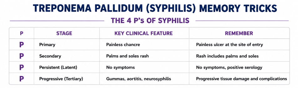
Painless chancre, Palms and sole rash
●Borrelia burgdorferi

●Mycobacterium tuberculosis
●General
■Acid-fast bacillus (AFB); slender rod
hemoptysis (coughing out blood)
