Lymphatic System and Immunity
Overview of the Lymphatic System
Lymphatic System Definition: A collection of cells and biochemicals that travel in lymphatic vessels. It consists of a network of vessels that assist in circulating fluids and is closely associated with the cardiovascular system.
Primary Functions of the Lymphatic System:
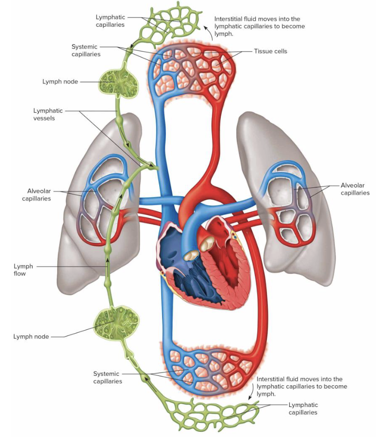
Fluid Transport: Transports excess interstitial fluid away from the interstitial spaces and returns it to the bloodstream.
Lipid Absorption: Absorbs lipids from the digestive system and transports them to the bloodstream. This is specifically accomplished by specialized lymphatic capillaries called lacteals.
Defense: Defends the body against diseases, allowing humans to live in a world populated by other organisms.
The Immune System: This term refers to the fact that many cells within the lymphatic system provide both defenses against disease and permanent immunity against future infections.
Lymphatic Pathways
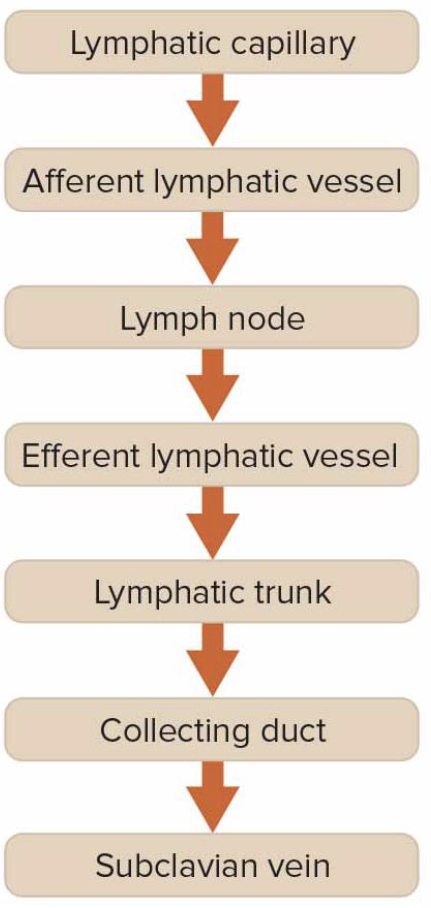
Pathic Sequence: Lymphatic capillaries > lymphatic vessels > lymph nodes > larger lymphatic vessels > lymphatic trunks > lymphatic collecting ducts > subclavian veins in the thorax.
Lymphatic Capillaries:
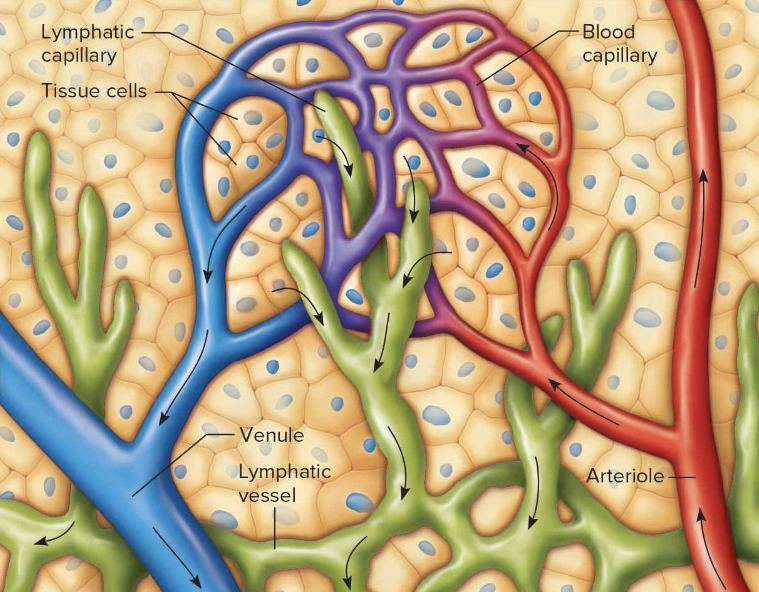
The vessels are microscopic, closed-ended tubes.
They form networks that parallel blood capillaries throughout the body.
The walls are thin and formed from simple squamous epithelium.
Tissue fluid (interstitial fluid) enters these capillaries; once inside, the fluid is called lymph.
These capillaries merge into larger lymphatic vessels.
Lymphatic Vessels:
The walls of these vessels are similar to veins but much thinner.
They are composed of distinct layers:
Inner Layer: An endothelial lining.
Middle Layer: Smooth muscle and elastic fibers.
Outer Layer: Connective tissue.
They contain semilunar valves, which ensure a one-way flow of lymph.
Larger vessels lead to lymph nodes and subsequently to larger lymphatic trunks.
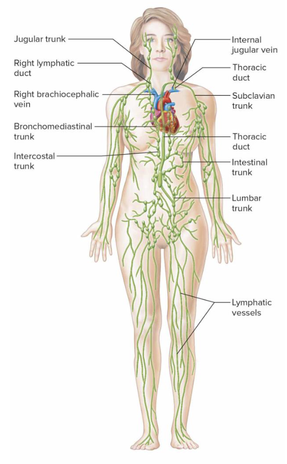
Lymphatic Trunks:
They drain lymph from the lymphatic vessels.
Trunks are named for the specific regions they serve:
Lumbar
Intestinal
Intercostal
Bronchomediastinal
Subclavian
Jugular
They drain into the lymphatic collecting ducts.
Lymphatic Collecting Ducts:
There are only collecting ducts for the entire lymphatic system:
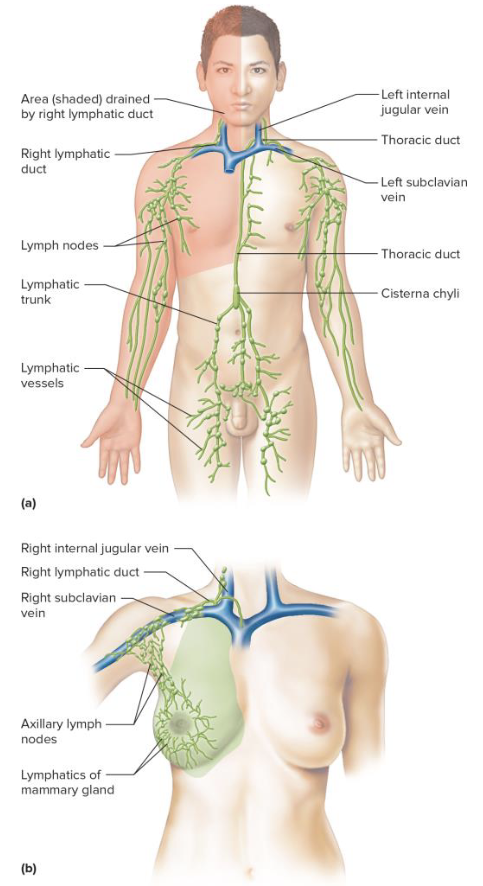
Thoracic Duct: The longer and wider of the two. It begins as a sac called the cisterna chyli and empties into the Left Subclavian Vein. It drains the majority of the body.
Right Lymphatic Duct: Much smaller than the thoracic duct. It begins in the left thorax and empties into the Right Subclavian Vein. It drains the upper left portion of the body.
Tissue Fluid and Lymph
Definition: Lymph is essentially tissue fluid that has entered a lymphatic capillary.
Tissue Fluid Formation:
Capillary blood pressure filters water and small molecules from the blood plasma.
The composition of tissue fluid is nearly identical to blood plasma, containing water, dissolved nutrients, gases, and hormones.
Critical Exception: Tissue fluid does not contain large plasma proteins. This is important as the absence of large proteins helps in maintaining osmotic balance in the interstitial space.
Formula:
Lymph Formation:
Filtration from plasma normally exceeds reabsorption, creating a net formation of tissue fluid.
This increases the tissue fluid hydrostatic pressure within the interstitial spaces, forcing fluid into the lymphatic capillaries.
This process is vital as it prevents edema (accumulation of excess tissue fluid).
Clinical Example of Edema: If a woman has axillary lymph nodes removed during breast cancer surgery, the lymphatic drainage from the upper limb is obstructed, resulting in localized edema.
Lymph Flow Mechanisms:
Lymph has low hydrostatic pressure, similar to venous blood, and requires assistance to flow.
Skeletal Muscle Activity: Contraction of skeletal muscles compresses vessels, pushing lymph forward.
Respiratory Process: During inspiration, low pressure is created in the thorax and high pressure in the abdomen, which sends lymph from the abdomen to the thorax.
Smooth Muscle: Smooth muscle in the larger lymphatic vessels contracts to aid flow.
Valves: Semilunar valves prevent backflow of lymph.
Exercise: Lymphatic flow reaches its highest levels during physical exercise.
Summary of Lymph Functions:
Absorption of dietary fats in the small intestine for delivery to the bloodstream.
Returning small proteins filtered by blood capillaries back to the bloodstream.
Collecting excess interstitial fluid and delivering it back to the bloodstream.
Delivering foreign particles to the lymph nodes for filtration.
Flap-like valves between the epithelial cells of lymphatic capillaries allow easy entry for tissue fluid.
Lymphatic Tissues and Organs
Lymphatic Tissue Content: Contains various cell types, predominantly lymphocytes and macrophages.
Mucosa-Associated Lymphoid Tissue (MALT):
Unencapsulated lymphatic tissue found in the digestive, respiratory, urinary, and reproductive tracts.
Tonsils and Appendix: Composed of lymphatic nodules, which are compact masses of lymphatic tissue.
Peyer’s Patches: Aggregates of lymphatic nodules located in the ileum (distal portion of the small intestine).
Lymphatic Organs: These consist of encapsulated lymphatic tissue and include the lymph nodes, thymus, and spleen.
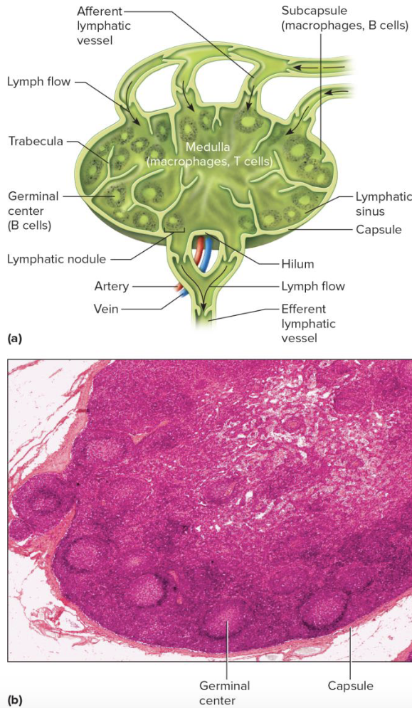
Lymph Nodes:
Typically bean-shaped and less than long.
Located in groups or chains along larger lymphatic vessels (except in the Central Nervous System).
Anatomy/Structure: Includes the capsule, subcapsule (contains B cells and macrophages), germinal center (contains B cells), medulla (contains T cells and macrophages), trabeculae, sinuses, and the hilum where vessels exit.
Afferent vs. Efferent: Lymph enters through afferent vessels and leaves through efferent vessels.
Functions: Filter potentially harmful particles from lymph and perform immune surveillance (monitoring body fluids via lymphocytes and macrophages). They are also centers for lymphocyte production alongside red bone marrow.
Major Locations: Cervical, axillary, supratrochlear, inguinal, pelvic, abdominal, and thoracic regions.
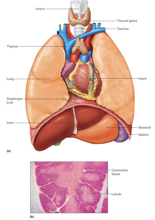
Thymus:
A soft, bilobed gland located in the mediastinum, posterior to the upper portion of the sternum.
Divided into lobules containing lymphocytes derived from red bone marrow.
Thymocytes: Most cells in the thymus are inactive.
T Lymphocytes (T cells): Some cells mature into functional T cells and leave the thymus to provide immunity.
Thymosins: Hormones produced by the thymus that stimulate T cell maturation.
Life-Span: Large during infancy and childhood, shrinks during puberty, and is small in adults. In the elderly, lymphatic tissue is replaced by adipose and connective tissue.
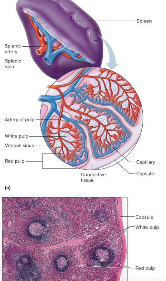
Spleen:
The largest lymphatic organ, located in the upper left abdominal cavity, inferior to the diaphragm and lateral to the stomach.
Contains venous sinuses filled with blood.
Red Pulp: Contains red blood cells, lymphocytes, and macrophages.
White Pulp: Composed of lymphocytes.
Functions: Filters blood (similar to how nodes filter lymph), breaks down worn-out red blood cells, and uses macrophages to destroy foreign particles.
Immunity: Innate (Nonspecific) Defenses
Immunity Definition: The ability of the body to prevent pathogen entry or destroy pathogens that enter.
Pathogens: Disease-causing agents like bacteria, viruses, protozoa, and fungi.
Innate Defenses: General defenses that protect against many types of pathogens.
Species Resistance: Certain species are resistant to diseases affecting others due to lack of specific receptors, temperature, or chemical environments.
Mechanical Barriers (First Line of Defense):
Skin and Mucous Membranes: Prevent entrance. Epidermis sloughing removes bacteria.
Ciliated Epithelium: In the respiratory tract, it traps and sweeps away pathogens.
Fluids: Tears, saliva, and urine wash away microorganisms.
Hair: Traps pathogens.
Chemical Barriers:
Enzymes: Pepsin (gastric juice) and lysozyme (tears) destroy microorganisms.
Interferons: Block viral replication, act against tumors, and stimulate phagocytosis.
Defensins: Peptides from neutrophils that cripple microbes by making openings in membranes.
Collectins: Proteins protecting against bacteria, yeast, and some viruses.
Complement: A group of plasma proteins that stimulate inflammation, attract phagocytes, and enhance phagocytosis.
Natural Killer (NK) Cells:
A small population of lymphocytes distinct from B and T cells.
Defend against viruses and cancer cells by secreting cytolytic substances called perforins that lyse cell membranes.
They also enhance inflammation.
Fever:
Triggered by infection stimulating lymphocytes to secrete Interleukin-1 (IL-1), also known as endogenous pyrogen.
IL-1 raises the thermoregulatory set point in the brain.
Benefits: Inhibits microbial growth by signaling the liver and spleen to sequester iron; also increases phagocytic activity.
Phagocytosis:
Neutrophils and monocytes are the most active phagocytes.
Chemotaxis: Chemicals from damaged tissue attract these cells.
Macrophages: Monocytes that leave the blood. They can be "free" or "fixed" in tissues.
Mononuclear Phagocytic System: Also called the reticuloendothelium; consists of all monocytes and macrophages in the body.
Inflammation
Signs of Inflammation:
Redness: Caused by vasodilation.
Swelling: Results from increased capillary permeability and fluid entry into tissues.
Heat: Derived from blood arriving from deeper body areas.
Pain: Stimulation of pain receptors.
Inflammatory Process:
Walled off infection site inhibited spread.
White blood cells gather to perform phagocytosis.
Exudates containing fibrinogen form a fibrin network.
Post-Infection: Phagocytes remove dead cells; cell division replaces lost tissue.
Summary of Actions:
Vessels dilate $\rightarrow$ Tissue becomes red, warm.
Permeability increases $\rightarrow$ Swelling and pain.
WBC invasion $\rightarrow$ Pus formation (accumulation of WBCs, bacteria, debris).
Fibroblasts arrive $\rightarrow$ Connective tissue sac formation.
Adaptive (Specific) Defenses (Third Line of Defense)
Basis: Ability to distinguish "self" from "non-self".
Antigens: Non-self molecules that evoke an immune response. Can be proteins, polysaccharides, glycoproteins, or glycolipids. Most effective are large, complex molecules.
Haptens: Small molecules that are not antigenic alone but become so when combined with a larger carrier molecule.
Lymphocyte Origins:
Produced throughout life, starting in fetal development from red bone marrow precursors.
T Lymphocytes (T cells): Specialize in the thymus. Account for to of circulating lymphocytes. Found in lymph nodes, thoracic duct, and white pulp of the spleen.
B Lymphocytes (B cells): Specialize in red bone marrow (named after the Bursa of Fabricius in chickens). Account for to of blood lymphocytes. Abundant in lymph nodes, spleen, and intestinal lining.
Cellular Immune Response
T Cell Activation: Requires an Antigen-Presenting Cell (APC/accessory cell).
APC Mechanism: Phagocytizes the antigen, digests it, and displays fragments on its membrane bound to Major Histocompatibility Complex (MHC) proteins (also called Human Leukocyte Antigens or HLA).
Specialized T Cells:
Helper T Cells: Activate other cells and stimulate B cells to produce antibodies.
Cytotoxic T Cells: Attack virally infected or cancerous cells directly.
Memory T Cells: Provide future immune protection.
Cytokines (Polypeptide secretions):
Colony-stimulating factors: Stimulate bone marrow to produce lymphocytes.
Interferons: Block viral replication, stimulate macrophages, and attack cancer.
Interleukins: Control lymphocyte differentiation and proliferation.
Tumor Necrosis Factor (TNF): Stops tumor growth, causes fever, and stimulates differentiation.
Humoral Immune Response
B Cell Activation: Occurs when an antigen fits its receptor; full activation requires cytokines from Helper T cells.
Proliferation: Activated B cells clone themselves.
Differentiation:
Memory B Cells: Provide future immunity.
Plasma Cells: Synthesize and secrete large globular proteins called antibodies (immunoglobulins).
Antibody Structure:
Y-shaped proteins composed of amino acid chains: heavy and light chains.
Chains are joined by disulfide bonds.
Each antibody has a unique variable region (antigen-binding site) at the tips of the Y.
Types of Immunoglobulins (Ig):
IgG (): Found in plasma and tissue fluid; acts against bacteria, viruses, toxins; activates complement.
IgA (): Found in exocrine secretions (tears, breast milk); defends against bacteria/viruses.
IgM (): Found in plasma; reacts with antigens on RBCs after mismatched transfusions; activates complement.
IgD (<1\%): Found on B cell surfaces; aids in B cell activation.
IgE (<1\%): Found in exocrine secretions; promotes inflammation and allergic responses.
Antibody Actions
Direct Attack:
Agglutination: Clumping of antigens.
Precipitation: Making antigens insoluble.
Neutralization: Covering toxic portions to render them harmless.
Complement Activation:
Opsonization: Coating antigen-antibody complexes to make them susceptible to phagocytosis.
Chemotaxis: Attracting macrophages and neutrophils.
Lysis: Rupturing cell membranes of pathogens via osmotic rupture.
Neutralization: Changing the molecular structure of viruses.
Localized Changes: Stimulation of inflammation to prevent spread.
Immune Response Timing and Classification
Primary Immune Response: Occurs on first encounter. Antibodies appear in to days. Produces IgM first, then IgG.
Secondary Immune Response: Subsequent exposure. High antibody concentration produced in to days by memory cells. Antibodies can remain for years.
Classifications:
Naturally Acquired Active: Exposure to live pathogens $\rightarrow$ disease symptoms and permanent immunity.
Artificially Acquired Active: Exposure to a vaccine $\rightarrow$ no symptoms, permanent immunity.
Naturally Acquired Passive: Antibodies from mother to fetus/newborn $\rightarrow$ short-term immunity.
Artificially Acquired Passive: Injection of antiserum/antitoxin $\rightarrow$ short-term immunity without stimulating an immune response.
Hypersensitivity and Autoimmunity
Type I (Immediate-reaction): Allergy; overproduction of IgE. Histamine release causes hives, asthma, or anaphylactic shock.
Type II (Antibody-dependent cytotoxic): Phagocytosis and complement lysis of antigens (e.g., mismatched blood transfusion).
Type III (Immune-complex): Antigen-antibody complexes deposit in tissues (e.g., Rheumatoid arthritis).
Type IV (Delayed-reaction): T cell-mediated; occurs after repeated skin exposure to allergens; takes about hours (e.g., dermatitis).
Autoimmunity: Immune system produces autoantibodies and cytotoxic T cells that attack "self" tissues.
Examples: Type 1 Diabetes (pancreatic beta cells), Multiple Sclerosis (myelin), Graves' Disease (thyroid), Rheumatoid Arthritis (joint linings), Systemic Lupus Erythematosus (connective tissue).
Transplantation and HIV/AIDS
Graft Types:
Isograft: Identical twin donor.
Autograft: From self.
Allograft: Same species (relative or matched donor).
Xenograft: Different species (e.g., pig heart valves).
Tissue Rejection: Major Histocompatibility Complex (MHC) matching is required; immunosuppressive drugs prevent rejection.
HIV/AIDS:
HIV attacks macrophages and Helper T cells.
Loss of Helper T cells prevents B cells from producing antibodies due to lack of cytokines.
Leads to opportunistic infections and cancer.
Transmission: sexual contact, needles, birth/milk, or infected blood/tissue.
Life-Span Changes
The thymus gland shrinks early in life, becoming only as powerful as it was in infancy.
Elderly individuals have a higher risk of infection and cancer.
T cell numbers decrease slightly; B cell numbers stay constant, but activity levels for both decline.
Antibody response slows; IgG and IgA levels increase, while IgM and IgE levels decrease.
Increased production of autoantibodies.