Anatomy of the Central Nervous System (CNS)
Anatomy of the Central Nervous System (CNS)
Page 1: Introduction to the Central Nervous System (CNS)
The Central Nervous System (CNS) consists of:
The brain
The spinal cord
The Peripheral Nervous System (PNS) encompasses sensory and motor nerves that connect the CNS to the rest of the body.
Page 2: Components of the CNS
The primary components that make up the CNS include the brain and spinal cord.
The PNS includes nerves that carry signals to and from the CNS, assisting in sensory and motor functions.
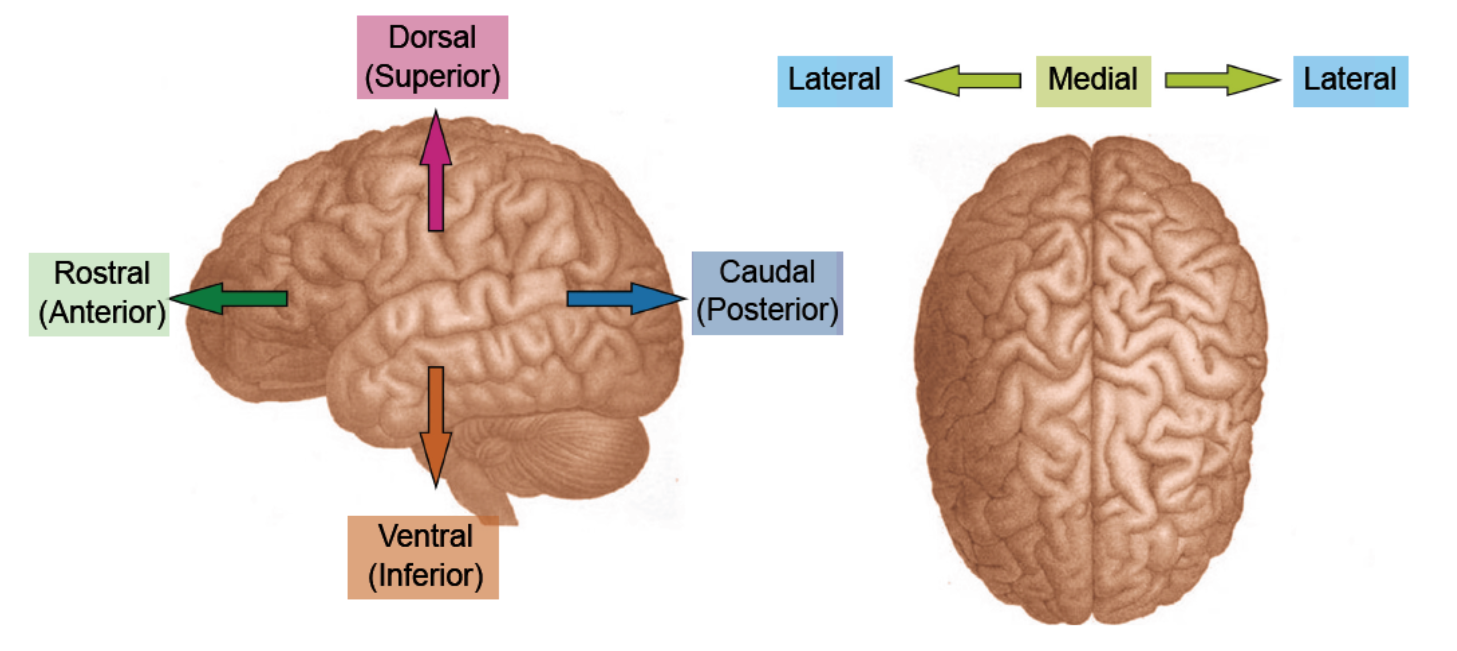
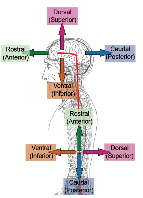
Page 3: Anatomical Terminology (Directional Terms)
Rostral (Anterior): Refers to the front of the brain or toward the nose.
Dorsal (Superior): Refers to the upper side or top of the brain.
Lateral: Refers to the sides of the brain, away from the midline.
Medial: Refers to the middle or center of the brain, toward the midline.
Ventral (Inferior): Refers to the bottom or lower side of the brain.
Caudal (Posterior): Refers to the back of the brain or toward the tail end.
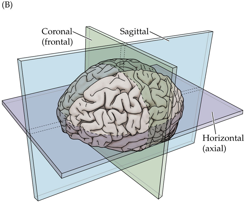
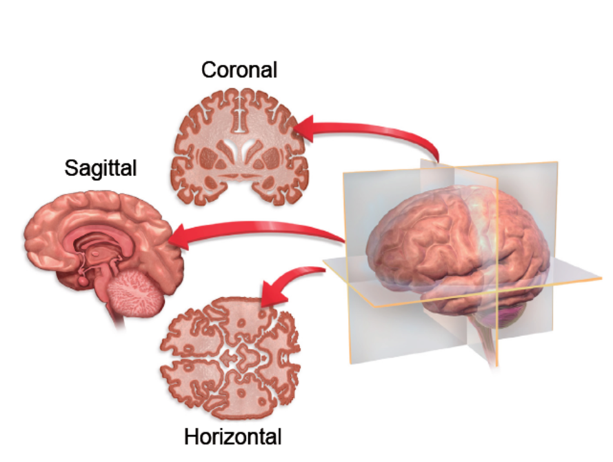
Page 4: Anatomical Terminology (Planes of Section)
Coronal (Frontal): A plane that divides the brain into front and back sections.
Sagittal: A plane that divides the brain into left and right sections.
Horizontal (Axial): A plane that divides the brain into upper and lower sections.
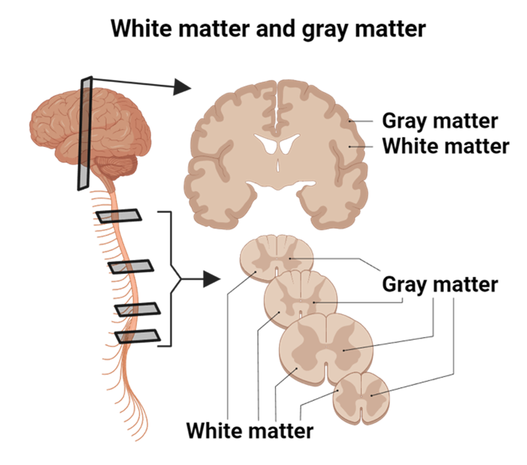
Page 5: Tissue Types in the CNS
Gray Matter:
Composed of cell bodies and dendrites.
Associated with processing and cognition.
White Matter:
Composed of myelinated axons that conduct signals between different regions of the CNS.
Corpus Callosum: The structure that connects the left and right hemispheres of the brain, composed of white matter.
Page 6: Further Details on Gray Matter
Gray Matter includes:
Cell bodies and dendrites which form networks for processing information.
Collections of cell bodies in the CNS are called nuclei.
In the PNS, collections of cell bodies are called ganglia.
Page 7: Further Details on White Matter
White Matter:
Primarily consists of myelinated axons which are crucial for communication between different brain regions and between the brain and spinal cord.
In the CNS, bundles of axons are referred to as tracts.
In the PNS, bundles of axons are referred to as nerves.
Page 8: Concept Check: Anatomical Planes
Identify the anatomical plane of images presented:
Images are labeled with measurements (100 cm, 120 mm).
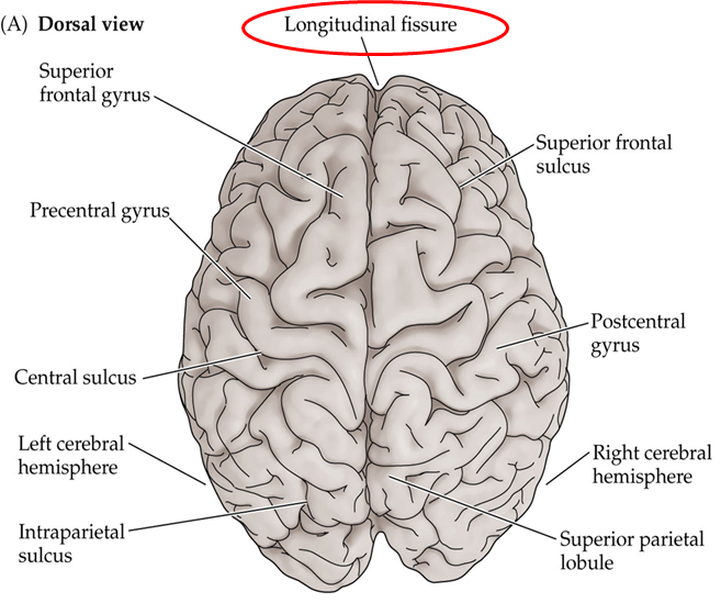
Page 9: Cerebral Cortex Identification
Key features of the cerebral cortex include:
Gyri: Elevated ridges or folds of cortex.
Sulci: Grooves or fissures between gyri.
Notable fissures include:
Longitudinal fissure: Divides the two hemispheres of the brain.
Central sulcus: Separates the frontal lobe from the parietal lobe.
Lateral (Sylvian) fissure: Separates the temporal lobe from the frontal and parietal lobes.
Parieto-occipital sulcus: Separates the parietal and occipital lobes.
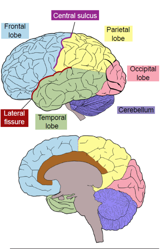
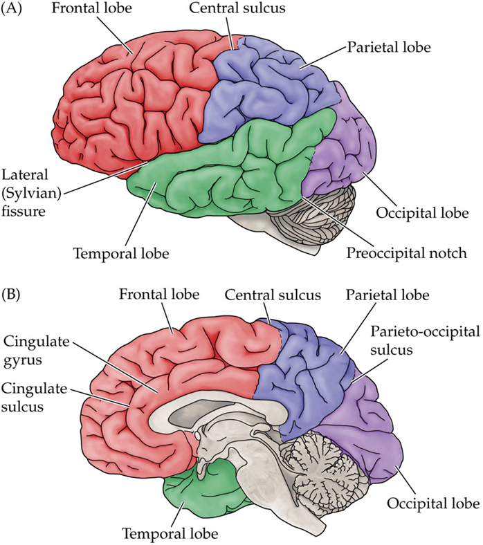
Page 10: Identification of Brain Lobes
Major lobes of the brain include:
Occipital Lobe: Located at the back, primarily involved in visual processing.
Temporal Lobe: Bordered by the lateral fissure, involved in auditory processing and memory.
Parietal Lobe: Just anterior to the occipital lobe; associated with sensory processing. Bounded by the central sulcus and parieto-occipital sulcus with the lateral fissure on the ventral side.
Frontal Lobe: Anterior part of the brain, involved in reasoning, planning, and motor control; bounded by the central sulcus posteriorly and the lateral fissure inferiorly. Includes the Somatosensory cortex and the Primary motor cortex.
]
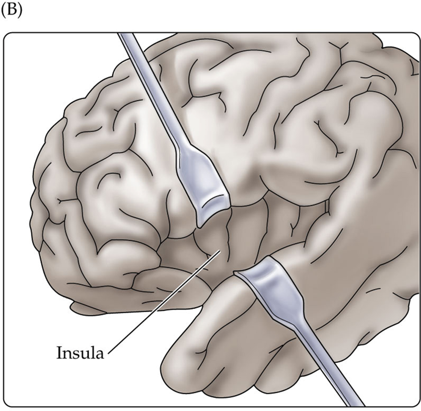
Page 11: Insula Cortex
The insula is a region of the cerebral cortex located deep within the lateral sulcus, involved in consciousness, emotion, and perception of bodily states.
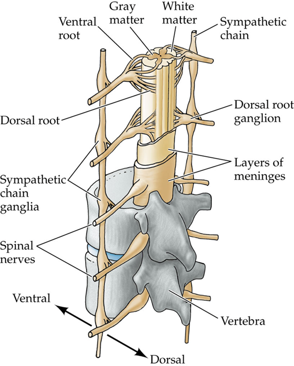
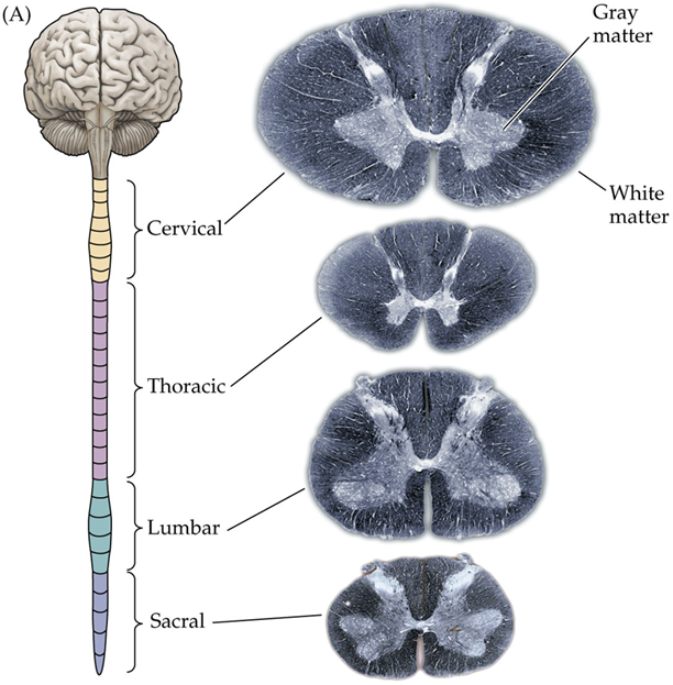
Page 12: Structures of the Spinal Cord
Regions of the Spinal Cord:
Cervical
Thoracic
Lumbar
Sacral
Orientation:
Rostral (anterior) and Ventral (inferior) orientations in the context of the spinal cord structure.
The relationship of spinal nerve roots to the spinal cord as seen in the vertebral column:
Ventral root: Carries motor neurons.
Dorsal root: Carries sensory neurons.
The Gray Matter includes the dorsal and ventral horns where sensory and motor neuron bodies reside, respectively.
The White Matter is organized into different tracts, while sympathetic chains are present in the surrounding area.
]
Page 13: Spinal Nerve Distribution
The spinal cord has 31 pairs of nerve bundles (many axons) that exit the vertebral column. They include:
Cervical nerves: 8 pairs (C1-C8)
Thoracic nerves: 12 pairs (T1-T12)
Lumbar nerves: 5 pairs (L1-L5)
Sacral nerves: 5 pairs (S1-S5)
Coccygeal nerve: 1 pair (Co1)
Notable enlargements of the spinal cord include:
Cervical enlargement: Related to arm nerve connections.
Lumbosacral enlargement: Related to nerve connections for the legs.
The Cauda Equina refers to the collection of nerve roots at the lower end of the spinal cord.
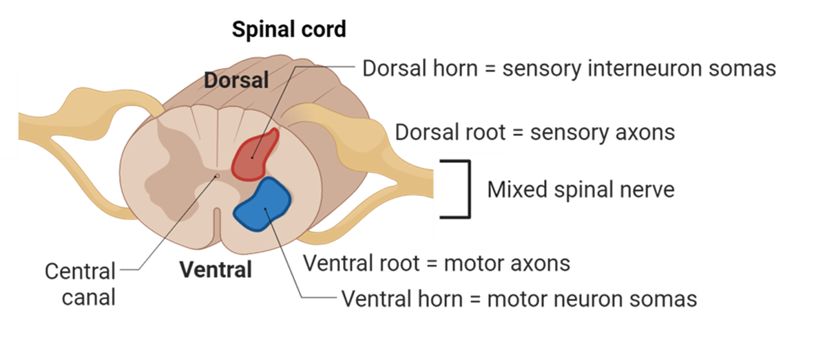
Page 14: Functional Organization of the Spinal Cord
The spinal cord is organized functionally with:
Dorsal Horn (Gray Matter): Houses sensory neurons, which receive sensory information from the body.
Ventral Horn (Gray Matter): Houses motor neurons, which send signals to muscles.
Axons emerge as bundles in dorsal and ventral roots and merge to form a mixed nerve, carrying both sensory and motor information.
Lateral Horn (Gray Matter): Contains autonomic neurons that innervate glands and smooth muscles, playing a crucial role in regulating involuntary bodily functions.
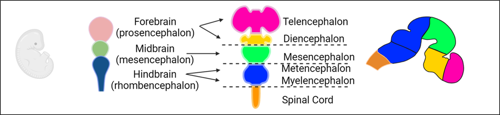
Page 15: Neurodevelopment Stages
**Embryonic Cell Layers: **
Ectoderm: Outer layer that differentiates into nervous system components.
Mesoderm: Middle layer; contributes to structures such as muscles and vertebrae.
Endoderm: Inner layer; forms internal organs.
Neural Development Process:
During the third week of embryogenesis, a neural plate forms from ectoderm cells; borders are established between the ectoderm and neural plate.
The neural plate bends dorsally, leading to the formation of the neural tube, critical for CNS development.
Once the neural tube closes, neural crest cells detach from the epidermis; these cells eventually differentiate into most of the PNS.
The notochord typically degenerates and exists primarily as the nucleus pulposus in intervertebral discs; mesodermal cells develop into somites, precursor structures for the axial skeleton and muscles.
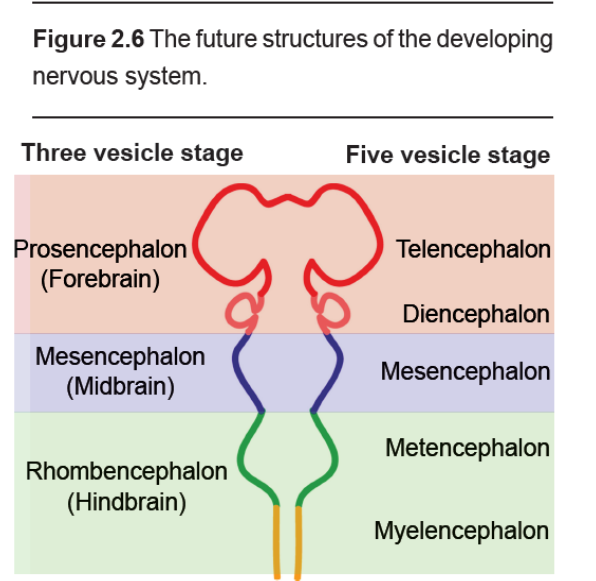
Page 16: Specialization of CNS Subdivisions
The CNS subdivides during weeks 3-11 of development and includes:
Prosencephalon (Forebrain):
Telencephalon: Develops into the cerebral cortex and basal ganglia.
Diencephalon: Develops into the thalamus and hypothalamus.
Mesencephalon (Midbrain): Contains structures such as the PAG (Periaqueductal Gray) and substantia nigra.
Rhombencephalon (Hindbrain):
Metencephalon: Develops into the pons and cerebellum.
Myelencephalon: Develops into the medulla oblongata and brainstem.
The spinal cord also originates from this embryonic structure.
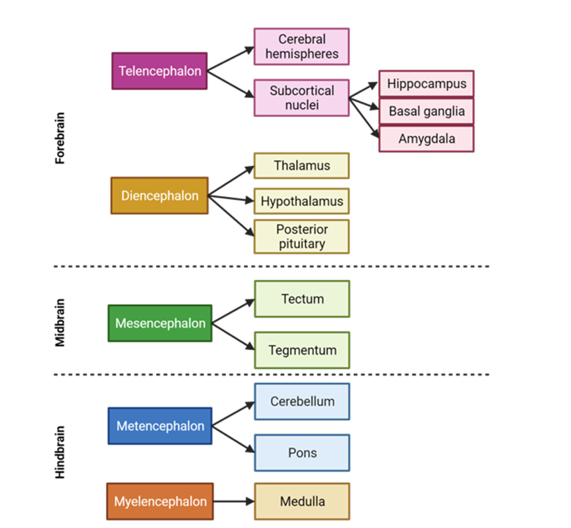
Page 17: Structural Overview of the CNS by Region
The brain can be classified into three major regions:
Hindbrain: Includes cerebellum, pons, medulla oblongata.
Midbrain: Contains structures essential for auditory and visual processing.
Forebrain:
Telencephalon: Includes the cerebral hemispheres and various nuclei such as the amygdala and basal ganglia.
Diencephalon: Contains the thalamus and hypothalamus, along with associated structures.
The adult brain retains these primary subdivisions derived from earlier developmental stages.
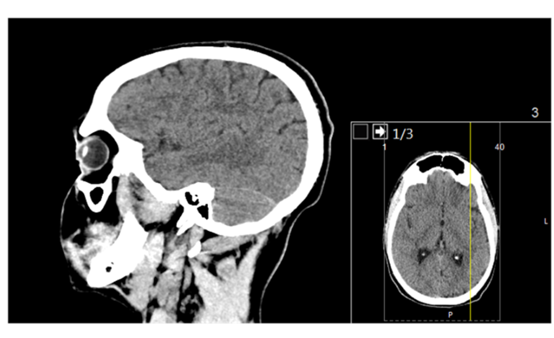
Page 18: Techniques for Visualizing the Brain
Two primary categories of imaging techniques exist for examining the brain:
Structural Techniques:
CT Scan (Computed Tomography): Employs X-ray imaging to produce cross-sectional views of the brain, providing structural data.
Functional Techniques:
PET (Positron Emission Tomography): Utilizes radiotracers to evaluate brain metabolism, measuring blood flow and abnormal protein deposits.
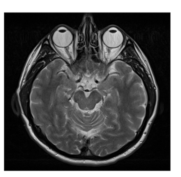
MRI (Magnetic Resonance Imaging): Employs magnetic fields and radio waves to create detailed images of brain anatomy.
Page 19: Continued Imaging Techniques
MRI Details:
Provides high-resolution images of the brain structures; aids in diagnosing various neurological diseases and conditions affecting brain integrity.
Page 20: Brain Structure Overview
Key brain structures include:
Brainstem: Controls basic life functions such as heart rate and breathing.
Cerebellum: Involved in motor control and coordination.
Various lobes:
Frontal Lobe: Associated with planning, personality, and voluntary movement.
Sylvian Fissure: Anatomical landmark that separates the temporal lobe from the frontal and parietal lobes.
Occipital, Temporal, and Parietal Lobes: Each contributing to specialized brain functions including vision, hearing, and sensory processing.
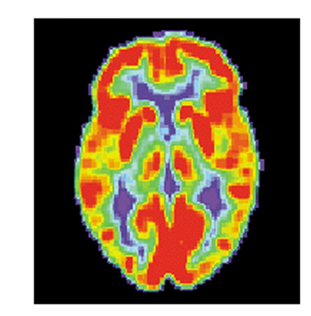
Page 21: Functional Brain Imaging Techniques
PET Scan Overview:
The PET scan evaluates physiological changes in the brain by measuring blood flow, allowing researchers and clinicians to infer areas of metabolic activity which correspond to neuronal function.
Page 22: Functional Imaging Studies
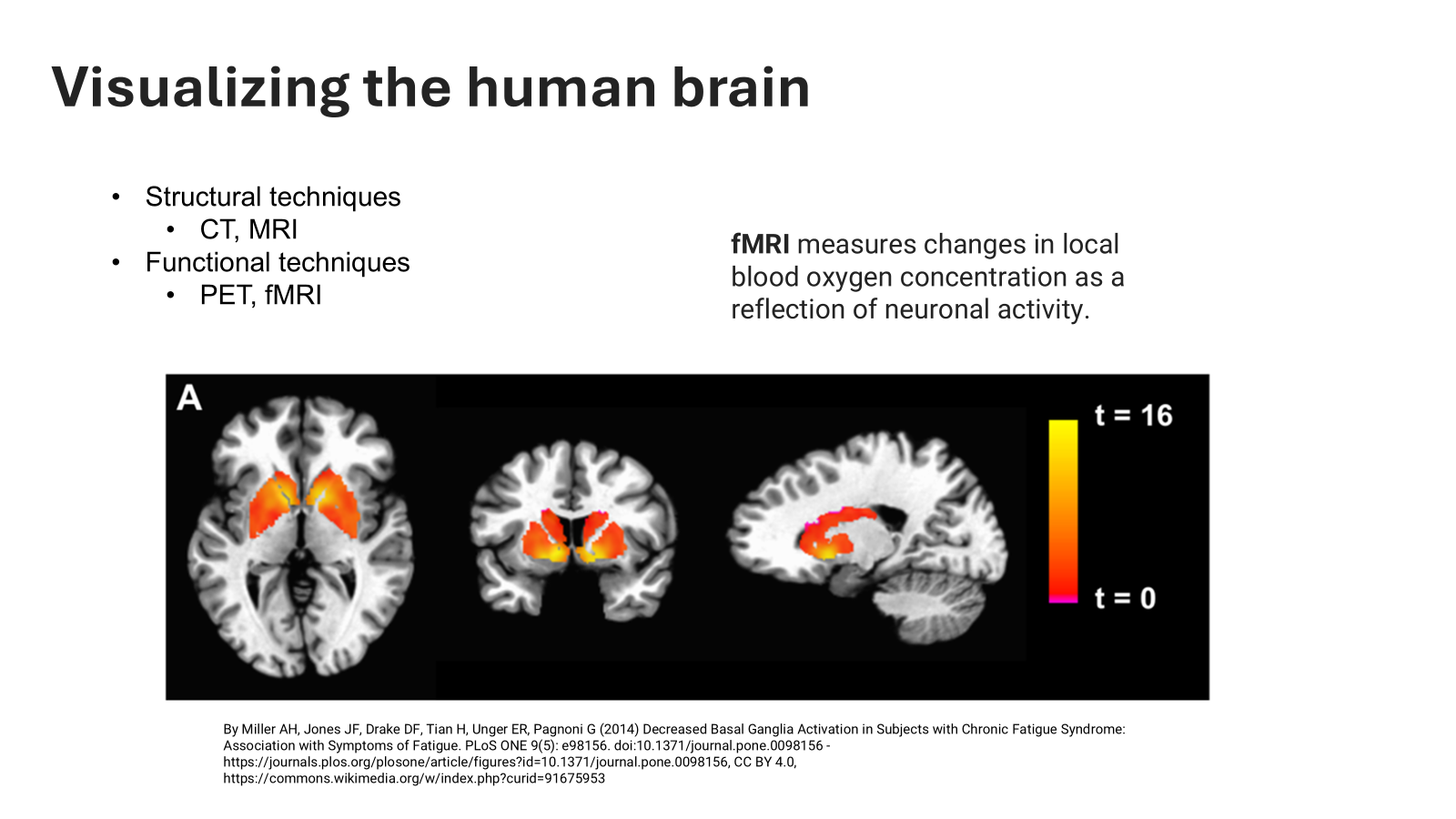
fMRI (Functional Magnetic Resonance Imaging) uses changes in local blood oxygen concentration as a measure of neuronal activity, revealing areas engaged in specific tasks or cognitive functions.
Recent studies highlight the effects of diminished basal ganglia activation in patients with chronic fatigue syndrome, relating it to fatigue symptoms.
These notes comprehensively summarize the material covered in the provided transcript, spanning the structure and function of the CNS, anatomical terminology, spinal cord organization, neurodevelopment, and techniques for brain imaging.