The Central Nervous System
Vocabulary
Gyri-folds on the surface of the cerebrum.
Sulci-grooves between the folds of the cerebrum.
Fissures–deep grooves between the folds of the cerebrum.
Diencephalon-area between the cerebrum and brainstem.
Cerebellum-portion of the brain in the posterior inferior region that processes information for coordination and movement.
Proprioception-position of joints.
Wernicke’s Area-specialized area in the brain that helps with understanding of speech.
Broca’s Area– specialized area in the brain that has to do with producing meaningful speech.
Nerves– a whitish fiber or bundle of fibers that transmits impulses of sensation to the brain or spinal cord, and impulses from these to the muscles and organs.
Cell body– The cell body carries genetic information, maintains the neuron’s structure, and provides energy to drive activities.
Dendrites– Dendrites are fibrous roots that branch out from the cell body, receiving and processing signals from the axons of other neurons.
Central Nervous System (CNS)- CNS is the processing center of the body consisting of the brain and spinal cord.
Neurotransmitters– a chemical substance that is released at the end of a nerve fiber by the arrival of a nerve impulse and, by diffusing across the synapse or junction, causes the transfer of the impulse to another nerve fiber, a muscle fiber, or some other structure.
Axon– the long threadlike part of a nerve cell along which impulses are conducted from the cell body to other cells.
Axon terminal– found at the terminal ends of axons, typically the sites where synapses with other neurons are found, and neurotransmitters are stored there to communicate with other neurons via these synapses.
Sensory (afferent) nerves– are the nerve fibers responsible for bringing sensory information from the outside world into the brain. Sensory information may involve special senses, such as vision, hearing, smell, or taste, as well as the sense of touch, pain, and temperature.
Motor (efferent) nerves– The efferent or motor division transmits impulses from the CNS out to the peripheral organs to cause an effect or action.
Somatic nervous system– The somatic nervous system (SNS), or voluntary nervous system is the part of the peripheral nervous system associated with the voluntary control of body movements via skeletal muscles.
Autonomic nervous system (involuntary)– The autonomic nervous system (ANS) is a component of the peripheral nervous system that regulates involuntary physiologic processes including heart rate, blood pressure, respiration, digestion, and sexual arousal. It contains three anatomically distinct divisions: sympathetic, parasympathetic, and enteric.
Skeletal muscle– a muscle that is connected to the skeleton to form part of the mechanical system which moves the limbs and other parts of the body.
Smooth muscle– muscle tissue in which the contractile fibrils are not highly ordered, occurring in the gut and other internal organs and not under voluntary control.
Synapse– The junction between two nerve cells, consisting of a minute gap across which impulses pass by diffusion of a neurotransmitter
Cardiac muscle– cardiac muscle, also called myocardium, is one of three major muscle types, found only in the heart. The rhythmic contraction of cardiac muscle is regulated by the sinoatrial node of the heart, which serves as the heart’s pacemaker.
Myofibrils– very fine contractile fibers, groups of which extend in parallel columns along the length of striated muscle fibers. The myofibrils are made up of thick and thin myofilaments, which help give the muscle its striped appearance.
Sarcomere– The sarcomere is the fundamental unit of contraction and is defined as the region between two Z-lines. Each sarcomere consists of a central A-band (thick filaments) and two halves of the I-band (thin filaments).
Actin– thin filaments of protein that forms (together with myosin) the contractile filaments of muscle cells, and is also involved in motion in other types of cell.
Myosin– thick filaments of fibrous protein that forms (together with actin) the contractile filaments of muscle cells and is also involved in motion in other types of cells.
The Central Nervous System
The Brain
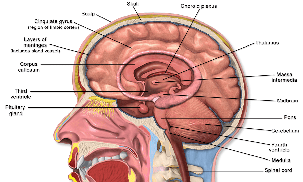
The major parts of the brain include the outer cerebrum and the inner diencephalon. The brain connects to the spinal cord by way of the brainstem.
The Cerebrum
The cerebrum is largest portion of the nervous system. The cerebrum consists of two hemispheres (right and left) connected by a white matter bridge called the corpus callosum. On the surface of the cerebrum are folds called gyri and grooves called sulci. Deep grooves are known as fissures. Each hemisphere is divided into lobes. The lobes are the frontal, parietal, temporal and occipital.
The frontal lobe processes information involving motor movements, concentration, planning and problem solving as well as the sense of smell and emotions. The parietal lobes process sensory information with the exception of hearing, smell and vision. The temporal lobes process information related to hearing, smell and memory as well as abstract thought and making judgments. The occipital lobe processes visual information.
Some lobes are divided by fissures. Along the superior aspect of the cerebrum lies the longitudinal fissure that divides the parietal lobes. The lateral fissure (Sylvian fissure) is located on the lateral aspect and separates the temporal from parietal lobes. One sulcus called the central sulcus is located midway on the lateral aspect of the cerebrum and extends from superior to inferior. The central sulcus separates the frontal from parietal lobes.
Deep in the lateral fissure is the insula which is often referred to as a fifth lobe of the cerebrum.

The Diencephalon
The diencephalon lies between the brainstem and cerebrum. Important structures include the thalamus and hypothalamus.
The thalamus is the largest part of the diencephalon. The thalamus carries all sensory information to the cerebral cortex except for the sense of smell which is carried directly to the frontal lobe of the cerebral cortex by the olfactory nerves. The thalamus is sometimes referred to as a relay station for sensory information. Examples of sensory information include auditory, visual and motor information. The thalamus is also intimately involved in emotions due to its connections to the limbic system.
The hypothalamus lies inferior and anterior to the thalamus. A stalk-like projection called the infundibulum projects anterior and inferior and connects to the pituitary gland. The hypothalamus is intimately connected with the endocrine system and helps to regulate hormones. The hypothalamus also regulates body temperature, thirst, hunger and sexual drive and is involved in processing emotions, mood, and sleep along with the reticular activating system.
Posterior to the diencephalon is the pineal gland which is a small endocrine gland that secretes the hormone melatonin. Melatonin helps to regulate sleep-wake cycles.
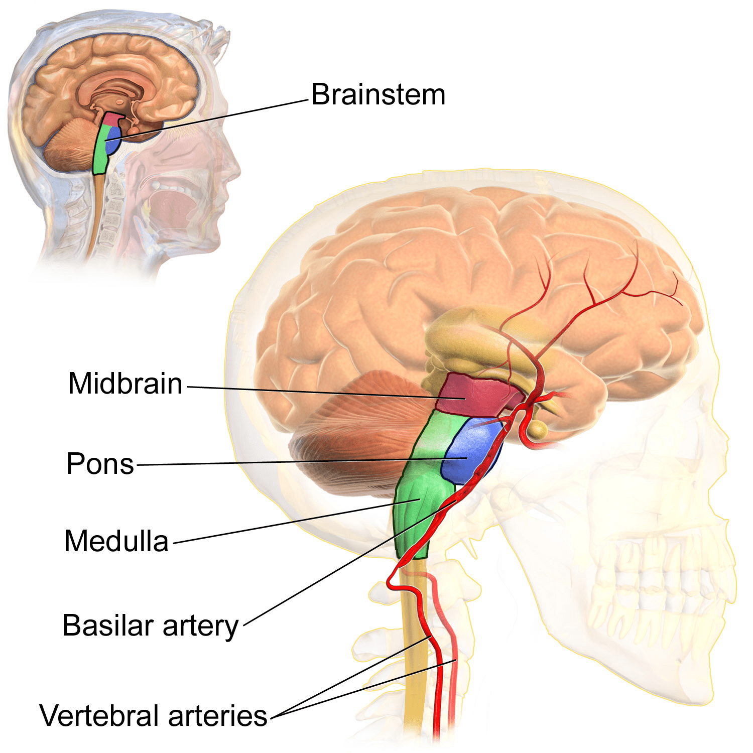
Brainstem
The brainstem lies between the cerebral cortex and the spinal cord. It consists of the midbrain, pons and medulla oblongata. The medulla oblongata is the most inferior portion of the brainstem and contains a number of centers for controlling heart rate, respiration, swallowing, vomiting and blood vessel diameter. Spinal pathways called tracts continue through the medulla connecting the spinal cord with the brain.
The pons is the middle section of the brainstem. The pons also contains spinal cord tracts as well as nuclei that help to control respiration and sleep.
The midbrain is the most superior portion of the brainstem and helps to process motor and hearing information.
The reticular formation is located throughout the brainstem and is primarily concerned with regulating sleep-wake cycles.
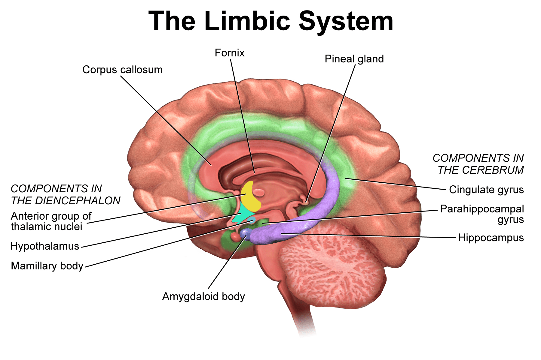
Limbic System
The limbic system consists of portions of both the cerebrum and diencephalon and is involved in the emotions as well as reproduction and memory. The limbic system contains the cingulate gyrus, portions of the thalamus, hypothalamus, mamillary and amygdaloid bodies, hippocampus, and nucleus accumbens.
The Spinal Cord
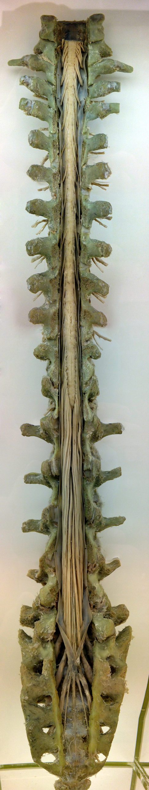
The spinal cord begins at the foramen magnum of the occipital bone and extends to the second lumbar vertebra. It ends in a cone-like structure called the conus medullaris. A structure known as the cauda equina extends from the inferior end of the spinal cord. The cauda equina (horse’s tail) consists of nerves that extend downward to exit the foramen of the lumbar and sacral vertebrae. The spinal cord consists of cervical, thoracic, lumbar and sacral segments.
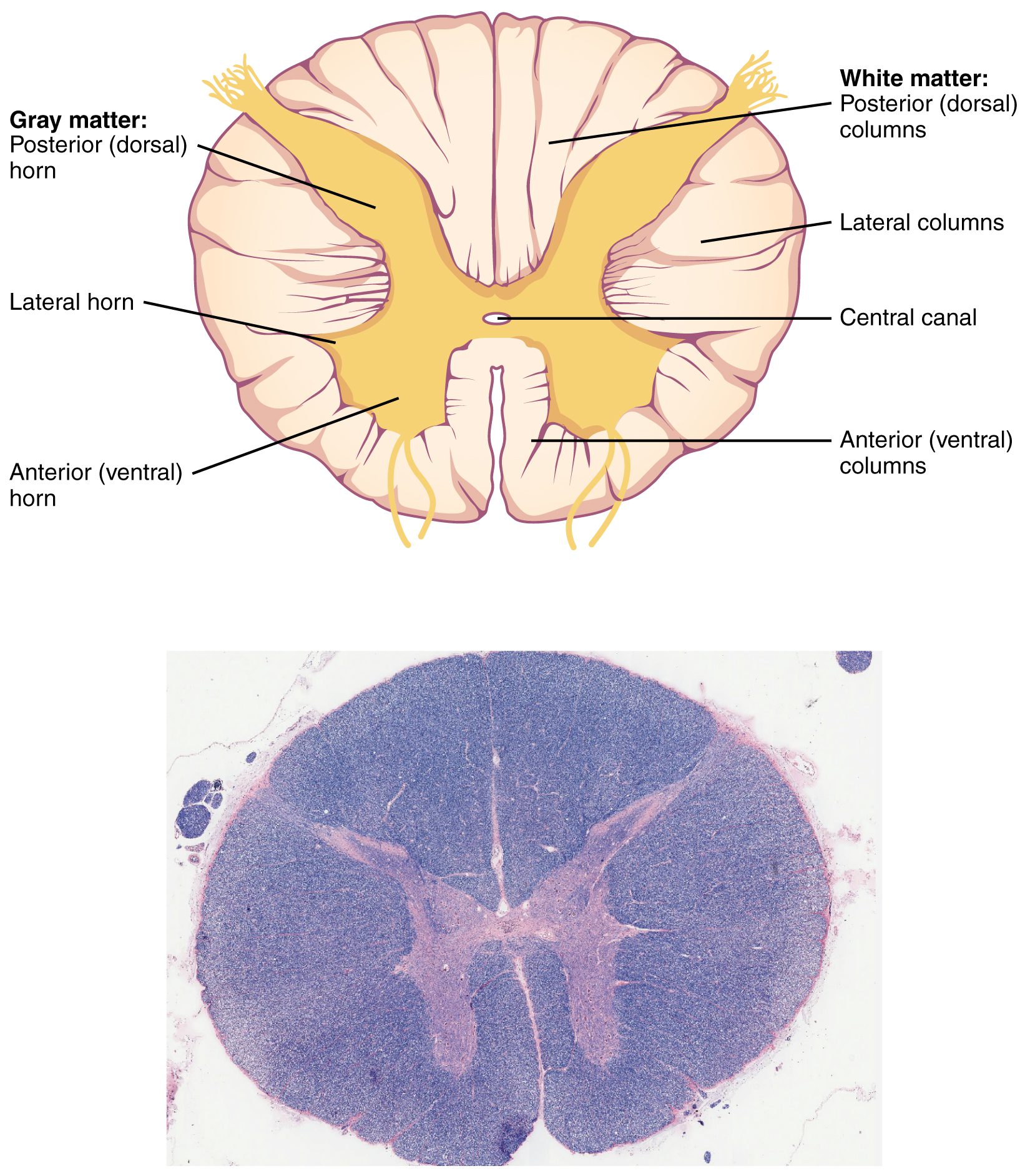
The spinal cord consists of a core of gray matter surrounded by white matter. The white matter is divided into sections called funiculi or columns. There are posterior, lateral and anterior columns. The gray matter is divided into horns, so there are posterior, lateral and anterior horns. Both sides of the spinal cord are connected by the posterior and anterior gray commissures and there is a central canal carrying cerebral spinal fluid in the center.
There is a large groove in the front called the anterior median fissure and a shallow groove in the back called the posterior median sulcus.
Both the brain and spinal cord are covered by the meninges. There are 3 layers in the meninges, these include the outer dura mater, the middle arachnoid mater and the inner pia mater. There is a space between the arachnoid and pia mater called the subarachnoid space which carries cerebral spinal fluid.
The space between the dura mater and the vertebrae is called the epidural space. Anesthetics are sometimes injected into this space (epidural injection).
Cerebral spinal fluid is produced by structures in the brain called choroid plexi which are located in hollow structures in the brain called ventricles. There are 2 lateral ventricles, a 3rd and 4thventricle which connect to the central canal of the spinal cord.
About 500 ml of cerebral spinal fluid is produced daily and there is about 125 ml in the nervous system at any given time. The CSF is absorbed by structures called arachnoid granulations or villi located in the arachnoid mater.
An abnormal buildup of CSF is called hydrocephalus which can cause a variety of brain symptoms. Hydrocephalus can be treated by surgery or a device called a shunt that drains the excess fluid into the chest or abdomen.

The Cranial Nerves
The cranial nerves are important in assessing the function of the nervous system, especially after trauma to the head. There are 12 pair of cranial nerves, and each nerve has a different function.
The cranial nerves are represented by Roman numerals, and each has a unique name.
Some nerves carry sensory information, some carry motor information and some carry both sensory and motor information.
Cranial nerve I, the olfactory nerve carries the sense of smell.
Cranial nerve II, the optic nerve carries the sense of vision.
Cranial nerves III, IV, VI, the oculomotor, trochlear, and abducens nerves work together to move the eyes.
Cranial nerve V, the trigeminal nerve carries sensation from the face and motor information to the chewing muscles or muscles of mastication which include the temporalis and masseter muscles.
Cranial nerve VII, the facial nerve carries sensory information for taste from the anterior 2/3 of the tongue and motor information to the muscles of facial expression.
Cranial nerve VIII, the auditory or vestibulocochlear nerve carries information for hearing, balance and equilibrium.
Cranial nerve IX, the glossopharyngeal nerve carries sensory information for taste from the posterior 1/3 of the tongue and motor information to the swallowing muscles.
Cranial nerve X, the vagus nerve carries sensory and motor information to and from the organs.
Cranial nerve XI, the spinal accessory nerve carries motor information to the sternocleidomastoid and trapezius muscles
Cranial nerve XII, the hypoglossal nerve carries motor information to the tongue.

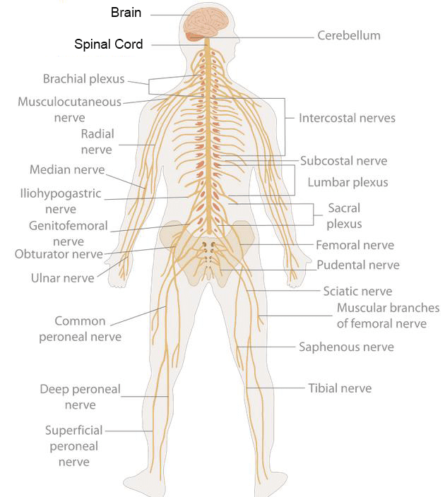
Spinal Nerves
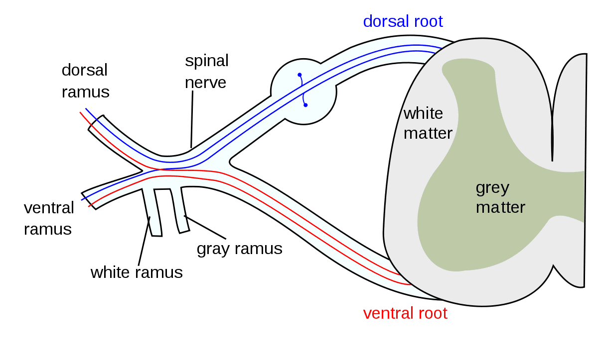
Spinal nerves emerge from the spinal cord. There are 31 pair of spinal nerves and these are named after where they originate in the spinal cord. The spinal nerves include 8 cervical, 12 thoracic, 5 lumbar, 5 sacral and 1 coccygeal.
Spinal nerves are formed by dorsal and ventral nerve roots. The nerves branch to the body with some nerves traveling to the autonomic nervous system.
Spinal nerves carry both sensory and motor information.
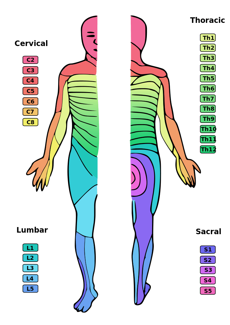
Dermatomes
The body can be divided into sections called dermatomes. Each dermatome represents a specific area of the body innervated by a spinal nerve.
Dermatomes are important in helping to localize peripheral nervous system injuries.
Autonomic Nervous System
The autonomic nervous system can be thought of as an “automatic” system because it works to maintain homeostasis in the body even when it is in an unconscious state. The autonomic nervous system (ANS) can control respiratory, cardiovascular, urinary, digestive and reproductive functions. It works to maintain balance of fluids, electrolytes, blood pressure, nutrients, and blood gasses. The ANS does this by sending motor impulses to viscera, cardiac and smooth muscle. Since it sends motor impulses to viscera, the ANS is also known as a visceral motor system.
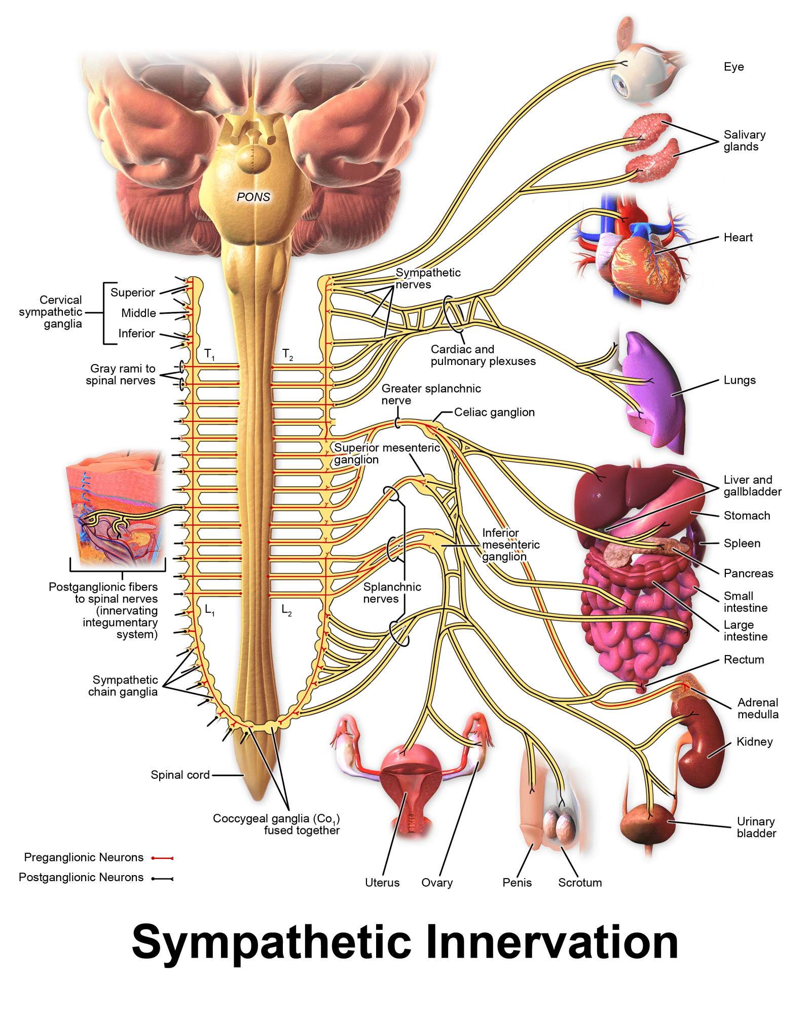
The ANS is divided into two subdivisions. The sympathetic is often referred to as the
“fight or flight” system. It is located in the thoracic and lumbar spines and sends fibers to the organs. The parasympathetic division begins in the cervical and lower lumbar spines and sends fibers to the same organs as the sympathetic. The sympathetic and parasympathetic divisions typically have the opposite effect on organs and thus work to maintain balance based on the body’s needs. For example the sympathetic system can increase heart rate while the parasympathetic system decreases it.
The sympathetic nervous system works to increase heart rate, dilate air passages, increase activity of sweat glands, increase glucose levels in the blood, dilate the pupils, and decrease digestive activity. It can increase the amount of blood moving to the cardiac and skeletal systems while decreasing blood flow to the skin. It also decreases urinary activity.
The parasympathetic division of the ANS originates in the brain and sacral region of the spinal cord. It is sometimes referred to as the craniosacral division of the ANS.
The parasympathetic division is sometimes called the rest and digest division and has the opposite effects of the sympathetic nervous system such as pupil constriction, deceasing the heart rate and breathing and increasing digestion.

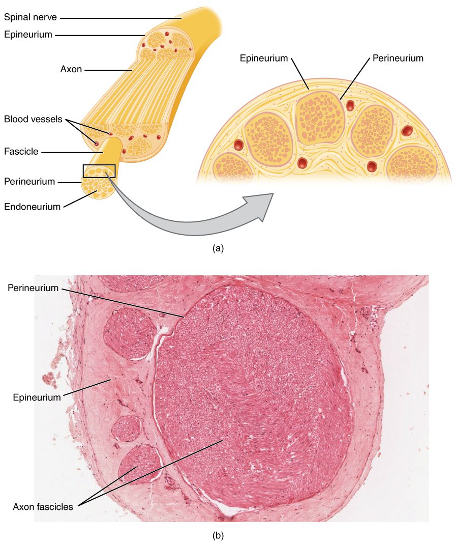
Nerves
Nerves are bundles of fascicles, which are further composed of individual nerve fibers (axons). It is important to realize that since nerves contain numerous fibers some of these fibers can carry sensory information while others carry motor information. Therefore one nerve can carry both sensory and motor information. This type of nerve is known as a mixed nerve.
The outer layer of a nerve consists of the epineurium. The epineurium consists of dense connective tissue that surrounds and protects the nerve. Inside the nerve the fibers are bundled in fascicles with each fascicle surrounded by a sheath called a perineurium. Inside the fascicles are bundles of neurons each surrounded by a thin layer of loose connective tissue called the endoneurium.
Nerve Plexus
Spinal nerves can combine to form structures called plexi. There are four major plexi in the human body. The cervical plexus (C1-C4) innervates the posterior head and skin of the neck. The phrenic nerve (C3-4-5) emerges from the cervical and brachial plexi and runs through the thorax to innervate the diaphragm.
The brachial plexus (C5-T1) consists of the ventral rami from spinal nerves C5-T1 and form the axillary, radial, musculocutaneous, ulnar and median nerves.
The lumbar plexus consists of spinal nerves L1-L4. The sacral plexus consists of the ventral rami from spinal nerves L4-S4. Sometimes both plexi are referred to as the lumbosacral plexus. The major nerves exiting the lumbosacral plexus include the obturator, femoral, and sciatic.
Nervous System Cells
The 2 types of cells in the nervous system include neurons and neuroglia.
Neurons are the cells that transmit and store information in the nervous system and neuroglia are support cells.
Some examples of neuroglia include the star-shaped astrocytes that help to regulate electrolytes, oligodendrocytes that produce myelin, empendymal cells that produce cerebrospinal fluid and microglia that clean up debris.
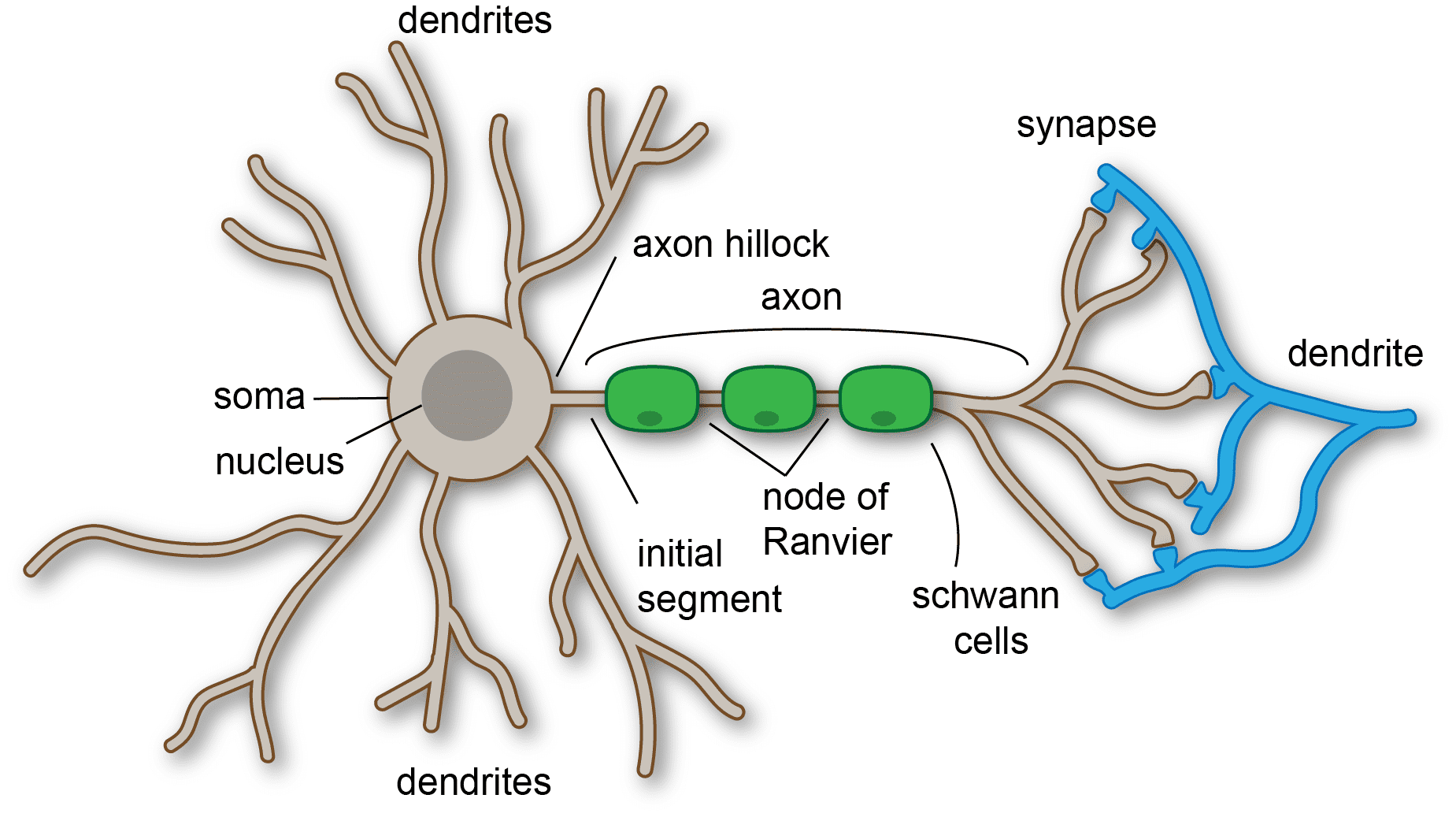
The parts of a neuron include the dendrites, cell body, axon hillock, axon, and axon terminal.
Neurons can connect and communicate with other neurons. The connection between neurons is called a synapse and the space between neurons is called the synaptic cleft.
Neurons communicate with each other by sending chemical packets called neurotransmitters across the synaptic cleft. So, the presynaptic neuron sends a neurotransmitter across the synaptic cleft to the post-synaptic neuron.
There are 2 essential messages that neurons send. One is an excitatory message that stimulates the post-synaptic neuron and the other is an inhibitory message that keeps the post synaptic neuron from sending additional messages.

The Neuron Action Potential
Neurons, like many other cells in the body, do not exist at equilibrium with their surroundings. In fact there is a net negative charge on the inside of the neuron with respect to the outside. This negative charge exists mostly because of differences in membrane permeability to different electrolytes. The cell membranes of neurons are slightly permeable to sodium and potassium. Although they are permeable to both sodium and potassium they are slightly more permeable to potassium.
There are also a number of negatively charged ions inside of the neuron’s cell membrane. These include phosphates, sulfates, ATP, RNA and proteins. These negatively charged ions cannot leave the cell. So if potassium (which is positively charged) is allowed to move out of the cell, then the inside of the cell becomes more negative (due to the presence of the negative ions) than the outside of the cell.
As this ionic gradient increases some positive ions are attracted back into the cell. Eventually the cell reaches a steady state by which potassium diffuses out of the cell at the same rate that it moves into the cell via the ionic gradient.
There is much more sodium outside of the cell than inside. The neuron’s cell membrane is not very permeable to sodium so just a little sodium moves into the cell via its concentration gradient. We also have the sodium-potassium pump working to maintain both sodium and potassium gradients by moving sodium out of the cell and potassium into the cell.
Remember that the sodium-potassium pump requires energy in the form of ATP. The nervous system has a lot of these pumps in order to function. In fact about 70% of the energy used by the nervous system is used by the sodium-potassium pumps.
So, if we put all of these effects together we end up with a net negative charge on the inside of the cell with respect to the outside. This negative charge is approximately -70 millivolts (mV) and is called resting membrane potential.
Depolarization
Neurons communicate by sending chemical messages from one neuron to another. These chemicals are called neurotransmitters. The neurotransmitters move from one neuron to another across an area known as the synaptic cleft. The neuron sending the message is called the presynaptic neuron. The neuron receiving the message is called the post-synaptic neuron.
Once the neurotransmitter travels across the synaptic cleft it attaches to a receptor on the post-synaptic neuron. There are two possible messages carried by neurotransmitters. One is to trigger the post-synaptic neuron to send another message. This essentially moves the information forward. The other possible message is to inhibit the post-synaptic neuron (hold the information back).
In order to trigger the post-synaptic neuron the neurotransmitter will cause the opening of sodium gates on the post-synaptic neuron. In other words the presynaptic neuron is said to be excitatory. When the sodium gates open sodium rushes into the neuron. This changes the potential by making it less negative due to the positive sodium ions rushing into the cell. We say the cell is depolarizing. Remember the resting membrane potential is negative (-70mV). In other words the cell is polarized to begin with. Once the sodium gates open causing the cell to become less negative there is less polarization. So the cell is depolarizing with the opening of sodium gates.
Threshold
If the stimulus is strong enough to cause enough of a change in potential to reach a certain level the neuron will react by opening more sodium gates and depolarizing at a rapid rate. In neurons the level is at about -55mV. In other words if a stimulus is great enough to cause a neuron to depolarize to -55 mV then we say that it has reached the threshold. Once the neuron reaches the threshold it will continue to depolarize to about +30 mV. The rapid change in potential from -55 mV to +30mV is called an action potential. This is called the all or none principle which means that once the threshold is reached the neuron continues through the cycle of depolarization and repolarization to resting membrane potential.
Action Potentials
Action potentials are generated at the axon hillock of neurons. There are a large number of sodium gates that react to changes in membrane potential. These sodium gates are called voltage-gated sodium channels because they open in response to a change in membrane potential. When a stimulus causes depolarization to the threshold the voltage-gated sodium channels open causing more voltage-gated channels to open resulting in a large influx of sodium into the cell. The action of sodium channels causing more sodium channels to open is a positive feedback system.
Voltage-gated potassium channels also open at the same time as the sodium channels. The potassium channels work more slowly than the sodium channels. The result is that some potassium diffuses out of the cell but much more sodium diffuses in.
After the maximum depolarization is reached at about +30mV-+40mV the sodium gates close and the potassium gates remain open allowing potassium to diffuse out of the cell. This causes the membrane potential to become more negative. This occurs until the resting membrane potential is reached.
Neurotransmitters
Neurotransmitters
Neurotransmitters are like tiny packets of messages that are sent from one neuron to another. All of the activity in the brain is governed by neurotransmitters.
It is important to know about neurotransmitters because some medications mimic or block these. Also, a number of neuromuscular diseases are caused by problems with neurotransmitters.
Let’s use a 2 neuron model to demonstrate how neurotransmitters work. The first neuron is called the pre-synaptic neuron and the second is the post synaptic neuron. The neurotransmitter moves across the synaptic cleft from the pre to the post synaptic neuron.
There are excitatory neurotransmitters that promote the opening of sodium channels on the post synaptic neuron and inhibitory neurotransmitters that promote the opening of potassium channels.
Excitatory neurotransmitters include epinephrine and norepinephrine (adrenaline and noradrenaline). Both are secreted by sympathetic neurons, so they cause the effects of the sympathetic nervous system on the body.
Epinephrine and norepinephrine are considered adrenergic-like adrenaline so they connect to adrenergic receptors on other neurons.
Dopamine is generally considered excitatory, but it can be inhibitory depending on the neurons. Dopamine plays a role in pleasure and motivation. Drugs like methamphetamine attach to dopamine receptors to simulate the effects of dopamine. Cocaine blocks the proteins involved in recycling dopamine so the result is more dopamine in the synaptic cleft.
Acetylcholine is also an excitatory neurotransmitter that is secreted by parasympathetic neurons and motor neurons attaching to muscles to stimulate muscle contraction. Acetylcholine is considered cholinergic, so it will connect to cholinergic receptors on neurons.
Inhibitory neurotransmitters include Gamma amino butyric acid (GABA). that plays a role in anxiety and promotes a calming effect. GABA also helps to decrease blood pressure and plays a role in pain and sleep.
Another inhibitory neurotransmitter is Serotonin, which plays a role in memory, learning and depression.
One important type of breakdown of neurotransmitters is called reuptake. Serotonin is broken down and recycled by reuptake. Serotonin is secreted by the pre-synaptic neuron then moves to the post-synaptic neuron to attach to receptors. The serotonin in the synaptic cleft will re-enter the pre-synaptic neuron through a transport protein. Once inside, it is broken down by monoamine oxidase.
Drugs such as selective serotonin reuptake inhibitors or SSRIs, block the transport protein causing more serotonin to remain in the cleft.
Reflexes
Reflexes
Reflexes are involuntary responses to stimuli that occur unconsciously. The deep tendon reflex consists of a muscle, nerve pathway and the spinal cord. The muscle contains a sensory receptor that senses changes in stretch of the muscle. This receptor is called a muscle spindle. The muscle spindle contains motor neurons called gamma motor neurons. These neurons begin in the spinal cord and extend to the muscle spindle.
When the tendon of the muscle is tapped by a reflex hammer the muscle spindle senses the change in length of the muscle and sends a message via a sensory neuron (usually in a spinal nerve) to the spinal cord. There it synapses with a motor neuron (again in a spinal nerve) that sends a message to the muscle to contract.
Spinal reflexes are also influenced by central nervous system neurons. These neurons have an inhibitory effect on reflexes. Peripheral nervous system neurons extend from the spinal cord to the muscle. One reason for eliciting reflexes is to differentiate an upper motor neuron versus lower motor neuron problem.
If the nervous system is intact then the reflex will look normal. This means the brain is providing an inhibitory effect on the reflex. In other words the brain is inhibiting the reflex so it appears normal. The reflex will look exaggerated with damage to upper motor neurons. This occurs in stroke victims.
Diminished or absent reflexes will result from problems with lower motor neurons. In other words the pathway between the spinal cord and muscle is damaged so the signal cannot get through. This occurs in peripheral nerve problems such as spinal disc ruptures, spinal stenosis and demyelinating disorders.
The Babinski reflex is a good example of an exaggerated reflex produced by the removal of inhibition by the central nervous system. The Babinske reflex is present in newborns because myelination is not fully completed and the infant brain is not fully developed, so the normal inhibition is missing.
The presence of a Babinski reflex is normal in newborns, but is not in adults. The presence of a positive Babinski reflex in adults indicates central nervous system damage. We say the Babinski reflex is a pathological reflex since it indicates damage to the central nervous system.


Saltatory Conduction
Saltatory Conduction
Myelin is a lipid substance created by special cells called Schwann cells. The myelin wraps around the axon. Myelin is what makes white matter white. Gray matter axons don’t have myelin.
There are gaps in the myelin sheath called Nodes of Ranvier. The gaps are important because they contain a large number of voltage gated sodium channels.
Remember the action potential is generated at the axon hillock. This action potential moves down the axon and reaches a node of Ranvier. At the node, a large number of sodium channels open allowing sodium to rush into the axon and the axon depolarizes.
This has the effect of giving the action potential a boost, like a relay.
If you were to observe this phenomenon, it would look like the action potential jumps from node to node.
We call this saltatory conduction.
Saltatory conduction in myelinated axons is much faster than in unmyelinated axons, such as in gray matter.
White matter is faster than gray matter.
We can see this in the animation below. The neuron on the left is unmyelinated so the action potential travels much slower than the myelinated neuron on the right.
So, why is this important?
This is important because of demyelination. Some disease and disorders actually cause neurons to lose their myelin sheaths.
When myelin is lost, neurons can’t transmit as much information.
Some of these disorders include peripheral neuropathies like diabetic neuropathy, carpal tunnel syndrome and neuropathy of pregnancy.
The loss of myelin can cause loss of sensation, motor function and reflexes.
