Matrix Biology: Composition, Functional Properties, and Biological Roles of the Extracellular Matrix (ECM) in Health and Disease
Introduction
This section provides an overview of Matrix Biology, focusing on the Extracellular Matrix (ECM). The ECM is a complex and dynamic network of molecules secreted by cells that provides structural and biochemical support to surrounding cells and tissues. We will explore:
The diverse molecular composition of the ECM.
The structure and function of major ECM components including collagens, other structural proteins (elastin, laminins, fibronectin), proteoglycans, and glycosaminoglycans.
The regulatory roles of matricellular proteins and ECM-modifying enzymes.
The multifaceted functions of the ECM, from providing structural support to regulating cell behavior.
The importance of the ECM in tissue homeostasis and its involvement in various disease states, including genetic disorders and fibrosis.
Overview of Tissues and the Extracellular Matrix
Tissues and Cellular Environments: The human body is a complex organization of diverse organ systems, which are themselves intricate assemblies of different tissue types (e.g., epithelial, connective, muscle, nervous). Tissues are composed of specific cell types that synthesize and secrete a variety of molecules. These molecules assemble to form diverse cellular (micro)environments.
Extracellular Matrix (ECM): ECM molecules are key components of these cellular microenvironments.
The ECM is a highly specialized, dynamic, three-dimensional scaffold in which cells reside within tissues.
It is composed of a variety of fibrillar and non-fibrillar molecules.
The ECM reciprocally influences cellular function, which in turn modulates diverse fundamental aspects of cell biology in normal tissue homeostasis and in the progression of disease.
Essentially, cellular microenvironments (the interplay between cells and the ECM) determine and preserve normal function and influence an organ's response in disease.
ECM molecules play a central role in maintaining normal function and tissue homeostasis, and changes in the ECM often influence the development of disease.
Major Components of the Extracellular Matrix
The ECM is biochemically and structurally diverse, comprising several major classes of molecules:
1. Collagens
Abundance and Distribution: Collagens are the most abundant proteins in the ECM, with widespread distribution in virtually all tissues.
Function: They are crucial structural proteins that construct fibers, networks, and filaments within the ECM, providing tensile strength and structural integrity.
Structure:
Formed from polypeptide alpha-chains.
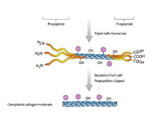
Three alpha-chains associate to form a trimeric triple helix (either homotrimeric, with three identical alpha-chains, or heterotrimeric, with different alpha-chains).
Typical collagenous domains within alpha-chains consist of repeating triplets of amino acids: Gly-X-Y, where X is often Proline and Y is often Hydroxyproline. Glycine, being the smallest amino acid, is essential for the tight packing of the triple helix.
Synthesis:
Individual alpha-chains are synthesized as procollagen molecules, which have N-terminal and C-terminal propeptides.
Hydroxylation of proline and lysine residues, and glycosylation, occur post-translationally.
Three procollagen alpha-chains assemble to form a procollagen triple helix within the cell.
After secretion from the cell, the propeptides are cleaved by specific proteinases to form a mature collagen molecule.
These mature collagen molecules then self-assemble into larger supramolecular structures like fibrils.
Diversity (Superfamily of 28 Types): There are 28 different types of collagen, encoded by at least 46 genes. They are categorized into several subclasses based on their structure and supramolecular assembly:
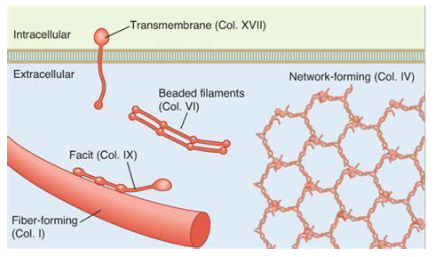
Fibrillar Collagens (most common): e.g., Type I, II, III, V, XI. These form long, cable-like fibrils with a characteristic banded pattern.
Heterotypic Fibrils: Fibrillar collagens often co-assemble into fibrils containing mixtures of different collagen types.
Dermis, tendon, bone: Primarily Type I, with Types III and V.
Cartilage, vitreous humor (eye): Primarily Type II, with Types IX and XI.
Network-forming Collagens: e.g., Type IV (major component of basement membranes), Type VIII, Type X. They form mesh-like networks.
Fibril-Associated Collagens with Interrupted Triple Helices (FACITs): e.g., Type IX, XII, XIV. They associate with the surface of fibrillar collagens and link them to other ECM components. Their triple helical domains are interrupted by non-collagenous domains.
Self-assembled forming Anchoring Filaments: e.g., Type VII (forms anchoring fibrils that link basement membranes to the underlying stroma).
Self-assembled forming Beaded Filaments: e.g., Type VI. Forms flexible, beaded filaments.
Membrane-Associated Collagens with Interrupted Triple Helices (MACITs): e.g., Type XIII, XVII (transmembrane collagens).
Multiplexins (Collagens with Multiple Triple Helix Domains and Interruptions): e.g., Type XV, XVIII.
2. Other ECM Structural Proteins
Elastin:
Provides elasticity and resilience to tissues, allowing them to stretch and recoil (e.g., in skin, blood vessels, lungs).
Synthesized as a soluble precursor, tropoelastin, which is composed of alternating hydrophobic domains (conferring elasticity) and cross-linking domains (rich in lysine).
Tropoelastin monomers undergo initial assembly into spherical nanoparticles (~200nm) and then larger tropoelastin droplets (~2-6µm), eventually forming mature coacervates.
These are then extensively cross-linked by lysyl oxidase enzymes, forming an insoluble, elastic fiber network. Desmosine and isodesmosine are characteristic cross-links in mature elastin.
The structure involves α-helical regions connected by β-spiral regions, allowing for extension and recoil.
Laminins:
Large, heterotrimeric glycoproteins that are major components of basement membranes.
Composed of α, β, and γ chains that assemble into a cross-like or T-shaped structure with globular domains.
Involved in cell-cell assembly, cell adhesion, migration, differentiation, and maintaining tissue architecture.
Specific regions interact with cell surface receptors (e.g., integrins, dystroglycan), other basement membrane components (e.g., collagen IV, nidogen, perlecan), and sulfated glycolipids.
Fibronectin:
A large, dimeric glycoprotein found in the ECM and plasma.
Functions as an adhesion molecule, binding to cell surface receptors (integrins) and other ECM components like collagen, fibrin, and heparin/heparan sulfate.
Plays roles in cell adhesion, migration, wound healing, and embryogenesis.
3. Proteoglycans (PGs) and Glycosaminoglycans (GAGs)
These are the second most abundant ECM molecules, characterized by their ability to bind large amounts of water, creating hydrated, gel-like matrices.
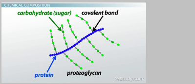
Basic Structure of Proteoglycans: Consist of a core protein covalently attached to one or more glycosaminoglycan (GAG) chains.
Glycosaminoglycans (GAGs):
Long, unbranched polysaccharide chains composed of repeating disaccharide units. One sugar is usually an amino sugar (N-acetylglucosamine or N-acetylgalactosamine), and the other is usually an uronic acid (glucuronic acid or iduronic acid).
Highly negatively charged due to sulfate and carboxyl groups (except hyaluronan which is non-sulfated). This attracts cations (like Na+) and water, leading to high hydration and swelling pressure, which resists compression.
Four main types of GAGs attached to protein cores in proteoglycans:
Chondroitin Sulphate (CS): Repeating units of glucuronic acid and N-acetylgalactosamine (often sulfated).
Dermatan Sulphate (DS): Similar to chondroitin sulphate but contains iduronic acid in place of some glucuronic acid residues.
Heparan Sulphate (HS): Repeating units of glucuronic acid or iduronic acid and N-acetylglucosamine (with variable sulfation). Heparin is a highly sulfated form of heparan sulphate, primarily found intracellularly in mast cells.
Keratan Sulphate (KS): Repeating units of galactose and N-acetylglucosamine (sulfated).
Hyaluronan (Hyaluronic Acid, HA):
Unique among GAGs: it is non-sulfated and not covalently attached to a protein core to form a proteoglycan (though it can associate non-covalently with proteoglycans like aggrecan).
Synthesized directly at the plasma membrane by hyaluronan synthase (HAS) enzymes, which extrude the growing chain into the extracellular space. Precursors are UDP-glucuronic acid (GlcA) and UDP-N-acetylglucosamine (GlcNAc).
A very large molecule, forms viscous solutions.
Found in synovial fluid (lubrication), vitreous humor, skin, and cartilage, where it contributes to hydration and tissue turgor.
Major Families of Proteoglycans:
Hyalectans (Lectin-containing PGs): Large aggregating proteoglycans found in the ECM, e.g., aggrecan (cartilage), versican (blood vessels, skin), neurocan, brevican (nervous system). They typically bind to hyaluronan.
Small Leucine-Rich Proteoglycans (SLRPs): e.g., decorin, biglycan, lumican, fibromodulin. Involved in collagen fibrillogenesis, growth factor binding, and cell signaling.
Pericellular Matrix PGs (Basement Membrane PGs): e.g., perlecan, agrin, collagen XVIII. Large multi-domain molecules that are key components of basement membranes.
Cell Surface PGs:
Syndecans: Transmembrane proteoglycans with HS (and sometimes CS) chains. Act as co-receptors for growth factors and modulate cell adhesion and signaling.
Glypicans: Attached to the cell surface via a glycosylphosphatidylinositol (GPI) anchor. Primarily carry HS chains.
Intracellular PGs: e.g., Serglycin. Found in secretory granules of hematopoietic cells, where its GAG chains (often heparin or highly sulfated CS) help package and store proteases and other granule contents.
4. Matricellular Proteins (Glycoproteins)
Definition: A large and heterogeneous family of non-structural (glyco)proteins in the ECM that modulate cell function by interacting with:
Cell-surface receptors (e.g., integrins)
Proteinases
Hormones and growth factors
Structural matrix proteins (e.g., collagens)
Other bioactive molecules.
They do not primarily serve a structural role but rather regulate cell activities.
Examples:
Thrombospondins (e.g., TSP-1, TSP-2): Involved in angiogenesis, cell adhesion, and growth factor activation.
SPARC (Secreted Protein Acidic and Rich in Cysteine; also known as osteonectin or BM-40): Modulates cell adhesion, proliferation, and ECM deposition.
Tenascins (e.g., Tenascin-C, Tenascin-X): Large, modular proteins involved in cell adhesion (often anti-adhesive), migration, and tissue remodeling.
Fibulins: A family of calcium-binding glycoproteins that associate with elastic fibers and basement membranes.
Osteopontin: Involved in bone mineralization, cell adhesion, and immune responses.
Cartilage Oligomeric Matrix Protein (COMP): Found in cartilage, tendons, and ligaments; involved in ECM organization.
CCN family proteins (e.g., CYR61, CTGF, NOV): Regulate cell proliferation, adhesion, migration, and angiogenesis.
Regulatory Role: Matricellular proteins play essential regulatory roles and are typically induced during:
Tissue remodeling (e.g., development, wound healing)
Tissue repair
Many disease states (e.g., inflammation, fibrosis, cancer).
They influence a wide range of cellular processes, including cell adhesion, migration, growth, and differentiation.
5. Enzymes: Components and Modifiers of the ECM
The ECM is not static; it is constantly being remodeled by various enzymes.
Matrix Metalloproteinases (MMPs): A large family of zinc-dependent endopeptidases that collectively can degrade all ECM components. They are crucial for tissue remodeling, wound healing, and angiogenesis, but their dysregulation is implicated in many diseases (e.g., arthritis, cancer metastasis).
ADAM (A Disintegrin And Metalloproteinase) and ADAMTS (ADAM with Thrombospondin motifs) Proteinases: Membrane-anchored or secreted proteinases involved in cleaving cell surface proteins (shedding) and processing ECM components (e.g., aggrecan degradation by ADAMTSs).
Serine Proteases: e.g., plasmin, elastase. Also contribute to ECM degradation.
Cathepsins: Lysosomal proteases that can also function extracellularly to degrade ECM.
HA-degrading enzymes (Hyaluronidases): Degrade hyaluronan.
Heparanases: Cleave heparan sulphate chains, releasing growth factors and affecting cell signaling.
Functions of the Extracellular Matrix
The ECM performs a multitude of critical functions:
Structural Support:
The ECM provides mechanical support and maintains the physical form of tissues and organs.
The biomechanical properties required of a tissue (e.g., tensile strength, elasticity, compressive resistance) are determined by the specific composition and organization of its ECM molecules.
Examples:
Tissues requiring high tensile strength (resistance to pulling forces) are enriched in fibrillar collagens (e.g., tendons, ligaments, bone, dermis).
Tissues requiring compressive properties are enriched in proteoglycans (especially aggrecan) and hyaluronan, which create a hydrated, resilient gel (e.g., cartilage, intervertebral discs, connective tissue of the placenta).
Tissues requiring elastic properties and recoil are enriched in elastin (e.g., skin, walls of large arteries, ligaments, lungs).
Regulation of Cell Behaviour and Tissue Homeostasis:
The ECM is not just an inert scaffold but actively influences cell behavior and tissue function.
It supplies cells with appropriate chemical and mechanical signals that regulate and maintain tissue homeostasis.
Matrix-stored chemical signals: The ECM can sequester and release growth factors and cytokines, modulating their availability and activity.
The ECM plays a role in:
Cell proliferation
Cell adhesion and migration (e.g., via integrin binding to fibronectin, laminin)
Cell differentiation
Inflammation
Angiogenesis (formation of new blood vessels)
Wound repair
Bone turnover
Cell death (apoptosis).
ECM in Disease
Changes in ECM composition, structure, or turnover are hallmarks of many diseases. The study of genetic disorders affecting ECM components has been central to understanding the ECM's role in health and disease.
Genetic Disorders Affecting ECM
Ehlers-Danlos Syndromes (EDS):
A group of inherited connective tissue disorders characterized by joint hypermobility, skin hyperextensibility, and tissue fragility.
Caused by mutations in genes encoding collagens (e.g., COL5A1, COL3A1) or enzymes involved in collagen processing (e.g., lysyl hydroxylase, ADAMTS2).
Some forms involve mutations in MMP genes that regulate collagen organization.
Hallmarks include elastic skin, hyperflexible joints, and in some severe types, valvular heart disease and arterial rupture.
Disorders of Ageing and ECM Degeneration
Intervertebral Disc (IVD) Disease:
A common disorder of ageing that impacts mobility and can lead to decreased workplace productivity, affecting nearly everyone at some point.
Hallmarks: Attributed primarily to the loss of glycosaminoglycans, particularly the proteoglycan aggrecan, from the nucleus pulposus of the disc. This leads to:
Decreased hydration of the disc.
Reduced compressibility and ability to withstand loads.
Increased risk of disc herniation and chronic back pain.
Pathological Changes in GAGs in IVD Disease:
Fewer and shorter chondroitin sulphate (CS) chains on aggrecan and versican, leading to decreased compressive resistance.
Can involve neovascularization and nerve ingrowth into the disc.
Potential for increased Ca2+ mineralization.
Aberrant synthesis of heparan sulphate proteoglycans (HSPGs) can alter growth factor and morphogen signaling, affecting nucleus pulposus cell phenotype.
Fewer and shorter keratan sulphate (KS) chains on aggrecan and fibromodulin can lead to abnormal collagen cross-linking and fibril formation.
Changes in hyaluronan can impair its aggregation with aggrecan and versican, and affect IVD development.
Cellular Changes in IVD Disease: Can include hypoxia, inflammation, and autophagy within disc cells.
Fibrosis: A Hallmark of End-Stage Organ Disease
Definition: Fibrosis is the damaging process of excessive formation and accumulation of connective tissue (scarring) in an organ or tissue in response to chronic injury or inflammation.
Pathogenesis:
Initiation: Chronic injury to epithelial and/or endothelial cells (from infections, toxins, autoimmune reactions, genetic disorders, etc.) leads to cell death and activation of innate immunity cells (e.g., macrophages, NK cells, PMNLs) and coagulation pathways (platelet activation, thrombin).
Inflammation and Mediator Release: Injured cells and activated inflammatory cells release growth factors (GFs like TGF-β, PDGF), cytokines, chemokines, reactive oxygen species (ROS), and other mediators.
Myofibroblast Activation: Persistent injury and these mediators lead to the activation and proliferation of myofibroblast precursor cells (which can originate from resident fibroblasts, epithelial/endothelial cells via EMT/EndoMT, or bone marrow-derived cells).
Excess ECM Deposition: Activated myofibroblasts are highly contractile cells that secrete large amounts of ECM components, particularly fibrillar collagens, leading to the formation of a dense, stiff, fibrotic matrix. This process becomes uncontrolled.
ECM in Fibrosis: The fibrotic ECM is qualitatively and quantitatively different from normal ECM:
It is highly crosslinked (less degradable, stiffer).
It has an altered composition of specific components.
It can store and release fibrotic growth factors, perpetuating the process.
Consequences: The excessive ECM deposition disrupts normal tissue architecture and function, leading to organ failure.
Common Diseases Exhibiting Fibrosis:
Pulmonary fibrosis (lungs)
Hepatic fibrosis/cirrhosis (liver)
Renal scarring/fibrosis (kidneys)
Cardiac fibrosis
Summary and Future Directions
Changes in the composition of extracellular matrix molecules within cellular (micro)environments elicit changes in cell metabolism and behavior.
Abnormal cell metabolism involving extracellular matrix components leads to changes in a tissue/organ that can affect its function (structural or cellular).
These ECM changes are significant contributors to the development and progression of many diseases.
Understanding the role of the extracellular matrix in health and disease offers potential for:
Discovering matrix-related abnormalities that cause disease.
Developing novel diagnostic tools (biomarkers of ECM turnover or damage).
Designing novel therapeutics, including approaches like tissue engineering that aim to regenerate or repair tissues by manipulating cells and ECM components.
Further Reading
The extracellular matrix (ECM) is a dynamic 3D network of interconnected macromolecules that provides structural support and regulates cellular functions in health and disease (Karamanos, 2019; Karamanos et al., 2021). Composed of proteins, glycoproteins, proteoglycans, and glycosaminoglycans, the ECM influences cell signaling, adhesion, migration, and differentiation (Theocharis et al., 2016; Naba et al., 2015). ECM composition and structure vary among tissues and undergo constant remodeling, with dysregulation associated with various pathologies, including cancer and fibrosis (Theocharis et al., 2019; Dzobo & Dandara, 2023). The ECM's mechanical and biochemical properties play crucial roles in tissue homeostasis and disease progression, with cells sensing these cues through receptors like integrins (Hynes & Yamada, 2012; Freedman et al., 2015). Recent advances in high-throughput technologies have accelerated ECM research, leading to the identification of potential diagnostic markers and therapeutic targets (Naba et al., 2015; Karamanos, 2019).