Neuro Anatomy 5 (Somatic Motor)
Organizational Structures
Primary motor cortex (precentral gyrus) Homunculus:
Determined by: number of motor units. Thus, areas that have more motor units take up a larger portion of the processing power of the precentral gyrus.
EX: Areas with more motor units, such as the hands, tongue, and face take a massive work from the precentral gyrus (resulting in more precise movements), while areas with less motor units, such as the glutes or back take less work from the precentral gyrus (resulting in less precise movements), but stronger
Upper Motor Neurons: Located in the CNS.
Lower Motor Neurons: Synapse with upper motor neurons in the spinal cord in order to deliver motor information to skeletal muscles.
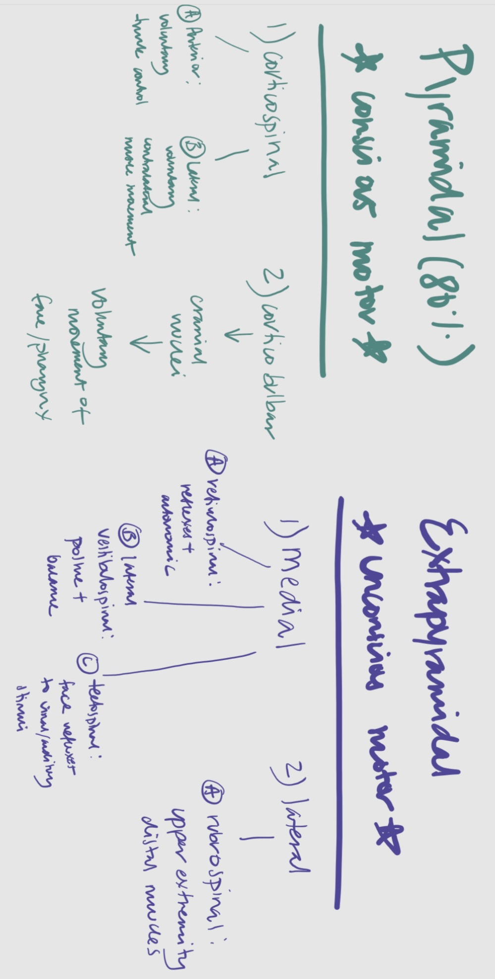
The Pyramidal System
These are tracts that travel through the “pyramids” of the medulla (90% of the motor nerve fibers) and prefrontal gyrus (motor)
Functions as a two-neuron chain responsible for precise, voluntary movements.
Components:
1) Corticospinal Tract: direction is from brain —> spinal cord —> muscles to control discrete voluntary skilled/precise movements.
Primarily composed of axons from the motor cortex.
Signals cross in the medulla, divided into:
a) Lateral Corticospinal Tract: Controls gross motor functions of contralateral limbs (however, injury would result in ipsilateral loss). This is 80-90% of the corticospinal tract composition (compared to anterior corticospinal part)
b) Anterior Corticospinal Tract: Conducts voluntary motor impulses from the precentral gyrus to motor centers of the spinal cord, mainly for trunk control.
2) Corticobulbar Tract: Provides conscious control over skeletal muscles of the eye, jaw, face, neck, and pharynx. Information is carried to motor neurons of cranial nerve nuclei rather than the spinal cord.
Pyramidal Signal Pathway
1) Upper motor neurons from the motor area in the cerebral cortex descend through the internal capsule
2) Crosses at the pyramids of the medulla.
3) They continue through either a) lateral or b) anterior corticospinal tract (in white matter “highway”) to the anterior horn of the gray matter in the spinal cord, synapsing with lower motor neurons.
4) Lower motor neurons exit via the anterior root (no ganglion) of the spinal nerve to innervate skeletal muscles or travel through the corticobulbar tract to cranial nerve nuclei.
Extrapyramidal system
Responsible for involuntary reflexes and modulation of movements (i.e., coordination). Aka, subconscious processing.
It indirectly controls the ventral horn cells
Chiefly found in the RAS of the pons and the medulla (specifically basal ganglia and substantia nigra)
Breaks down into:
1) Medial Pathways: Controls trunk/proximal limbs, specifically:
Reticulospinal Tract: Reflex activity + autonomic function
Lateral Vestibulospinal Tract: Posture + balance.
Tectospinal Tract: Modulates involuntary head and neck movements in response to visual/auditory stimuli.
2) Lateral Pathways:
Ruborospinal Tract: Controls muscle tone and movements of distal muscles of the upper limbs.
Abnormalities
Damage to the upper motor neurons results in hypertonia and spastic paralysis
Damage to the lower motor neurons results in hypotonia and flaccid paralysis
Damage to the pyramidal pathway results in spasticity (looks like hypertonia, loss of precise/fine movements)
Damage to the extrapyramidal pathway results in rigidity (looks like involuntary movements, such as tremors; think of Parkinsons)
Higher Centers
The headquarters is located at the cerebral cortex. Motor commands can be issued in the absence of a sensory stimulus.
Basal Nuclei: Modify voluntary and reflexive motor patterns at the subconscious level.
Hypothalamus: Controls stereotyped patterns related to eating, drinking, sexual activity; modifies respiratory reflexes.
Cerebral Cortex: Plans and initiates voluntary motor activity.
Thalamus and Mesencephalon: Control reflexes in response to visual and auditory stimuli.
Cerebellum: Coordinates complex motor patterns.
Pons and Superior Medulla Oblongata: Control balance and complex respiratory reflexes.
Brain Stem and Spinal Cord: Manage simple cranial and spinal reflexes.
Inferior Medulla Oblongata: Regulates basic respiratory reflexes.