Anatomy: Bone Tissue
Anatomy: Bone Tissue
Cells of Bone tissue
Osteogenic Cell 🡪 Osteoblast 🡪 Osteocyte
Supportive Connective Tissue
Extracellular Matrix
- 25% Water
- 25% Protein or organic matrix
- 95% Collagen fibers
- 5% Chondroitin sulfate
- 50% crystalized mineral salts
- Hydroxyapatite (calcium phosphate)
- Other substances (lead, gold, strontium, plutonium, etc.)
Types of Bone (2)
- Compact Bone
- Spongy Bone
Compact Bone
- Arranged in units called osteons or haversian systems.
- Osteons contain blood vessels, lymphatic vessels, nerves.
- What surrounds the Osteon canal: concentric rings of osteocytes along with the calcified matrix.
- Osterons are aligned in the same direction along lines of stress.
- These lines can slowly change as the stresses on the bone changes
Histology of Compact Bone
- Osteon: concentric rings (lamellae) of calcified matrix surrounding a vertically oriented blood vessel.
- Osteocytes are found in spaces called lacunae
- Osteocytes communicate thru canaliculi fixed with extracellular fluid that connect one cell to the next cell.
- Interstitial lamellae represent older osteons that have been partially removed during tissue remodeling.
The Trabeculae of spongy bone
- Latticework of thin plates of bone called trabeculae oriented along lines of stress.
- Spaces in between the trabeculae are filled with red marrow where blood cells develop.
- Spongy bone is found in ends of long bones and inside flat bones such as the hipbones, sternum, sides of skull, and ribs.
Spongy Bone
- Also known as “cancellous bone”.
- Does not contain osteons but consists of trabeculae surrounding many red marrow filled spaces.
- Forms most of the structure of short, flat, and irregular bones, and the epiphyses of long bones.
- Spongy bone tissue are light and supports/protects the red bone marrow.

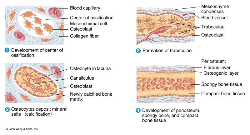
Bone Formation
- All embryonic connective tissue begins as mesenchyme.
- Also known as “Osteogenesis” or “ossification”.
- Begins when mesenchymal cells provide the template for subsequent ossification.
- Two types of Ossification:
- Intramembranous Ossification: formation of bone directly from or within fibrous connective tissue membranes.
- Endochondrial ossification: formation of bone from hyaline cartilage models.
Intramembranous Ossication (in depth)
- Also called “dermal ossification” because it normally occurs in the deeper layers of connective tissue of the dermis of the skin.
- Roofing bones of the skull:
- Frontal bones
- Parietal bones
- Occipital bone
- Temporal bones
- Mandible
- Clavicle
Endochondral ossification
- Developing bones are deposited as a hyaline cartilage model and then this cartilage is replaced by bone tissue
- All bones except roofing bones in the skull, mandible, and clavicle.
Growth at Epiphyseal plates
- Zones of resting cartilage
- Anchors growth plate to bone
- Zone of proliferating cartilage
- Rapid cell division (stacked coins)
- Zone of hypertrophic cartilage
- Cells enlarged and remail in columns
- Zone of calcified cartilage
- Thin zone, cells mostly dead since matrix calcified.
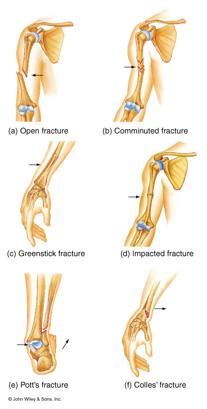
Bone Fractures
- Closed/open
- Partial/complete
- Displaced/nondisplaced
- Simple/compound
Subluxation: an incomplete or partial dislocation of a joint or organ.
Luxation: a complete dislocation of a joint or organ
Steps in Facture Repair
- Formation of a fracture hematoma
- Fibrocartilaginous callus formation
- Bony callus formation
- Bone remodeling
Fibrous Joints: Sutures
- Allow for brain growth
- Allow for passage through the birth canal
Symphyses
- Fibrocartilage unites bone
Bone Disorders
Arthritis:
- DJD: Degenerative joint disease
Inflammatory Joint Disease
- Rheumatoid Arthritis
- May be caused by transient infection that results in autoimmune attacks against collagen in the bones at joints.
- Swain Neck deformity.
Infectious Arthitis
- Lyme Disease
- Bull’s Eye rash
- Gonorrhea
- Sometimes no symptoms
Scoliosis: Abnormal curve of the spine.
Acromegaly
- Body produces too much Growth Hormone
- Only in Adults
- Tissues grow larger than normal
- Excessive growth can cause serious disease and even premature death.
Gout
- Results from overload of Uric Acid in the body
- Leads to the formation of urate crystals that deposit in the joints
- Causes recurring attacks of joint inflammation (arthritis)
- May cause joint destruction, decreased kidney function, and kidney stones.
Spina Bifida
- Birth defect
- Incomplete development of the spinal cord or its coverings
- Prevent with folic acid
- Spina bifida means “split” or “open” spine
- Is usually detected before a baby is born and treated right away.
Myeloma: Cancer in which abnormal cells collect in the bone marrow and form tumors.
- Starts in the bone marrow where blood cells are
- Bone marrow starts to overproduce abnormal white blood cells
- Leukemia cells don’t do the work of normal WBC’s and they don’t stop growing when they should.