Topic 3A
Overview of Cells
Cells are the fundamental life unit, essential for understanding health and disease.
Structure and function are interrelated:
Biochemical functions depend on cell shape and subcellular structures.
Importance of cell membranes for physiological processes:
Permeability is crucial in treatments and understanding overall cell function.
Cell Abundance and Diversity
Human body comprises trillions of cells (over 200 distinct cell types).
Variances in size, shape, and subcellular components dictate different functions.
Common Structures of Human Cells
Plasma Membrane:
Flexible outer boundary regulating substance entry and exit.
Cytoplasm/Cytosol:
Intracellular fluid and organelles.
Nucleus:
Control center housing DNA.
Extracellular Materials
Substances located outside cells, categorized into:
Extracellular fluids: e.g. Blood plasma, cerebrospinal fluid (CSF).
Cellular secretions: e.g. saliva, mucus.
Extracellular matrix: Holds cells together, functioning like glue.
Plasma Membrane Functionality
Acts as a dynamic barrier segregating intracellular from extracellular fluids.
Comprised of:
Membrane lipids (mainly phospholipids) form a lipid bilayer.
Critical role in cellular function through selective permeability.
Plasma Membrane Structure
Hydrophilic polar headgroup attached to two hydrophobic non-polar fatty acid tails
Fluid mosaic model:
Proteins float within a fluid lipid bilayer, constantly changing configurations.
Glycocalyx:
Coating of sugars that facilitates cell identification.
Glyco means attached to sugars
eg Glycolipid: Sugar joined to a lipid
Composition of Membrane Lipids
Lipid bilayer structure:
Phospholipids: Amphiopathic molecules, with hydrophilic heads and hydrophobic tails.
Phospholipids are modified triglycerides
Key to membrane’s selective permeability and structure.
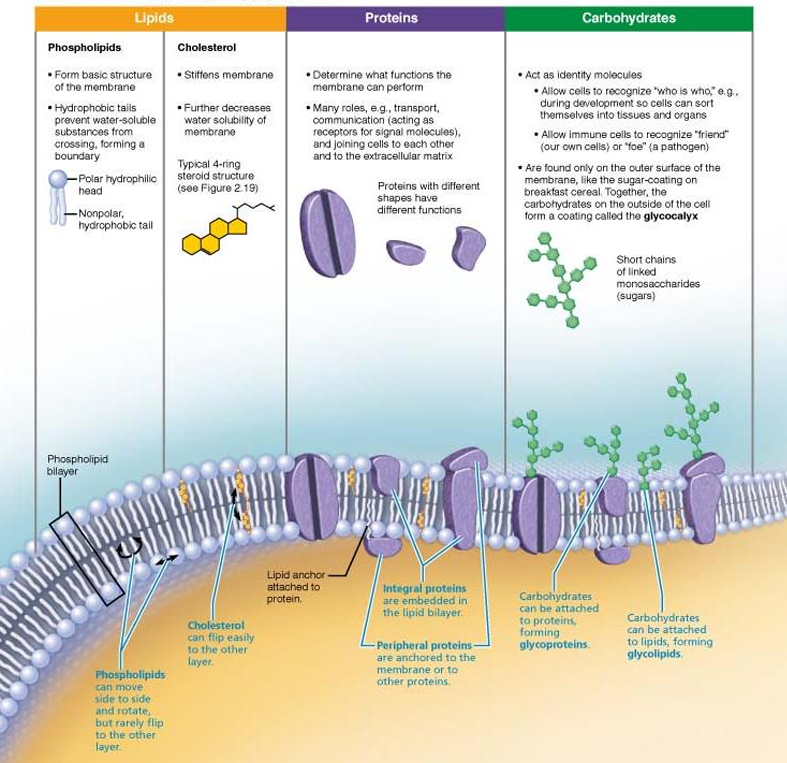
Cholesterol
Increases membrane stability
Glycolipids
Lipids with sugar groups on the outer membrane surface
Membrane proteins
Integral or peripheral proteins
Membrane proteins
Integral or peripheral proteins
Glycocalyx
Consists of sugars (carbohydrates) sticking out of cell surface (extracellular)
Attached to proteins (glycoproteins) and lipids (glycolipids)
Every cell has different patterns of this sugar-coating
Functions as specific biological markers for cell-to-cell recognition
Membrane Construction
Integral Proteins:
Firmly inserted into the membrane with most being transmembrane proteins meaning parts of the proteins are on both sides of the membrane
All transmembrane proteins are integral but not all integral proteins are transmembrane
Has both hydrophobic and hydrophilic regions
Hydrophobic in the cell membrane
Hydrophilic outside the cell membrane
Involved in transport and signalling processes.
Peripheral Proteins:
Not fully embedded in the membrane
Function as
enzymes
Include filaments on the intracellular surface used for plasma membrane support
Motor proteins for shape change during cell division
Cell-to-cell connections for signalling
Membrane Proteins Functions
Transport:
Create hydrophilic channels or act as carriers.
ATP is used as an en energy source to actively pump substances across the membrane
Receptors:
Membrane proteins exposed to the outside of the cell may have receptors that fit specific chemical messengers known as ligands
Ligands can include hormones, neurotransmitters, growth factors, etc
When bound, chemical messengers cause a shape change in the protein that initiates a chemical reaction
Enzymatic Activity:
Catalyze metabolic reactions.
Cell Recognition:
Glycoproteins serve as identification tags for cell recognition.
Cell-to-Cell Connections:
Link adjacent cells through various junctions.
Cytoskeleton Attachment:
Maintain cell shape and assist in movement.
Fixes the location of certain membrane proteins
Cell Junctions Types
Most cells are bound together to form tissues and organs
Some cells such as red blood cells and lymphocytes are free
Tight Junctions:
Integral proteins fuse to from an impermeable junction that prevents molecules and fluid from passing through adjacent cells.
Encircle the whole cell
On the apical surface
Visualized as a ziplock
Desmosomes:
Formed when linker proteins called cadherins of adjacent cells interact
Anchoring junctions linking cells through linker proteins
Linker proteins are anchored through thickened plaques on the cytoplasmic surface.
Visualized as velcro
Gap Junctions:
Transmembrane proteins called connexions allow small molecules to pass between adjacent cells for communication.
Used to spread ions, simple sugars, or other molecules between cells
Allows electrical signals to be passed quickly from one cell to the next
Visualized as playground tubes
Membrane Transport Overview
Selective Permeability:
Some molecules pass easily while others do not, determined by transport processes.
Passive Transport: No energy required.
Active Transport: Requires energy (ATP).
Passive Membrane Transport
Diffusion Types:
Simple diffusion (natural)
Molecules move from high concentration to low concentration
Eg. smoke filling a room, dye in water
Concentration gradient: difference in concentration
Speed of diffusion depends on…
Size of molecule
Temperature
Concentration
Diffusion in cells
Plasma membranes stop the diffusion of most solutes
Small lipid-soluble solutes can freely pass via simple diffusion
Eg. O2, CO2, fatty acids, some steroid hormones
Facilitated diffusion (carrier and channel-mediated), osmosis.
Molecules are transported passively down the concentration gradient with the assistance
Carrier-mediated facilitated diffusion
Carriers: transmembrane integral proteins that transport large polar molecules such as sugars and amino acids
Substances bind to protein carriers in membrane
Binding of molecule causes carrier to change shape, moving the molecule
Carriers can become saturated, leading to a maximum rate of transport that cannot be exceeded, regardless of the concentration gradient.
Channel-mediated facilitated diffusion
Channel: aqueous-filled transmembrane protein
Transports small, lipid-insoluble molecules down a concentration gradient
Two types
Leakage channels (always open)
Gated channels (controlled by chemical or electrical signals)
Channel-mediated osmosis
Channels called aquaporins allow for large quantities of water to diffuse across membranes
Aqua = water, Porin = channel
Aquaporins are important in kidneys, blood cells, other tissues and they regulate balance
Filtration:
Movement across capillary walls relying on pressure gradients. (kidneys)
Osmosis
Osmolarity: Measure of total concentration of solute particles
Water moves by osmosis from low-solute (high water) to high-solute (=(low water) concentration regions.
Solutes move from high concentration to low concentration
Movement occurs until equilibrium is reached
Understanding tonicities:
Isotonic: a solution with the same solute concentration as another solution, resulting in no net movement of water across the cell membrane.
Hypertonic: a solution with a higher solute concentration than another solution, causing water to move out of the cell and leading to cell shrinkage.
Hypotonic: a solution with a lower solute concentration than another solution, resulting in water moving into the cell and potentially causing it to swell or even burst.
Movement of water causes pressures
Hydrostatic: pressure of water inside cell pushing on membrane
Osmotic: the tendency of water to move into cell by osmosis
Active Membrane Transport Mechanism
Active Transport Processes:
Use ATP, aiding transport of substances against concentration gradients.
For individual molecule transport
Require carrier proteins such as a uniporter, symporter, and antiporter
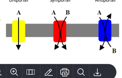
Types of Active Transport:
Primary
Required energy comes directly from ATP hydrolysis
Energy causes the transport protein shape
Shape change causes solutes (ions) bound to protein to be pumped across membrane
Example of pumps: Na+ -K+, hydrogen (proton) pumps
Sodium-potassium pump
An enzyme that pumps Na+ out of the cell and K+ into the cell
NOKIA
3 Na+ move outside, 2 K+ come inside
Secondary (cotransport)
Required energy is obtained indirectly from ionic gradients created by primary active transport
energy stored in gradients is used indirectly to drive transport of other solutes
Vesicular Transport
Transport of macromolecules or fluids in vesicles (bulk transport).
Endocytosis
Involves formation of protein-coated vesicles
Usually involve receptors; therefore, can be a selective process
Substance being pulled in must be able to bind to its unique receptor
Once vesicle is pulled inside cell, it may fuse with lysosome or undergo transcytosis
Types
Phagocytosis - “cell eating”
Pseudopods form and engulf particles
Formed vesicle is called a phagosome
Phagocytosis is used by macrophages and certain other white blood cells
Pinocytosis - “cell drinking”
Plasma membrane folds in, fluids enter cell
Used by some cells to sample environment
Nurtrient absorption in small intestine
Membrane components recycled back to membrane
Receptor-mediated - specific endocytosis and transcytosis
Receptors on plasma membrane to bind to specific molecules
Both receptor and attached molecules are internalized
Exocytosis:
Ejection of substances such as hormones, mucus, cellular water, and neurotransmitters from the cell, often in secretion processes.
The substance being ejected is enclosed in the secretory vesicle
The secretory vesicle contains the substance to be removed from the cell
The secretory vesicle is coated by a protein called v-snare which finds and hooks up to target t-snare proteins
Transcytosis: transport into, across, and then out of the cell
Vesicular trafficking: transport from one area or organelle in cell to another
Summary of Active Transport Processes
Descriptions and examples are provided, highlighting the importance of transport proteins and energy utilization in maintaining cellular functions.