Chapter 9
New Designs for Living
Zoologists recognize 32 major phyla of living multicellular animals
Survivors of about 100 phyla that appeared 600 million years ago during Cambrian explosion, the most important evolutionary event in geological history
All major body plans evolved with a few million years due to extensive selection and adaptation processes
Basic uniformity of all life derives from common ancestry and similar cellular construction
Hierarchical Organization of Animal Complexity
Five major grades of organization are recognized
Each grade is more complex than the previous grade
The five grades are
Protoplasmic
Cellular
Cell-tissue
Tissue-organ
Organ-system
Protoplasmic Grade of Organization
Life is organized from simple to complex
Each hierarchical grade is more complex than the preceding one
Five grades of organization of organismal complexity
Protoplasmic grade of organization
Unicellular groups are the simplest eukaryotic organisms
Perform all basic function of life within the confines of a single cell
Protoplasm contains organelles with specialized functions and diversity among groups is due to varying subcellular components and structures
Cellular Grade of Organization
Form metazoans - multicellular organisms such as volvox
Have greater structural complexity by combining cells into larger aggregates
Cells are specialized parts of the whole organism but cannot live alone
Cells demonstrate division of labor and perform specialized tasks earlier accomplished by subcellular components of unicellular organisms
Cell Tissue Grade of Organization
Cells grouped together into definite patterns or layers to perform a common function as a coordinated unit called tissue
Most cells can still be scattered all around the body
Animals such as jellyfish represent this group and are called eumetazoans
Due to the unique structure of sponges, some scientists still classify them at the cellular level rather than the cell-tissue level
Tissue Organ Grade of Organization
Aggregated tissue now assembled into larger functional units called organs
Organs can be composed of more than one kind of tissue and have specialized functions
The heart is surrounded by connective tissues
Represented by flatworms
Organ system grade of organization
Several organs work together to perform a common function for the survival of the animal
Considered the highest level of organization and associated with most complex animal phyla such as nemerteans, crabs, and chordates
Several organs work together to perform a common function for the survival of the animal
Considered the highest level of organization
Associated with most complex animal phyla such as annelids, arthropods, and chordates
Animal Body Plans
Animal body plans are different in
Grade of organization
Body symmetry
Symmetry is balance of proportions and the correspondence of size and shape of parts on opposite sides of a median plane
Number of embryonic layers
Number of body cavities
Types of animal symmetry
Spherical: ball shaped
Radial: tube- or vase-like
Bilateral: right and left sides
Spherical and Radial Symmetry
Spherical symmetry
Any plane passing through the center and divides the body into mirrored halves
Best suited for floating and rolling
Found in unicellular forms but rare in large animals
Radial symmetry
Body divided into similar halves by more than two planes passing through longitudinal axis
Found in sponges, jellyfishes, sea urchins, and related groups
End of tubular body forms called oral surface while the opposite end forms basal attachment disc called aboral surface
Biradial Symmetry
Variant form of radial symmetry
Have part that is single or paired rather than radial
Only two planes passing through the longitudinal axis that produces mirrored halves
Usually sessile, freely floating, or weakly swimming animals like ctenophores
No anterior or posterior end
Can interact with the environment in all directions
Organism divided along a saggital plane into two mirror portions forming right and left halves
Much better fitted for directional (forward) movement which is advantangeous to an animal moving through its environment head first
Associated with cephalization, which is the differentiation of a head region and the concentration of nervous tissues and sense organs in the front area
Also has mouth in front to allow for more efficient feeding detection of prey
Regions of Bilaterally Symmetric Animals
Anterior - head end; posterior - tail end
Dorsal - back (upper) side; ventral - front (belly) side
Medial - midline of body; lateral - right and left sides
Distal - parts farther from the middle of the body
Proximal - parts nearer the middle of body
Frontal plane (coronal plane) - divides body into dorsal and ventral halves
Sagittal plane - divides body to right and left
Transverse plane (cross section) - divides body into anterior and posterior halves
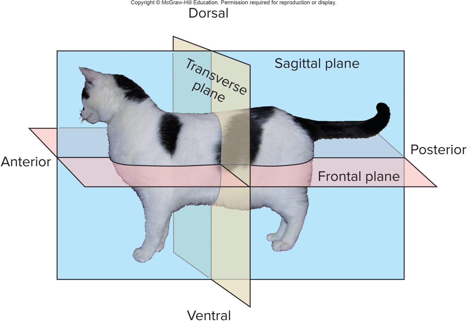
Body Cavities
Internal space represented by guy cavity and fluid-filled body coelom that cushions and protects internal organs
Body cavity is dependent on mesodermal pouch formation during gastrulation
Types of Body Cavities
Acoelomate: no body cavity
Pseudocoelomate: partial body cavity
Coelomate: true body cavity
Variations in Body Cavity
Sponges (cellular grade of organization) are acoelomate
After blastula formation, cells reorganize to form adult body but do not form gastrula
Cells grow and surround a chamber called spongocoel
Bastula has no external opening to gut cavity forms
Other animal phyla
Development proceeds from blastula to gastrula
Opening to the archenteron is the blastocoel; becomes the mouth or the anus
Embryo now has two cavities - gut and blastocoel
Inside gut is lined by endoderm, outer layer of cells is ectoderm, middle area lined with mesoderm
Protostome Mesoderm Formation
Mesoderm forms as endodermal cells near blastopore migrate into the bastocoel
Three body plans are possible
Acoelomate plan
Mesodermal cells completely fill the blastocoel so no body space is formed
Gut is only body cavity
Region between ectoderm and endoderm is filled with spongy mass of parenchyma cells that are from embryonic connective tissue and are important for transport and disposal of metabolic wastes
Pseudocoelomate
Mesodermal cells line with the outer edge of the blastocoel only partially lined with mesoderm
Pseudocoelom is a false body cavity
Two body cavities formed where persistent blastocoel forms pseudocoelom with a gut cavity
Schizocoelous coelomate plan
Mesodermal cell fill blastocoel and then splits to form a space called a coelom
A true body cavity that is complete lined by mesoderm
Two body cavities are formed: gut and coelom
Developmental Origins of Body Plans in Triploblastic
Triploblastic animals follow one of several major developmental pathways
Most common pathways are by spiral or radial cleavage
Radial cleavage
Typically accompanied by three traits
Blastopore becomes the anus and the new opening becomes the mouth
Coelom formation is by enterocele
Cleavage is regulative
Animals with these features are called deuterostomes
Protostome Cleavage Pathways
Spiral Cleavage
Produces embryos whose developmental pattern contrast with those of deuterostomes
Blastopore becomes the mouth
Cleavage is mosaic
May be acoelomate, pseudocoelomate, coelomate via schizocoely
Animals with these features are called lophotrochozoan protostomes
Ecdysozoan protostomes
Exhibit a range of cleavage patterns including spiral and superficial cleavage
Can be coelomate or pseudocoelomate
Blind and Complete Gut
A few diploblastic and triploblastic form (incomplete) gut cavity
Same opening for entrance for food and exit of wastes
Most common animal groups form a complete gut
Allows for one-way flow of food from mouth to anus
Tube-within-a-tube design is very adaptive to various types of food
Segmentation
Metamerism (segmentation) is a common feature of metazoans
Serial repetition of similar body segments along longitudinal axis of body
Each segment is a metamere or somite that contains internal and external structures of several vital organ systems
Segments can be seen during early development and also appear as superficial ectodermal and body wall features in adults
Permits greater body mobility and complexity of structure and function
Found in annelids, arthropods, and chordates
Components of Animal Body
Animal bodies consist of cellular components derived from the germ layers
Extracellular Components
Two categories of extracellular components
Body fluids that fill up two compartments
Intracellular space: within body cells
Extracellular space: outside of body cells like blood plasms and interstitial fluids
Extracellular structural elements
Support connective tissues, cartilage, and cuticle as storage depot and mechanical protection
Cellular Components
Derived from germ layers
Four types of tissues form during embryonic development
Epithelial tissue
Sheet of cells that covers an internal or external surface of the animal body
Internally, function as lining for all organs and ducts as well as all passageways
Allow the transport for secretions and various materials to all cells
Has modifications to produce lubricating mucus and specialized hormones and enzymes
Externally, used for protection
Epithelial cells are classified based on form and number of layers
Blood vessels never penetrate epithelial tissues, cells depend on diffusion
Simple Epithelia
Single layer of cells; occur in all metazoans
Classified based on shape and function
Squamosal: flattened cells forming continuous lining blood capillaries, lungs, and surfaces allowing diffusion and transport
Cuboidal: short box-like cells lining the ducts and tubules of kidneys, salivary glands, and other secretory systems
Columnar: taller than cuboidal with elongate nuclei and many small finger-like projections called microvilli for increased absorption along the intestines
Stratified Epithelia
Two or more cell layers and are mostly restricted to vertebrates
Adapted to withstand mechanical abrasion and distortion
Line the oral cavity, esophagus, anal canal, and skin that has high keratin proteins
Separated from underlying tissues by a basal layer that continues to divide and produce new cells, which are pushed toward the surface where they are sloughed off and replaced
Keratinized skin cells found in reptilian, bird, and mammalian skin are mostly stratified squamosal epithelium
These cells die, lost nuclei, and form scale-like layers that are waterproofed to protect the deeper layers of living cells
Transitional Epithelia
Type of stratified epithelia great for stretching
Found in urinary tract and bladder of vertebrates to allow enlargement while storing wastes
In relaxed state appears to have many cell layers
When stretched appears to have only a few layers of extremely flattened cells
Connective tissue
Widespread in body but contains relatively few cells, many fibers, and a ground substance or matrix that suspend fibers
Two types of connective tissue proper occur in vertebrates
Loose connective tissue
Also called areolar connective tissue
Serve as “packing material”
Anchors blood vessels, nerves, and organs
Consists of fibroblasts that synthesize fibers, ground substance, and wandering macrophages that destroy pathogens and damaged cells
Dense connective tissue
Characterized by densely packed fibers and little matrix
Forms tendons, ligaments, and fasciae arranged as sheets that surround skeletal muscles
Tendons have collagen fibers
Most abundant protein of great tensile strength found in all animals that require flexibility and resistance to stretching
Other specialized connective tissues include blood, lymph, adipose tissue, cartilage, and bone.
Cartilage and Bone
Semi - rigid tissue of firm matrix cells (chondrocytes) with collagen and elastin
Lacks blood supply so nutrients and wastes have to diffuse through the ground substance such that it heals very slowly after damage
Bone is strongest connective tissue may be calcified matrix (osteocytes) around collagen fibers
Blood vessels flow through tiny channels called canaliculi and large canals that allow for continuous repair called remodeling
Muscular tissue
Muscular tissue is the most abundant tissue in animals
Originates from mesoderm
Two types of muscle cells: straited and smooth
Straited - transversely striped muscle
Skeletal - long cylindrical fibers and multi-nucleated
Voluntary muscle in vertebrates; invertebrates can have stimulatory and inhibitory control
Cardiac - short, branched network, and uninucleate
Connected by intercalated discs
Involuntary muscle
Smooth muscle tissue
Smooth - non visceral muscle
Long, tapering, unbranched, one central nucleus, and involuntary control
Vertebrates and invertebrates have ultrastructural differences
Invertebrates commonly have them as body wall muscles and around ducts and sphincters
Vertebrates have them around blood vessels and surrounding internal organs like intestines and the uterus
Nervous tissue
Does not repair itself
Nervous tissue is specialized to receive stimuli and conduct impulses from one region to another
Two basic cell types
Neuron
Structural and functional unit of the nervous system
Parts of a neuron
Soma: nucleated body
Dendrites: Extensions from the some that receive signals from receptors
Axon: carries signal away from nerve body - also called nerve fiber - to other cells and effector organs
Myelin (insulating) sheath - increases speed of signals during transmission
Synapses - specialized nerve junction between each neuron or effector organ
Neuroglia
Non-nervous cells that insulate neurons and support nervous functions
All animals and their organ systems are made from these four basic tissue types
Histology is the study of types of tissues
Complexity and Body Size
More complex grades of metazoan organization permit and promote large body sizes
Problems of large body size
As body increases in size, there is less surface area compared to volume
Surface area increases as the square of body length and volume increases as the cube of body length
Therefore, large animals have inadequate surface area to provide respiration and nutrient flow to cells deep in the body
Solutions for Large Body Size
Solutions to the surface to volume issue of large body size
The folding and invagination of body surfaces to maximize surface area with minimal size
The flattening of body shapes to allow all cells to have internal spaces not far from the surface
Most large organisms use the development of internal transport mechanisms to move nutrients, wastes, and gases between cells and the external environment
Results in more complex and more specialized organ systems
Benefits of Large Body Size
Buffers against environmental fluctuations
Provides protection against predators and promotes offensive tactics
Even through large animals need more energy and oxygen, the cost of maintaining body temperature is less per gram of body weight than in small animals
Energy costs of moving a gram of body weight over a given distance is less for larger animals than for smaller animals
Therefore, ecological opportunities are different for larger animals compared to smaller ones and result to extensive adaptive diversification