9c
9.4 Involuntary Movement Through Reflexes
automatic patterned response to a stimulus
Stretch reflex
Withdrawal reflex
Crossed-extensor reflex
Reflex Arc
neural pathway for reflex
Components:
Sensory Receptor: Detects the stimulus.
Afferent Neuron: Transmits sensory information to the CNS.
Integration Center: Processes the information (located in CNS).
Efferent Neuron(s): Carries commands away from CNS.
Effector Organ: Muscle or gland that produces response.
Types of Reflexes
Cranial or Spinal Reflexes: Depending on their origin.
cranial takes longer
spinal is short
Somatic or Autonomic (Visceral) Reflexes: Classifies if they involve voluntary (somatic) or involuntary (autonomic) systems.
Innate or Conditioned Reflexes: Innate reflect reflexes that are genetically programmed, while conditioned are learned.
Monosynaptic or Polysynaptic Reflexes: Monosynaptic has one synapse, while polysynaptic involves multiple neurons.
Most monosynaptic reflexes are for activation
most of the multisynaptic reflexes are for inhibituion
Classification Recap: Every reflex can be identified within both categories above.
Muscle Spindle Stretch Reflex
Mechanism:
Stimulation of Receptors: Muscle spindle detects the stretch in the muscle.
Afferent Pathway: Afferent neuron transmits sensory information from the muscle to the spinal cord.
Integration: In the spinal cord, an interneuron may be involved.
Efferent Pathway: Efferent neurons send impulse back to the muscle causing contraction.
Reciprocal Inhibition: Inhibition of flexor muscles allows extensor muscles to contract.
MUSCLE REFLEXS KALTURA
Pupillary Light Reflex
Classification: It is a cranial reflex, which involves cranial nerves II (Optic) and III (Oculomotor).
Nature: It is an autonomic, innate, polysynaptic reflex.
Reflex Arc Composition: Includes photoreceptors → Afferent neurons (C II) → Midbrain nuclei → Efferent neurons (C III) → Iris for pupillary size adjustment.
9.5 Voluntary Motor Control
Overview: Involves the conscious control of movement from the inception of a movement idea to the execution through precise muscle activation and feedback.
Process Steps:
Formation of movement ideas.
Programming of motor commands.
Correct muscle activation during actions.
Feedback mechanisms ensuring smooth execution.
Innervation of Skeletal Muscle
Efferent Neuron Origin: Typically arises from the ventral horn of the spinal cord or cranial nerve nuclei in the brainstem.
Each skeletal muscle is controlled by one motor neuron, termed the lower motor neuron.
Neuromuscular Junction: Always excitatory; it directly activates muscle fibers.
Consequences of Activation:
Activation of motor neuron causes muscle contraction.
Non-activation leads to muscle atrophy or failure to move.
Pyramidal and Extrapyramidal Neurons
decending motor pathways
Pyramidal System: Functions for direct voluntary control of skeletal muscle.
Extrapyramidal System: Modulates involuntary movement, posture, muscle tone, and coordination of involuntary movements.
Pyramidal Tracts and Voluntary Movement
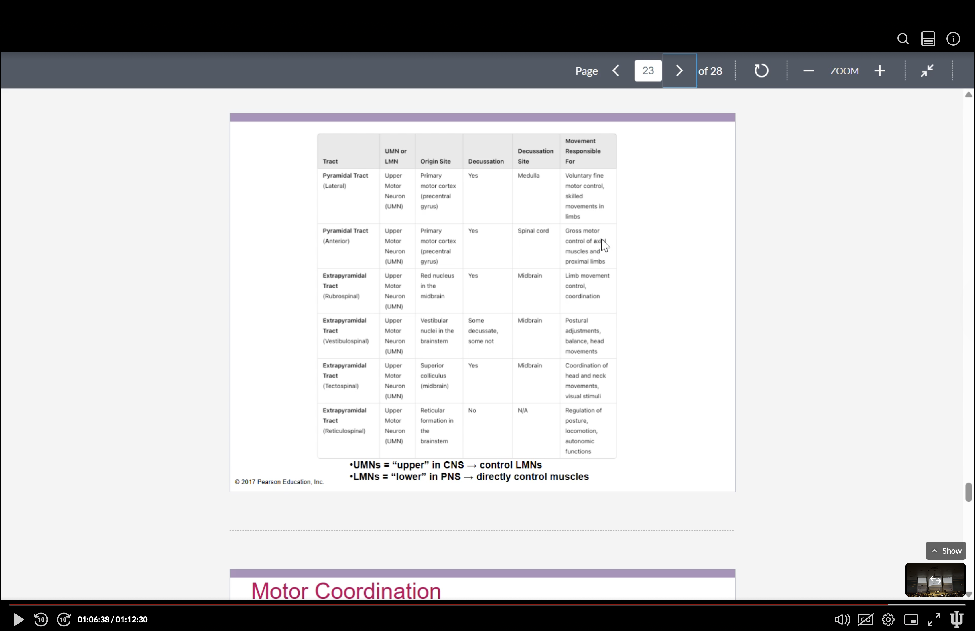
pyramidal tract: fine control of voluntary movements, predominantly of distal extremities.
Upper Motor Neurons: Originate in the primary motor cortex.
Input: Directly influences motor neurons; some inputs go through interneurons.
Collaterals: Can send signals to additional motor areas including brainstem nuclei.
Decussation: Refers to the crossing over of nerve fibers to the opposite side of the central nervous system during their descent; leads to contralateral control of limbs.
crossing from right to left or left to right
Types of Pyramidal Tracts:
Lateral Tract: Controls voluntary movement of distal extremities; decussates at the medulla.
Anterior Tract: Involved with voluntary movement of proximal and axial muscles; decussates at the spinal cord.
Extrapyramidal Tracts
Definition: Encompasses all motor pathways that lie outside the pyramidal tracks, providing support to voluntary movements especially for proximal muscles.
Extrapyramidal Tracts for Voluntary Movement
Rubrospinal Tract: Originates in the red nucleus of the midbrain; it modulates voluntary movements, especially flexion in the upper extremities; it decussates at the midbrain, operates contralaterally.
Ventromedial Pathways: These pathways can be categorized into several tracts that manage both voluntary and involuntary movements:
Vestibulospinal Tract: Responsible for posture and balance, originating in vestibular nuclei; maintains balance through involuntary adjustments.
Tectospinal Tract: Involuntary movements of the head in response to visual and auditory stimuli, originating in the superior colliculi; operates contralaterally after decussation at the midbrain.
Reticulospinal Tract: Involves the regulation of muscle tone and posture, originating from the reticular formation; divided into:
Pontine or Medial Tracts: Help stimulate leg extensors for upright posture.
Medullary or Lateral Tracts: Inhibit leg extensors for appropriate posturing.
Motor Unit Control
Upper Motor Neurons (UMNs): Located in the CNS, they control lower motor neurons (LMNs).
Lower Motor Neurons (LMNs): Located in the PNS, they directly manage muscle contraction and action.
Motor Coordination - Cerebellum and Basal Nuclei
Cerebellum Functions:
Coordinates voluntary movements ensuring timing and precision.
Balances and maintains posture.
Supports motor learning by adjusting movements through feedback.
Critical in fine-tuning movements to avoid tremors and ensure smooth action.
Basal Nuclei Functions:
Initiates and inhibits voluntary movements.
Regulates muscle tone necessary for smooth and purposeful movements.
Controls automatic, learned motions, choosing purposeful over unwanted movements by regulating excitatory and inhibitory signals.
Disorders of the Basal Nuclei
Huntington’s Chorea: A genetic degenerative disorder affecting the basal nuclei resulting in loss of motor coordination, exaggerated movements, and cognitive decline in advanced stages.
Parkinson’s Disease: A neurodegenerative disorder marked by dopamine deficiency in the substantia nigra, characterized by motor rigidity, tremors at rest, and gait disturbances leading to cognitive decline in late stages.