Synaptic Signaling
Vocabulary:
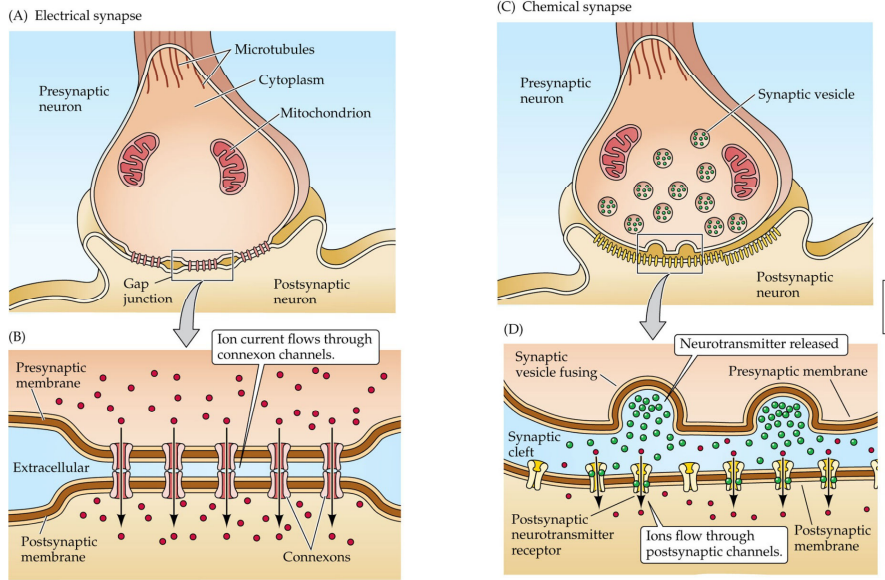
Electrical synapse: Between two neurons with cytoplasmic connection, via a connexon. Has bidirectional flow, typically with excitatory currents, secondary messengers, and metabolites/ions. Rapid coordinated transmissions have synchronized effects. Commonly dendritic or with astrocytes. Used in the brainstem, interneurons, and with cognition/memory/perception (discovered via stimulation in mice with very little temporal loss). Quicker than chemical synapses, synchronize neural circuit function
Connexon (hemichannel): Made up of 6 connexins, line up across from each other on two connecting neurons and dock to form a gap junction from 2 connexons, last about 2 hours. This pore is wider than it is for voltage-gated channels, allowing secondary messengers
Presynaptic neuron: The neuron with an action potential being sent at a synapse, contains the active zone
Postsynaptic neuron: The neuron receiving current at a synapse
Brainstem: Contains neurons that control breathing
Chemical synapse: Neurons are separated by the synaptic cleft, utilize neurotransmitters for signaling. Can amplify signals by activating enzymes, lead to the integration of signaling
Synaptic cleft: Separates presynaptic and postsynaptic neurons that utilize chemical transmission, contains interstitial fluid and filaments. Where neurotransmitters are released
Vesicle: Typically stores neurotransmitters
Active zone: Where “small molecule” neurotransmitters are released from at a synaptic cleft
Neuropeptide: A subtype of neurotransmitter that is released from distinct locations that aren’t the active zone of the synaptic cleft
Neurotransmitter: A signaling molecule released during chemical synaptic transmission, diffuse across a synaptic cleft and bind to receptors on the postsynaptic neuron. Typically stored within a vesicle but not always
Postsynaptic density: A portion of the post synaptic neuron where neurotransmitters bind at excitatory synapses
Otto Loewi: Proved chemical neurotransmission, removed 2 frog hearts with only one attached to the vagus nerve (one denervated). He recorded the beating of both hearts, and stimulated the vagus nerve for it to slow. He collected the perfusate and applied it to the denervated heart, where beating there started to slow as well. Determined the nerve was releasing bioactive substances (now known as acetylcholine)
Katz: Did work with neuromuscular junctions, used intracellular recordings of a frog neuromuscular junction to record both resting and APs. Wanted to determine the basis of electrical charge passing through the NMJ and how it occurred. Noticed that small depolarizations occurred even when not stimulated, shaped like endplate potentials, and called them miniature endplate potentials (MEPPs). Found a drug (Curare) that blocked ACh receptors. He lowered levels of Ca2+, and after stimulating the skeletal muscle, found the EPP diminish while MEPPs stayed the same, leading to the conclusion that MEPPs were caused by spontaneous and discrete ACh release and that it came in “packets” with Ca2+ playing a role in their release
Boyd & Martin: Put a cathode into a NMJ with low levels of Ca2+, recorded spontaneous potentials at 0.4 mV that disappeared when curare was applied, then stimulated a neuron with low levels of Ca2+ and saw EPPs and MEPPs normally, concluded that motor neuron AP synchronization is in distinct released of ACh
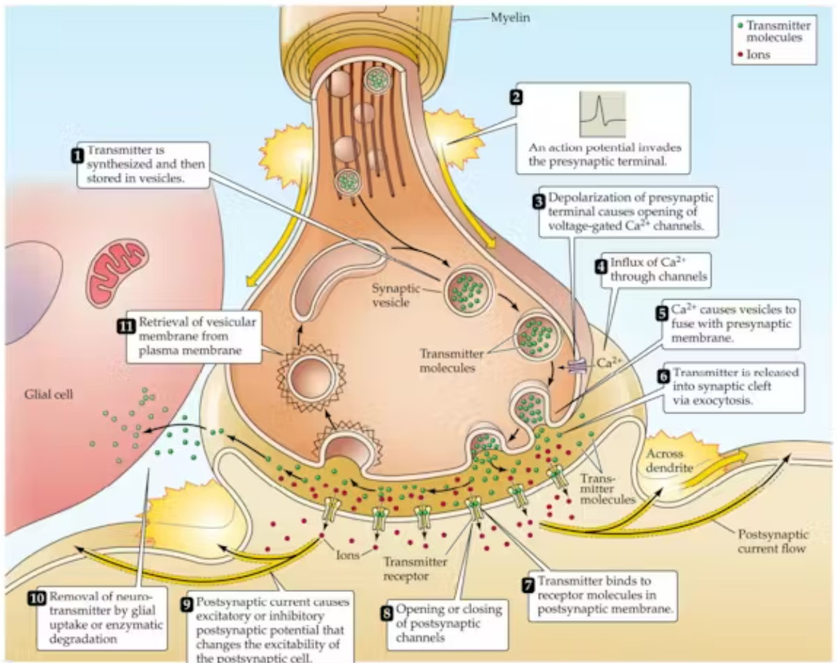
Voltage-gated Ca2+ channels: Influx of calcium through these causes eventual neurotransmitter release
Neuromuscular junction (NMJ): A chemical synapse between a neuron and a muscle fiber, where acetylcholine, (ACh) is released to be excitatory, has synaptic delay
Motor endplate: The receiving part of the NMJ on a muscle fiber, contains receptors for Na+ and K+. When Ca2+ is injected the postsynaptic cell will depolarize
Endplate potential (EPP): Depolarization of the motor endplate, will almost always cause an AP leading to contraction of the muscle fiber
Heuser & Reese: Hypothesized that exocytosis released vesicle contents, used electron microscopy to visualize fusion of from vesicles at NMJ and recorded EPPs
4-amino pyrideine: Blocks voltage-gated K+ channels to prolong presynaptic action potentials and increase the release of neurotransmitters
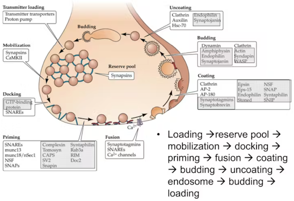
Vesicle cycle: The process of making a vesicle that is eventually fused to the membrane to release something, and it’s breakdown
Loading: Uses energy, proton pump exchanges
Reserve pool: Vesicles filled with neurotransmitters that are tethered away from the axon terminal as to not release, tethered by synapsin
Mobilization
Docking: Moving to the axon terminal
Priming
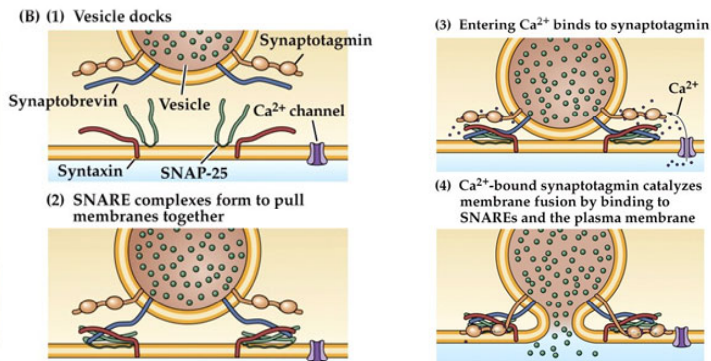
Fusion: Happens after SNARE proteins entwine an bring membranes into opposition, Ca2+ will bind to synaptotagmin and enhance curvature of the plasma membrane
Coating: Process of getting proteins around the vesicle to stabilize it and provide function
Budding
Uncoating: Done by HSC70 (a chaperone), auxilin (co-chaperone) and synaptojanin (phosphatase)
Endosome
Budding
Loading (start again)
Synapsins: Tether vesicles to the reserve pool, phosphorylated by CAMKII to be released to the axon terminal
NSF: An ATPase which helps activate SNAREs to cause entwining
SNAPs: A soluble NSF attachment protein that associates the NSF with the SNARE
SNAREs: SNAP Receptors, for complexes with SNAPs, entwine to physically cause the vesicle to fuse (requiring NSF).
SNAP-25, in the plasma membrane and part of t-SNARE
Synaptobrevin: A type of SNARE, vesicle-associated membrane protein, part of v-SNARE
Syntaxin: In the plasma membrane, part of t-SNARE
munc13 + munc18: Stabilize entwined SNAREs
Synaptotagmin: A Ca2+ sensor for SNARE complexes, vesicle associated and causes exocytosis when bound to Ca2+. Not a SNARE. Bound to the vesicle, when bound to by Ca2+ will release
Clathrin triskelia: Different adaptor proteins that assemble clathrin, include AP-2 and AP-180, make a sphere to start endocytosis
Dynamin: A GTPase, acts like molecular scissors
Myasthenia: A disorder characterized by muscle weakness, Lambert-Eaton myasthenia syndrome (LEMS) is an autoimmune version of this where Ca2+ cannot come in
Botulinum toxin: Will cleave SNAREs in motor neurons of NMJs, and cause flaccid paralysis and inability to contract
Tetanus toxin: Cleaves SNAREs in inhibitory spinal cord interneurons causing spastic paralysis
Latrotoxin: Simulates vesicle exocytosis at the NMJ to cause muscle spasms and pain
Exocytosis: The process of a vesicle with neurotransmitters inside fusing and releasing them, using NSF, SNAPs, SNAREs, and synaptotagmin
Endocytosis: The process of a cell receiving a vesicle or it getting recycled, involves clathrin, adaptor proteins, dynamin, and HSC70/auxilin/synaptojanin
Ionotropic: When a receptor opens to ligand bonding, rapid and brief effects, lets in ions
Metabotropic: When ligand binding activated an enzyme cascade, has slower and more prolonged effects
G protein-coupled receptors (GPCRs): Metabotropic receptors, a neurotransmitter binds to a receptor causing a conformational change to occur and allow GTP to bind to a G-protein
Endplate current (EPC): A small inward current in an NMJ, excitatory
I(ion X) = g(ion X) * (Vm-Ex)
Post synaptic potential (PSP): Amount of change in Vm produced by a neurotransmitter binding to a receptor, alter the probability that an AP will fire. Have a greater contribution the closer they are to the axon initial segment (meet less resistance)
Excitatory post synaptic potential (EPSP): Depolarizing, caused by ion flow of Na+ and K+
Inhibitory post synaptic potential (IPSP): Hyperpolarizing, caused by ion flow of Cl-
Glutamate: A neurotransmitter that frequently opens ionotropic Na+ receptor channels
GABA: A neurotransmitter that frequently opens ionotropic Cl- receptor channels
Reversal potential: The most important factor for determining if an IPSP or EPSP will occur, essentially Ex and where ion flow switches directions. With depolarizing, above threshold. With hyperpolarizing, under a threshold
Temporal summation: Integration of IPSPs and EPSPs over time, high frequency stimulation at the same synapse
Spatial summation: Integration of IPSPs and EPSPs for a neuron, simultaneous stimulation at multiple locations of the neuron
Glial synaptic transmission: Some neurotransmitters bind to glial receptions which can change membrane potentials and activate intracellular signaling cascades, especially with Ca2+ that can propagate between glia and stimulate secretion of signaling molecules
Glial signaling molecules: Play roles in modulating neurotransmitter release, postsynaptic responses, plasticity related to Ca2+, and pruning synapses
AP → Axon terminal → Open voltage gated Ca2+ channels → influx of Ca2+ → Triggers fusion of vesicles with plasma membrane → neurotransmitters diffuse across the synaptic cleft → bind to a receptor → open channels to increase/decrease membrane potential → removal of neurotransmitter