9.1 - Genitourinary System
LECTURE NOTES
SECTION 1
Functions and Anatomical Structure of Kidney
Excretion of metabolic products e.g. urea, uric acid, creatinine
Excretion of foregin substances e.g. drugs
Homeostasis of body fluids, electrolytes and acid-base balance
Regulates blood pressure
Secretes hormones, e.g. erythropoietin and renin
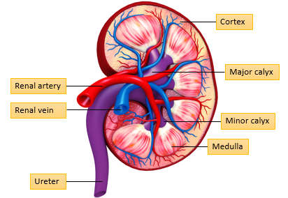
Renal artery brings blood to the kidney and renal vein takes blood away
Granular part is the cortex
From the medulla, urine drains into minor calyx. From minor into major calyx and then into the ureter
Urine is then carried from ureter to bladder and then excreted through urethra
Renal Bloody Supply to the Kidney
Renal Artery → Segmental Artery → Interlobar Artery → Arcuate Artery → Interlobular Artery → Afferent Arteriole → Glomerular Capillaries
Afferent arteriole brings the blood to the nephron
Peritubular capillaries functions
Reabsorption - things that are in the nephron are reabsorbed into these capillaries and re-enter blood supply to the body
Secretion - some substances in the capillaries need to be gotten rid of, and this is an opportuniy for secretion of these substances
Brings nurtrients and oxygen for nephrons to function to allow nephrons to respire and function properly
Bladder and Urethra (Males vs Females)
Detrusor muscle: contracts to build pressure in the urinary bladder to support urination
Trigone: stretching of this triangular region to its limit signals the brain aabout the need for urination
Internal sphincter: Involuntary control to prevent urinaiton
External sphincter: Voluntary control to prevent urination
In urinary incontinence generally there is a malfunctioning of one of these sphincters
Bulbourethral gland: Located in males, which produces thick lubricant added to watery semen to promote sperm survival
Nephron
Outline - glomerulus (blood supply), surrounded by the bowman’s capsule. This enters the proximal convoluted tubule → thin descending LOH, then thin ascending to thick ascending LOH, and then distal convoluted tubule, which then leads into the collecting duct.
Mitochondrial Concentrations in the Nephron
Proximal convoluted tubule - epithelial cells are rich in mitochondria
Descending and ascending Loop of Henele - low density of mitochondria
Thick ascneding Loop of Henle - epithelial cells rich in mitochondria
Distal convoluted tubule - epithelial cells rich in mitochondria
Collecting duct - principal cells have low density of mitochondria whereas intercalated cells are rich in mitochondria
Types of Nephrons
There are two types of nephrons - superficial and juxtamedullary
Based around the length of the Loop of Henle
Superficial nephron
Bowman’s capsule sits to the outer cortex and loop of Henle is shorter, extending to within the outer medulla
Juxtamedullary nephron
Complex sits towards the border of the cortex and medulla
Loop of Henle is longer and extends deep within the inner medulla
Cortex vs Medulla
Cortex is granular whereas medulla has striated appearance
Tubes such as loop of henle and collecting duct sit within the medulla giving it this striated appearance.
Complexes like Bowman’s capsule sit within the medulla giving it the lumpy and granular appearance.
Juxtaglomerular Apparatus
Macula densa
Part of distal convoluted tubule, just at the start of DCT
Extraglomerular mesangial cells
Sitting between the DCT and arteriole
Juxtaglomerular cells.
Sit within the afferent arteriole
Functions: GFR (glomerular filtration regulation) and renin production (bp regulation)
SECTION 2