Lecture 3: Micropipettes, Centrifuges, and Spectrophotometers
Laboratory Basics: Glassware and Decontamination
Glassware Cleaning:
* Wash with soap or Alconox.
* Scrub thoroughly with a brush.
* Rinse sequence: Hot water (approximately 3 times), followed by deionized water (approximately 3 times).
* Contamination Warning: Traces of detergent or acetone can negatively impact biological samples in molecular biology labs.
Sterilization: Glassware and media can be sterilized using an autoclave.
Decontamination Protocols:
* Benches: Cleaned with Ethanol.
* Cells: Treated with Bleach.
Liquid Transfer Volumetric Standards
Transferring liquids is one of the most common steps for molecular/biochemical experiments
Accurate/consistent transfers = accurate results
>2 L: graduated cylinder, beaker
25 ml to 2 L: graduated cylinder
1 to 25 ml: glass or plastic pipettes
0.0001 (1/10th ml) to 1 ml: micropipettes
Types of Glass/Plastic Pipettes
Mohr Pipette: Defined as "To Contain" (TC). It should not be blown out. It features a dead space below the last graduation (usually > 1\,ml) that must not be used. It is more accurate for multiple dispenses from a single aspiration.
Serological Pipette: Defined as "To Deliver" (TD). This pipette must be blown out to deliver the exact volume specified.
Volumetric Pipettes: Calibrated to deliver a single, specific volume with high precision.
Transfer (Pasteur) Pipettes: Uncalibrated and disposable; intended for use when precision is not required.
Pipette Aids:
* Mechanical aids include pipette fillers, rubber pumps, motorized pipettes, and transfer pipettes.
* Contamination Risk: Most aids are not disposable and can cause sample contamination if used improperly (e.g., through excessive aspiration).
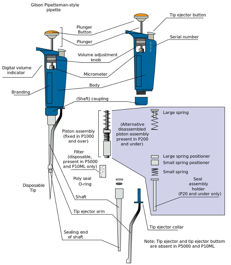
Micropipette Components and Models
Standard Components: Plunger, Tip ejector, Volume adjustment, Volume indicator, and Disposable plastic tip.
Types of Micropipettors:
* Single-channel: Manual porting for individual tubes; Eppendorf or Denville
* Multichannel Pipette: For high-throughput work (e.g., 96-well plates).
* Liquid Handler: Automated systems (e.g., Beckman Biomek 4200).
P# = max number of uLs
Eppendorf Reading
read from top to bottom
line is decimal place
Denville Reading
read from top to bottom
Red at the Top: Put the decimal after the ticks at the end (e.g., 0 3 2¦ = 320 uL).
Red at the Bottom : Put the decimal before the last red number (e.g., 1 0 5 = 10.5 µL).
Two Reds at the Bottom (P2/P10 Exception): Put the decimal right after the single black number at the top (e.g., 0 5 0 = 0.50 µL).
Micropipette Usage
Operational Steps:
1. Select the pipette closest to the target volume (e.g., use P10 for ).
2. Fit the tip tightly.
3. Push to the first stop.
4. Immerse in liquid and release the plunger slowly to aspirate.
5. Place in receiving tube and push past the second stop to dispense completely.
6. Eject the tip and release the plunger.
Sources of Error: Uncalibrated devices, dirt/debris, liquid on outside of tip, lack of mixing, poor technique, and systematic/random error.
Micropipette Care
Maintenance Warnings:
* Do not rotate beyond limits.
* Never use without a tip (prevents fluid entering the piston assembly).
* Do not immerse the barrel or hold the device horizontally with fluid in the tip.
* Do not snap back the control button.
* Do not flame the tip or jam the device into the tip holder (bends the metal tube).
Micropipetting Technique
Aspirate: draw or suck a specific volume of liquid into a pipette tip by creating negative pressure
* Temperature Equilibrium: Pipette, tips, and liquids should be at room temperature (). * Pre-Wetting: Aspirate and dispense the nominal volume 3 times to equilibrate temperature and humidify dead air space.
* Angle: Maintain a consistent angle (maximum ) to prevent varying hydrostatic pressure.
* Aspiration Depth: Immerse tip only below surface.
* Volume Range: Air displacement pipettes perform best between and of nominal volume.
* Dispensing: Discard the first and last dispense in a series of multiples. Use "Touch Off" methods (Side Wall or Surface) to remove residual droplets.
* Specific Liquids:
* Viscous: Use "Reverse Pipet" mode and slower speeds.
* Volatile: Pre-wet tips, pipette quickly, and use "Reverse Pipet" mode.
* Density: Recalibrate if the liquid density differs significantly from water.
History and Purpose of Centrifugation
Historical Context: First used in the mid-1800s for milk separation ().
Theodore Svedberg: Developed analytical ultracentrifugation in the early 1900s. He measured hemoglobin weight and predicted four subunits.
Svedberg Unit (S): (); relates to sedimentation rate.
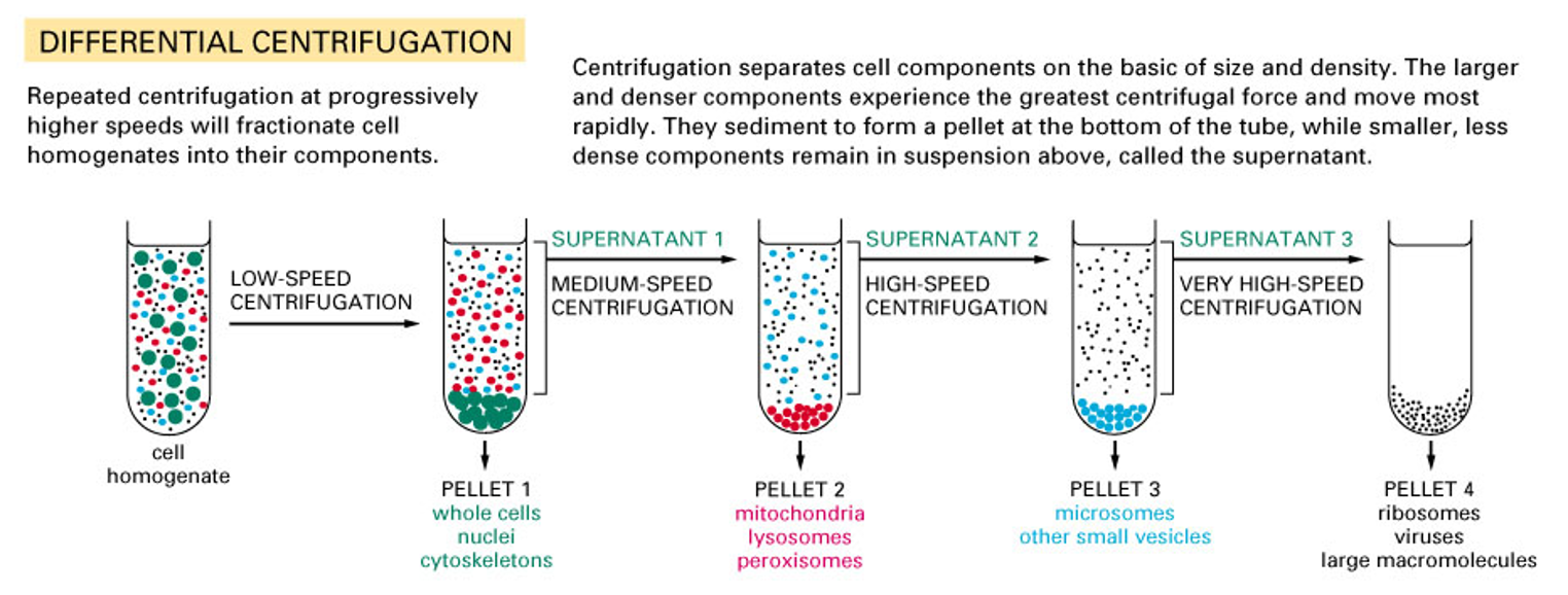
Separation Mechanisms: Processes include Homogenization, Differential sedimentation, and Density gradient centrifugation.
Specific Applications:
* Separating cells from media.
* Separating bacterial lysate into soluble and insoluble fractions.
* Isolating organelles or radioactive particles.
* Concentrating DNA, RNA, and proteins.
* Spin Down: Used to bring fluid from the lid/sides to the tube bottom, especially important for accurate concentration in frozen samples.
Physics and Calculation of Centrifugation
Sedimentation
Particles naturally sediment out of solution
Particles in solution are held in tubes or bottles which are placed in a rotor
Each particle sediments at a rate that is directly proportional to the applied centrifugal field (g), which is directed radially outward
Determined by the square of the angular velocity of the rotor
Radial distance of the particle from the axis of rotation (r, in centimeters)
Type of rotor (swinging bucket, fixed-angle)
Force Application: A centrifuge applies a force in multiples of Earth's gravity ().
Sedimentation Rate Factors: Proportional to the square of angular velocity, the radial distance (, in cm) from the axis, and the rotor type.
RPM vs. RCF:
* RPM: Revolutions per minute.
* RCF or g: Relative Centrifugal Force.
* Formula:
Comparative Example:
Rotor A Rotor B
Speed 14,000 rpm 14,000 rpm
Radius 5.98 cm 9.50 cm
Gravity 13,100 × g 20,817 × g
Reporting Requirements: Protocols must specify the rotor model (e.g., 45 Ti, JLA-8.1000), time, speed (rpm), force (rcf, g), and temperature.
Example:
G-actin was ultracentrifuged for 1 hour at 4 °C at 100,000 g
Centrifuge at 10,000 rpm in a Sorvall SS-34 rotor for 10 minutes
These are not standard across all rotors, due to differences in diameter
Report all data necessary to be able to reproduce the experiment
Categories of Centrifugation
Preparative Centrifugation:
Differential centrifugation
Small bench centrifuges
Large capacity refrigerated centrifuges
High speed refrigerated centrifuges
Ultracentrifuges
Preparative centrifugation is most common
Actual separation, isolation, purification
Large amounts of media with bacteria or yeast, blood, whole cells, organelles, plasma membranes, nucleic acids, proteins, viruses
Morphology, composition, biological activity
Analytical Centrifugation:
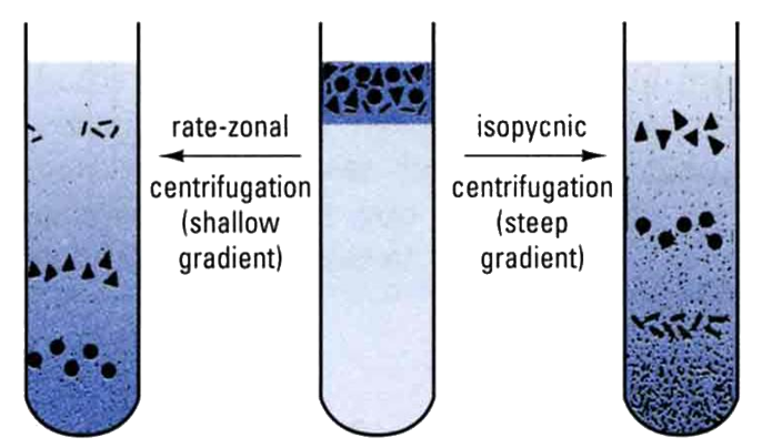
Density gradients via rate zonal (size) or isopycnic (density) centrifugation
Ultracentrifuges
Study the sedimentation characteristics of pure biological macromolecules and molecular structures
Requires only small amounts of material and utilizes specially designed rotors and detector systems to continuously monitor the process of sedimentation of the material
Determine:
Purity
Molecular weight/size
Conformational changes
Sedimentation and diffusion coefficients
Stoichiometry
Equilibrium constant
Thermodynamics
Absorbance, fluorescence
Device Classes:
* Small Bench Centrifuges: ; .
Simplest, least expensive centrifuges, many designs
Often refrigerated to prevent protein denaturation
Balances tubes within 0.25 g
Used to collect small amounts of material that rapidly sediment; 1 ml microcentrifuge tubes, 15 ml and 50 ml conical tubes
Pellet samples
Spin down solution on side of tubes
DNA/protein concentrating
Minipreps
* Large Capacity Refrigerated: ; . Holds to bottles; balances tubes within .
Collect substances that sediment rapidly, e.g. erythrocytes, precipitates, bacterial cells
Refrigerated to prevent protein denaturation
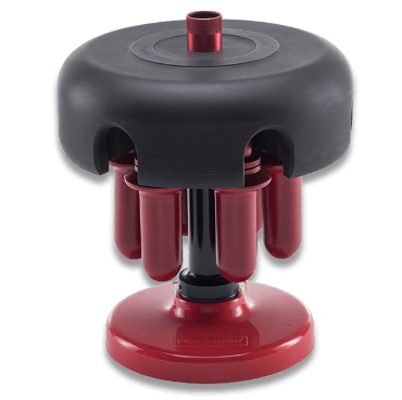
Capable of utilizing a variety of interchangeable swinging-bucket and fixed-angle rotors
* High Speed Refrigerated: ; . Collects microorganisms and cellular debris; balances within .
Used to collect microorganisms, cellular debris, larger cellular organelles and proteins precipitated by ammonium sulfate
Total capacity typically up to 1.5 L, although some cross over into the large capacity range
Refrigerated to prevent protein denaturation
Interchangeable fixed-angle and swinging-bucket rotors
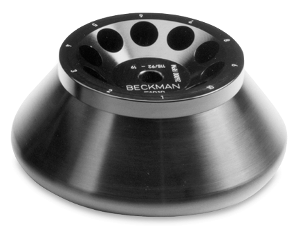
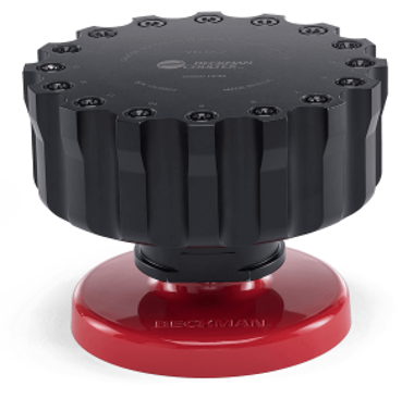
* Ultracentrifuges: ; . Evacuated chambers prevent heat from friction. Requires infrared sensors and heavy armor plating. Balances within .
Isolate viruses, organelles, membrane fractions, DNA, RNA, proteins
The rotor chamber is refrigerated, sealed, and evacuated, to minimize any excessive rotor temperatures being generated by frictional resistance between the air and the spinning rotor
Refrigerated to prevent protein denaturation
Safety features:
Infrared sensors measure temperature
Overspeed control system
Flexible drive shaft to absorb imbalances
Heavy armor plating
* Continuous Flow Centrifuges:
Media enters moving rotor; particles settle while cleared media overflows.
Used for harvesting from volumes of .
Rotor Types and Applications
Fixed-Angle: Angled, best for pelleting (bacterial pellets, DNA precipitation).
Swinging-Bucket: High g-forces; ideal for size and density purifications and analytical (e.g., Sucrose/Cesium Chloride gradients).
Zonal: For separating large volumes.
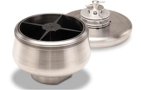
Vertical: DNA and virus purification.
Swinging Bucket Analytical Centrifugation
Meselson and Stahl Experiment
Used ultracentrifugation with (, ) to demonstrate the semi-conservative model of DNA replication by separating and DNA isotopes ( for ).
Saw that heavy DNA and light DNA separated into distinct bands within the gradient and found a heavy, hybrid, and light DNA bands
Density Gradient Separations
Used for Golgi body purification ( for ) and plant pigment analysis (e.g., Lutein knockout vs. WT).
Centrifuge Tubes and Balancing
Materials:
* Polypropylene: Opaque, good chemical/stress resistance.
* Polycarbonate: Clear, but less chemical resistance.
Pointed shape allows pellets to form on the side of the tubes
Balancing Scenarios (24-tube centrifuge):
* 1 Tube: Requires a balance blank.
* 2 Tubes: Place at opposite ends (1 & 13).
* 3 Tubes: Place at 1, 9, and 17.
* 5 Tubes: Can be balanced without blanks by specific distribution.
Note: Tubes must be balanced by mass, not just volume (e.g., of lysate may not balance exactly with of water due to density differences).
Spectroscopy and Spectrophotometry
Spectroscopy: The study of molecular structure and dynamics via the absorption, emission, and scattering of radiation.
Light, or visible radiation, can be resolved into its component wavelengths (as with a prism)
Study of molecular structure and dynamics through absorption, emission, and scattering
Spectrometry: The measurement of this interaction to produce spectra for qualitative or quantitative analysis.
Measurement of this interaction
Produces spectra that are used for theoretical studies on the structure matter, or concentration of a solution
Qualitative and quantitative analysis
Types of Spectroscopy
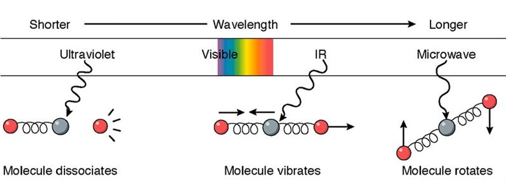
Electromagnetic
Visible, UV, infrared, fluorescence spectroscopy
Flame spectroscopy
X-ray spectroscopy (crystallography)
Nuclear magnetic resonance spectroscopy (NMR)
Circular dichroism
Non-electromagnetic
Ions (mass spectroscopy)
Electrons (electron spectroscopy)
Sound waves (acoustic spectroscopy)
Infared;
Similar to UV-Vis, but less precise, wavelength region ~1000-200,000 nm
Energy transitions due to changes in vibrational, rotational, and kinetic energy
Qualitative analysis of functional groups (carbonyls, alcohols, aromatics)
Fourier Transform Infrared (FTIR): analysis of plasma membranes, microorganism ID, secondary structure
Principles of Light Absorbance
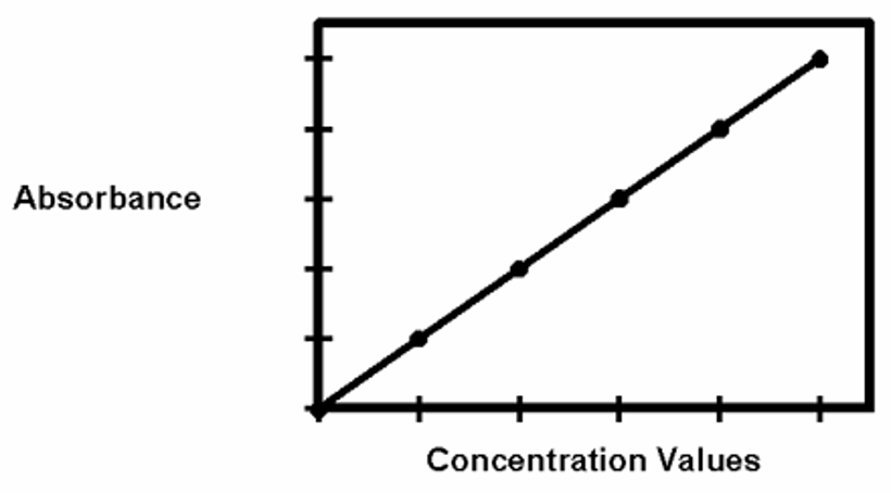
* Absorbance is a characteristic of a substance (like boiling point).
* It is directly proportional to concentration when the light path is constant.
* All substances in solution absorb light of one wavelength and transmit light of other wavelengths
* Absorbance is a characteristic of a substance, like melting point, boiling point, density, and solubility
* Because absorbance can be related to the amount of the substance in solution, absorbance can be used to quantitatively determine the amount of the substance in solution
Spectrophotometry
Spectrophotometry is an analytical method of measuring the amount of light absorbed by a substance in solution to determine identity, concentration, and other properties
Many substances absorb light at specific wavelengths within the ultraviolet (200-400 nm), visible (400-800 nm), and near infrared (800-1000 nm) spectra
Spectrophotometry measures the sample directly (no color change)
In colorimetry, samples are converted or conjugated to colored compounds via chromogenic reactions, and then measurements are made based on color
The absorbance of a solution is directly proportional to the concentration of the absorbing material when the light path is kept constant
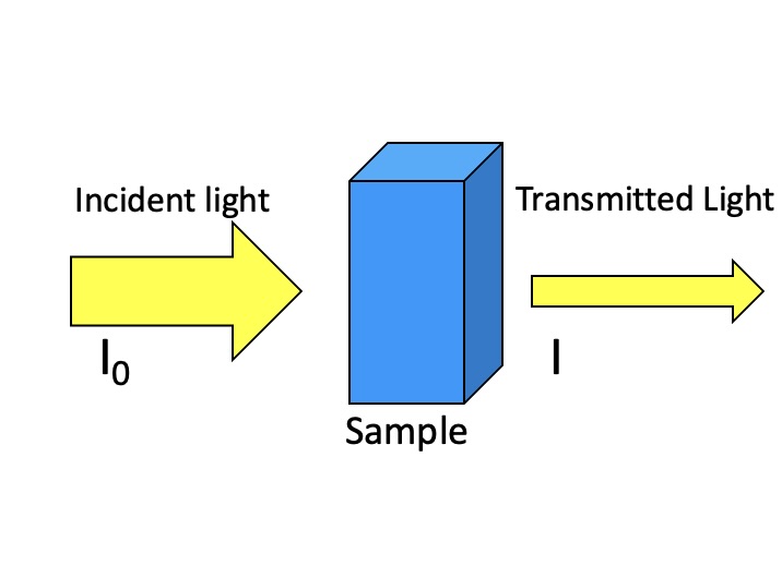
Spectrophotometers measure the intensity of a specific wavelength of light as it passes through a sample
They compare the amount of light transmitted through a blank sample (lacking the absorbing compound) to that transmitted through a sample
Spectrophotometer Components and Plate Readers
Light Source
* Tungsten Lamp: Visible region ().
* Deuterium Lamp: UV region ().
Monochromator: selects particular wavelengths of light for measurement
* Prism or a grating: disperses light, and a narrow exit slit selects only a small range of the dispersed spectrum
Sample compartment: holds cuvettes for light to pass through to the detector
Detector: photocell converts light into electrical current
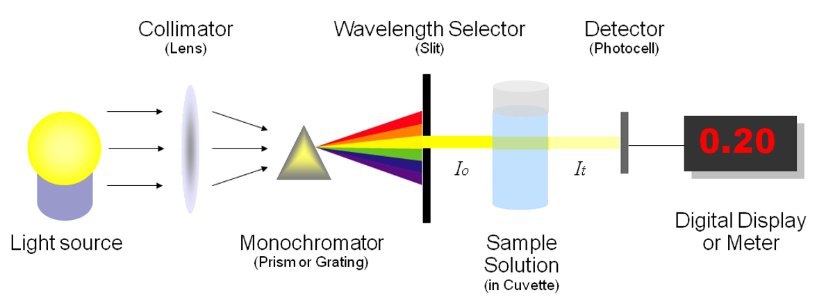
Plate Readers:
* Measure small volumes () in plates with typically 96 wells (up to 1536).
* Detects absorbance, fluorescence, luminescence, polarization, and scattering.
* Common in ELISAs, drug screenings, and enzyme assays.