Cardiac Interventions
Fall 2025 — Lecture Follow-Along Notes
Slide 1 — Invasive Cardiovascular Procedures
Coronary Artery Reperfusion
Percutaneous transluminal coronary angioplasty (PTCA)
Coronary artery stent
Cardiac Surgery
Coronary artery bypass graft (CABG)
Control of Dysrhythmias
Pacemaker
Defibrillator placement
Big picture: This lecture covers procedures used to restore coronary blood flow and devices/procedures used to manage abnormal heart rhythms.
Slide 2 — Coronary Artery Reperfusion
Main topic:
Restoring blood flow through a blocked coronary artery
This leads into PTCA/PCI.
Slide 3 — Percutaneous Transluminal Coronary Angioplasty
PTCA
Large cannula inserted into an artery
Femoral
Brachial
Second cannula threaded up to coronary arteries
Balloon-tipped catheter opens the blocked coronary vessel
Balloon:
Compresses atheroma/plaque
Stretches the vessel to >90%
Can also:
Laser atheroma
Cut atheroma
Place a stent
Followed by anticoagulants
Complications During Procedure
Dissection
Perforation
Abrupt closure
Vasospasm
Acute MI
Arrhythmias
Cardiac arrest
Post-Procedure Complications
Re-occlusion
Arterial occlusion
Bleeding
Insertion site
Retroperitoneal
AKI
Dye/contrast is used
Know: After PTCA, bleeding, perfusion and renal function are major concerns.
Slide 4 — Percutaneous Coronary Intervention
PCI = Percutaneous Coronary Intervention
This slide is mainly visual.
Think:
Blocked coronary artery → catheter/balloon → artery opened → blood flow restored
Slide 5 — Post-Procedure Care: Assessment & Analysis
History
Assess the patient's history.
Assessments
Insertion site
Are sheaths still in place?
Medications
Heparin
IIb/IIIa agent
Perfusion
Peripheral circulation
Signs of complications
While listening: If your professor talks about checking the affected extremity, this goes under perfusion/peripheral circulation.
Slide 6 — Collaborative Care Priorities: Planning
Nursing
Risk for:
Impaired perfusion/cardiac function
Impaired gas exchange
Acute pain
Bleeding
Fluid imbalance
Anxiety
Patient education:
Disease
Self-care
Collaborative
Monitor/manage:
Perfusion
Gas exchange
Bleeding
Vascular occlusion
Renal function
Slide 7 — Collaborative Care Interventions: Implementation
Nursing
Pain assessment and management
Follow protocol
Vital signs
ECG
Peripheral pulses
Site checks
Immobility
Head-of-bed positioning
Repositioning
Monitor I&O
Medication administration and outcomes
Explore anxiety
Education
Collaborative
Review test results/labs
Initiate protocols
Medications
Detect/manage complications
Transitional care needs
Slide 8 — Collaborative Goals & Outcomes: Evaluation
Patient:
Maintains perfusion
Maintains gas exchange
Maintains fluid balance
Remains pain-free
Shows no signs of bleeding
Reports no/decrease/lower anxiety
Identifies ways to manage anxiety
Remains free of complications
Discusses self-care program
Slide 9 — CABG
Next major section:
Coronary Artery Bypass Graft
Slide 10 — CABG
Indications
Uncontrolled angina
Left main coronary artery blockage >50%
Multivessel disease: meaning multiple blockages in different coronary arteries, which may require surgical intervention for effective management.
Unsuccessful PTCA → choice in intervention is calculated (not under our scope) based on the severity of the blockages, patient's overall health, and the potential benefits versus risks associated with surgical options.
What is CABG?
A blood vessel from another part of the body is grafted around the occluded coronary artery(ies)
Possible grafts:
Thoracic artery: A common choice for grafting, providing good blood flow due to its robust structure.
Saphenous vein : Often used for its accessibility, the saphenous vein can be harvested and utilized when the thoracic artery is not suitable.
Requires extensive:
Pre-op care
Intra-op care
Post-op care
Easy way to picture it: The blockage stays where it is. CABG creates a new route around it.
Slide 11 — Coronary Artery Bypass Grafts
Mainly visual.
Use this slide to remember:
Graft = new pathway around the coronary blockage
Slide 12 — CABG Procedure
General anesthesia
Surgical procedure
Sternotomy
Cardiopulmonary bypass (CPB) machine
Blood circulated through machine
Heart stopped
Vessels harvested and grafted
Chest tube placed
Epicardial wires placed
Slide 13 — Cardiopulmonary Bypass System
Mainly visual.
CPB
During cardiopulmonary bypass:
Blood circulates through CPB machine while heart is stopped.
Connect this slide back to the CABG procedure.
Slide 14 — Preoperative Management
Assessment & Analysis
CABG patients may be:
Older
Have multiple comorbidities: make sure these are addressed to optimize surgical outcomes and ensure comprehensive post-operative care.
Hypertension
Diabetes mellitus
Chronic obstructive pulmonary disease (COPD)
Medications That Improve Outcomes
Aspirin
Beta blockers
Statins
Testing
Chest X-ray
EKG
Labs:
Type & crossmatch
Coagulation studies
CBC
Metabolic panel
Assessment
Health history
Baseline assessment
Focused cardiovascular assessment
Glucose control: Maintaining optimal glucose levels is crucial in managing cardiac health, as uncontrolled diabetes can lead to increased rates of cardiovascular disease and complications.
Physiological:
Regular monitoring of heart function, including parameters such as blood pressure, heart rate, and rhythm, helps in early detection of potential issues and guides timely interventions.
Psychological:
Learning needs
Anxiety
Social:
Support available after surgery post operatively
Planning / Implementation / Outcomes
Patient education:
Reduce anxiety
Medication changes
Shower with antiseptic solution
What to expect during hospital course
Incentive spirometer
Exercises
Family/caregiver support
Transitional care
Slide 15 — Postoperative Management: Assessment & Analysis
Assessment minimum every 4 hours
Assess:
Neuro
Neurological status: Ensure alertness, orientation to person, place, and time, and evaluate pupil response and strength in all extremities.
Cardiac
Cardiac status: Monitor heart rate, rhythm, and blood pressure to assess cardiovascular stability and identify any abnormalities that may require intervention.
Respiratory
Respiratory status
Peripheral Vascular
Circulation/perfusion
Renal
Kidney function
Fluid/Electrolytes
Fluid status
Electrolytes
Family Needs
Assess family/support needs
Your professor has “WHY” on this slide, so listen for her explanation of why each body system needs to be closely monitored after CABG.
Slide 16 — Collaborative Care Priorities: Planning
Nursing
Impaired cardiac output
Impaired gas exchange
Risk for fluid/electrolyte imbalance
Risk for acute confusion
Acute pain
Risk for impaired renal function
Impaired thermoregulation
Risk of infection
Lack of knowledge
Collaborative
Watch for:
Fluid
Hypovolemia
Bleeding
Fluid overload
Cardiac
Cardiac tamponade
HTN
MI
Arrhythmias
Heart failure
Respiratory
Weaning from ventilator
Renal
AKI
Neuro
CVA
Other
Pain
Hypothermia
Infection
Slide 17 — Collaborative Interventions: Implementation
Collaborative
Monitor for complications
Ventilator settings
Wean from ventilator
ABGs
Follow protocols
Pacemaker settings
Labs
Warming devices
Physical therapy
Transitional care
Nursing
Monitor cardiac/perfusion:
VS
ECG
Hemodynamics
Signs of complications
Monitor pulmonary:
Ventilator
Chest tube care
Incentive spirometry
Monitor:
Labs
Urine output
Drainage
Electrolytes
Neuro/safety:
Manage pain
Monitor cognition
Infection/temperature:
Temperature
WBC
Drainage
Also:
Patient/family support
Education
Slide 18 — Collaborative Goals & Outcomes
Patient:
Maintains perfusion and cardiac output
Reestablishes/maintains gas exchange
Has controlled pain
Shows no signs of bleeding
Remains free of surgical-site infection
Is alert and oriented
Reestablishes thermoregulation
Remains free of complications
Has post-hospital care in place
Slide 19 — Pacemakers & Defibrillators
New section:
Implantable Cardiac Devices
Slide 20 — Think Back to Dysrhythmias
Your professor asks:
Why might someone need a permanent pacemaker?
To correct bradycardia: A condition characterized by an abnormally slow heart rate, which can lead to insufficient blood flow to the body.
To manage heart block: When the electrical signals in the heart are delayed or blocked, a pacemaker can ensure that the heart beats at a regular rhythm.
To treat certain types of heart failure: In cases where the heart's ability to pump blood is compromised, a pacemaker can help coordinate the heart's contractions.
Why might someone need a permanent defibrillator?
A permanent defibrillator may be necessary for individuals who are at high risk of life-threatening arrhythmias, as it can continuously monitor the heart's rhythm and deliver shocks to restore a normal heartbeat whenever dangerous irregularities are detected.
A person in afib
Slide 21 — Cardioversion & Defibrillation
Both treat tachyarrhythmias by delivering electrical current.
Electrical current:
Depolarizes a critical mass of myocardial cells
Essentially "resets" the heart
After repolarization, sinus node may recapture its role as pacemaker
Cardioversion
Synchronized with patient's ECG
Defibrillation
Unsynchronized
Main difference to memorize:
Cardioversion = synchronized
Defibrillation = unsynchronized
Slide 22 — Pacemakers
Definition
Electronic device that provides electrical stimuli to the heart muscle.
Used for:
Symptomatic dysrhythmias
Chronic dysrhythmias
Recurrent dysrhythmias
Dysrhythmias unresponsive to pharmacological therapy
Can be:
Transcutaneous
Transvenous
Implanted
Implanted: for men and women with atrial fibrillation or other cardiac arrhythmias such as:
, providing a long-term solution to manage heart rhythm effectively.
Below clavicle
Behind breast
Hardware
Generator
Circuitry
Batteries
Determines:
Heart rate
Strength of impulse from pacemaker to heart
Milliamperes
Sensitivity/detection of heart impulses
Millivolts
Electrodes
Attached to heart
Newer systems may be leadless
Slide 23 — Permanent Pacemaker
Mainly visual.
Use this slide to identify:
Generator
Leads/electrodes
Location of implanted device
Slide 24 — Pacemaker Programming
NASPE-BPEG Code
1st letter = Chamber PACED
A = Atrium
V = Ventricle
D = Dual
2nd letter = Chamber SENSED
A = Atrium
V = Ventricle
D = Dual
O = Off
3rd letter = TYPE OF RESPONSE from the pacemaker to what it senses
I = Inhibited
T = Triggered
D = Dual
O = None
4th = RATE ADAPTIVITY (dont really see)
O = None
R = Rate modulation
Also includes pacing-site capability and antitachycardia function.
Remember the order:

Paced → Sensed → Response → Rate
Slide 25 — Which Is Which? - PRACTICE FOR EXAM
This appears to be an identification/practice slide.
Use what you learned from the previous slide:
a paced- do not measure
a paced rhythm — an electrical signal is delivered to the heart to stimulate contractions, while you do not measure the output visually in the same way as a natural rhythm.
dual paced: firesand gets response from A and V to coordinate contractions, ensuring proper timing between the atria and ventricles for effective blood pumping.
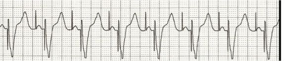
v paced: delivers electrical impulses to the ventricles, ensuring proper ventricular contraction and maintaining an adequate heart rate.

What chamber is paced?
What chamber is sensed?
What is the response?
Is there rate modulation?
Slide 26 — Pacemaker Complications
General/Insertion Complications
Dislodgement of pacemaker leads
Pneumothorax
Bleeding
Dysrhythmia
Infection
Device Problems
Electromagnetic interference
Can trigger alarms
Malfunction
Loss of capture/pacing
Under-sensing
Over-sensing
Patient
Medical alert ID!
Slide 27 — Implantable Cardioverter Defibrillator
ICD
Detects and terminates life-threatening episodes of:
V-tach
V-fib
Used for:
Post sudden cardiac death
CAD with LV dysfunction
EF <35%
Nonischemic dilated cardiomyopathy as listed on slide
Can be:
External/temporary
Internal/implanted
Can also provide: Pacing and Cardioversion
Slide 28 — Implantable Device Procedure Care
Assessment & Analysis
Assess:
Heart rate and Rhythm:
Incision site: infection risk
Chest X-ray: to make sure there is no pneumothorax or any other complications that could affect the procedure.
Medications:
Antiarrhythmics
Beta blockers
Diuretics
Anxiety
Depression
Anger
Body image: Negative body image can significantly impact a patient's willingness to participate in cardiac rehabilitation programs, potentially hindering recovery and overall quality of life.
Knowledge
Slide 29 — Collaborative Care Priorities: Planning
Nursing
Prevent infection
Pain
Bleeding
Mobility: not raising arm for about a week
Promote coping
Patient education
Collaborative
Cardiac output
Confirm lead placement
Device function
Protocols
Labs
Slide 30 — Collaborative Care Implementation
Nursing
Monitor:
Cardiac function
ECG
Perfusion: Assess peripheral pulses and capillary refill time, ensuring adequate oxygen delivery to tissues.
VS
Surgical site
Wound/dressing
Signs/symptoms of bleeding
Pain
Interventions:
Immobilize arm/use sling for about a week
Explore patient's perception of device
Patient Education
Teach:
Site care
Medications
Symptoms to report
Avoid electromagnetic interference (EMI)
Carry identification
Collaborative
Device settings
Chest X-ray
Labs
Medications
Slide 31 — Collaborative Care Goals & Outcomes
Patient:
Maintains cardiac output and perfusion
ECG reflects device settings
Remains free of infection
Remains free of bleeding
Is free of pain
Can discuss perceptions of device
Understands post-procedure precautions and care
End-of-Lecture Big Picture
For this PowerPoint, keep these four sections straight:
PCI/PTCA
→ Open the blocked coronary artery.
CABG
→ Create a new route around the blocked coronary artery.
Pacemaker
→ Provides electrical stimulation/pacing to the heart.
ICD
→ Detects and terminates life-threatening V-tach/V-fib.
And the electrical therapy distinction:
Cardioversion = synchronized
Defibrillation = unsynchronized