Respiratory System Anatomy
Vocabulary
Nasal cavity-The nasal cavity has a defined role in filtering and humidifying air for presentation to the lower airway.
Pharynx– throat, In the respiratory system, it receives air from the nose or the mouth and then moves the air past the epiglottis into the larynx. In the digestive system, it receives food from the mouth and moves it into the esophagus.
Trachea- large tube reinforced by cartilage rings that keep it from collapsing., Connects the larynx to the bronchi of the lungs.
Bronchi-Receive inspired air from the trachea and move it into the bronchioles.
Bronchioles-Connect the bronchi to the alveoli of the lungs, Bronchioles have a layer of smooth muscle which allows bronchoconstriction and bronchodilation, regulating the amount of air reaching the alveoli.
Alveoli– Site of gas exchange
Epiglottis– a flap of cartilage at the root of the tongue, which is depressed during swallowing to cover the opening of the windpipe.
Glottis- the part of the larynx consisting of the vocal cords and the opening between them. It affects voice modulation through expansion or contraction.
Larynx-The larynx is a hollow structure connected to the top of the trachea and is the passage through which inspired air moves into the bronchi of the lungs.
Diaphragm-skeletal muscle associated with quiet (normal) breathing that separates the thoracic and abdominopelvic cavities.
Pleura-serous double membrane that protects the lungs.
Ventilation– is the movement of respiratory gases between the atmosphere and the alveoli of the lungs → consist of two cyclic phases: inspiration & expiration.
Introduction
The respiratory system is essential in supplying the body with oxygen and removing carbon dioxide. It consists of a series of structures that allow for the passage of air into the body and the exchanges of gases with the blood. There are essentially two types of respiration. External respiration is the movement of gases into the body and blood. Cellular respiration is the use of oxygen and production of carbon dioxide by the cells.
Respiratory System Anatomy
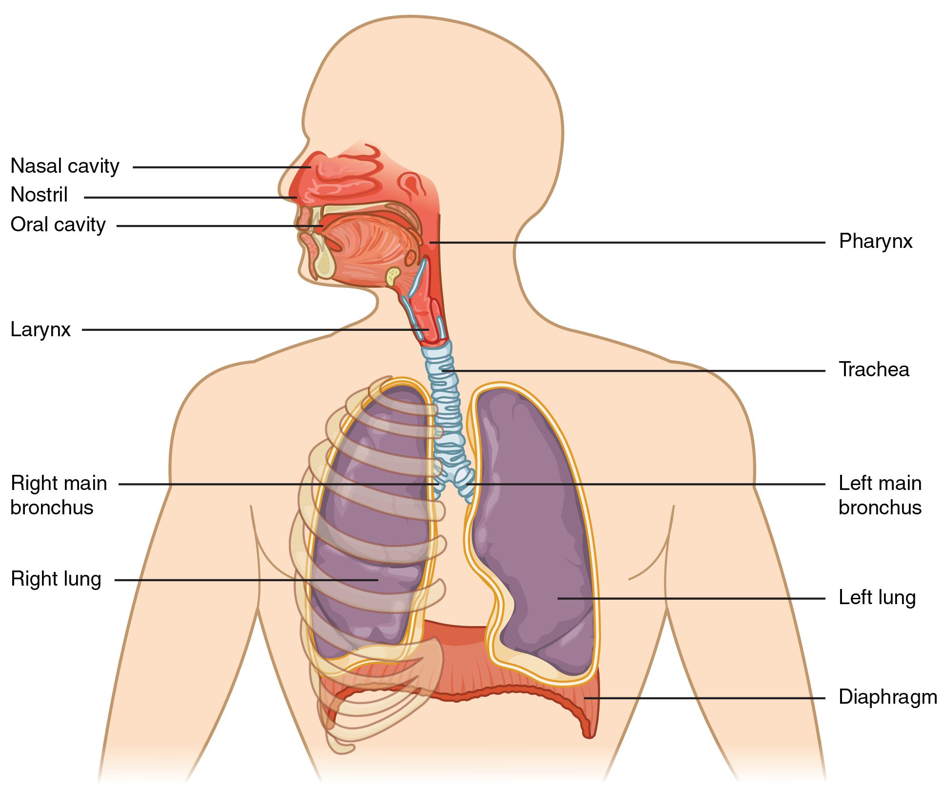
The respiratory system can be divided into the upper and lower respiratory systems. The upper respiratory system consists of the nose and nasal cavity, the sinus, pharynx, and the portion of the larynx above the vocal cords. The lower respiratory system consists of the portion of the larynx including the vocal cords and below, trachea, bronchi, bronchioles, lungs and alveoli.
Nasal Cavity
Air moves into the upper respiratory system through the nose at the nostrils to the nasal cavity. The epithelium lining the nasal cavity contains columnar and mucous secreting goblet cells. The nasal cavity contains bony protuberances called conchae. There are superior, middle and inferior conchae. The purpose of the conchae is to create turbulent flow of air. This works to warm the air and to provide more contact with the nasal mucosa and hairs so that particles can be picked up by the mucosa. The turbulent air can also reach the upper nasal cavity containing sensory receptors for smell.
Identify the majory respiratory structures in the image below.
Pharynx
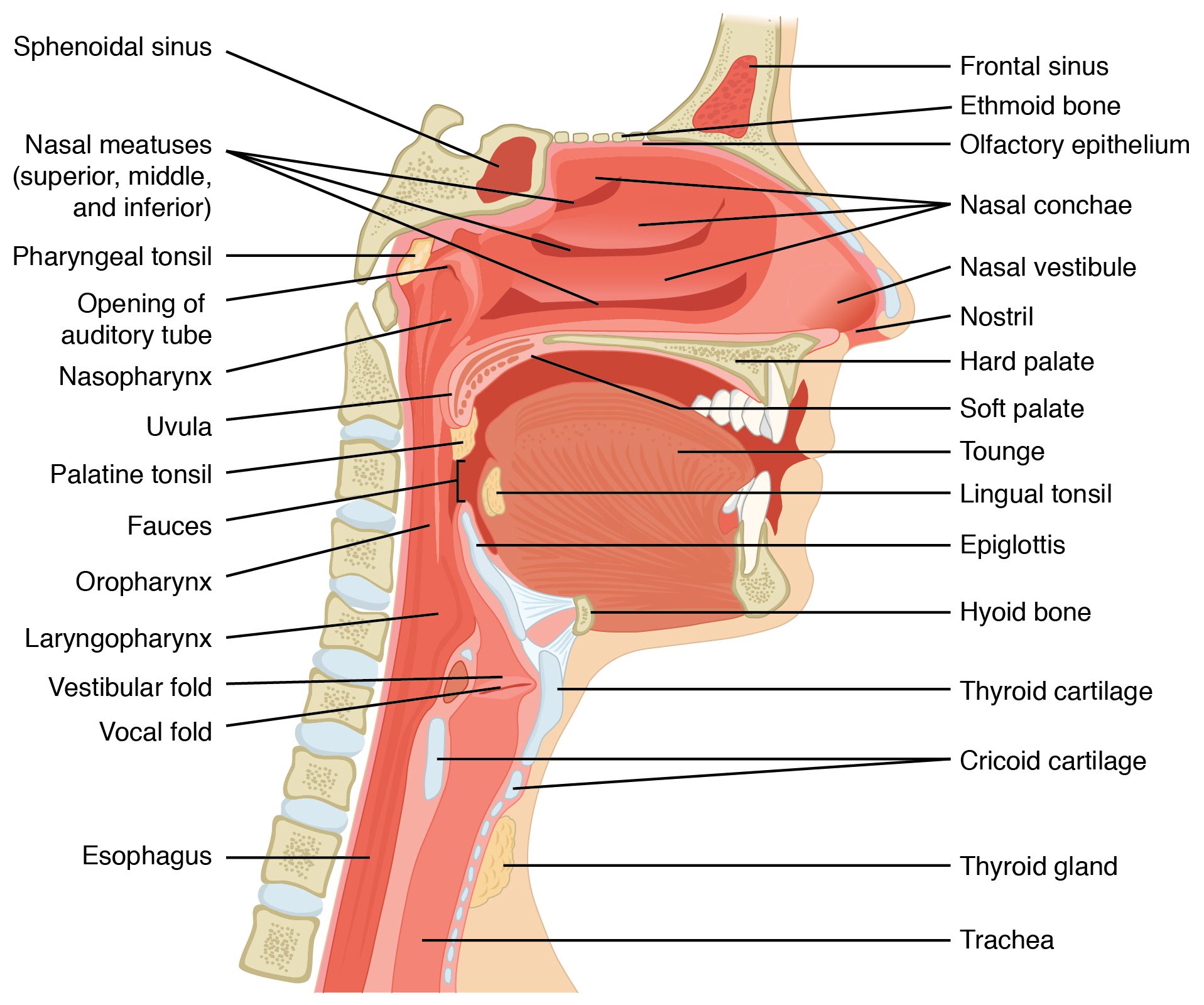
Air passes through the nasal cavity and enters the upper portion of the pharynx called the nasopharynx. The nasopharynx begins posterior to the conchae and extends inferiorly to the soft palate. The soft palate raises to close off the nasopharynx during swallowing to prevent substances from moving into the nasopharynx. The nasopharynx also contains connections from the Eustachian tubes.
Inferior to the nasopharynx is the oropharynx which extends from the soft palate to the epiglottis. The oropharynx is a shared passageway for air and substances on their way to the digestive tract. The most inferior portion of the pharynx is the laryngopharynx which extends from the tip of the epiglottis to the larynx. The laryngopharynx is also a shared pathway with the digestive tract.
Examine the structures of the pharynx and larynx in the image below.
Larynx
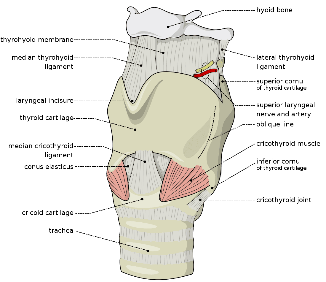
The larynx begins at the base of the tongue and extends to the trachea. The larynx contains cartilages, largest of which is the thyroid cartilage, commonly called the Adam’s Apple. Inferior to the thyroid cartilage is the cricoid cartilage. The epiglottis is an elastic cartilage flap that closes during swallowing to keep substances from moving into the trachea and air passages. Other cartilages include the arytenoids, corniculate and cuneiform cartilages. These cartilages are paired.
The vocal cords reside in the larynx and consist of two pairs of ligaments that extend from the arytenoid to the thyroid cartilages. One set of ligaments is called the false vocal cords. The other set is called the true vocal cords. When the vocal cords are relaxed they form a triangular space called the glottis.
Different pitches in the voice are produced by vibrations of the vocal cords. Vibration of smaller areas of the vocal cords results in higher pitches. Males typically have longer vocal cords than females that result in lower pitches.
Examine the cartilages of the larynx below. Pay particular attention to the thyroid and cricoid cartilages.
Lungs
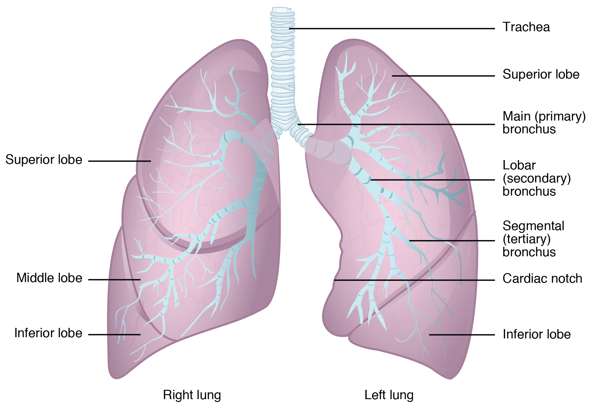
The lungs are two cone shaped structures residing in the thoracic cavity. The inferior portion of each lung reaches to the diaphragm. The superior portion extends about one inch above each clavicle. The right lung contains three lobes (superior, middle and inferior) and is larger than the left lung which contains two lobes (superior and inferior). The lobes are separated by fissures. The right lung includes a horizontal and oblique fissure while the left lung only contains an oblique fissure. The medial surface of each lung contains an area known as the hilum where vessels enter and exit. The left lung also contains the cardiac notch which is an indentation for the heart.
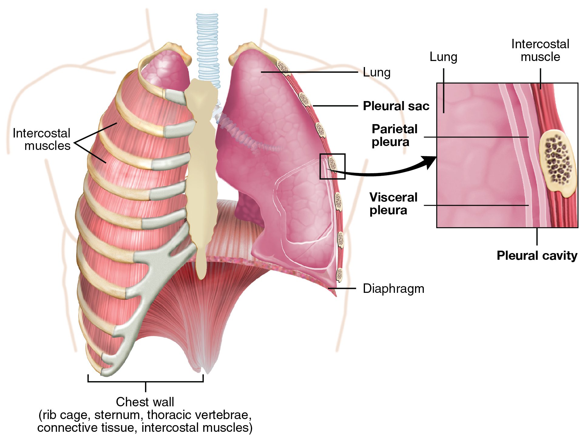
The lungs are surrounded by two pleural membranes. The surface of each lung contains a visceral pleural membrane that closely adheres to the lung’s surface. Lining the interior of the thoracic wall is the parietal pleural membrane. Both are serous membranes. A fluid known as pleural fluid is secreted by each membrane that reduces friction and helps to hold the membranes together.
Examine the structures of the lungs and pleural membranes in the images below.
Respiratory Physiology Part 1
Vocabulary
Diffusion- Process of gas exchange in the lungs, which is a passive transport mechanism, oxygen moves into the blood from lungs while carbon dioxide in the blood moves into lungs.
Partial Pressure-pressure of a single gas in a mixture of gasses.
Boyle’s Law-volume is inversely related to pressure.
Ventilation- is the movement of respiratory gases between the atmosphere and the alveoli of the lungs → consist of two cyclic phases: inspiration & expiration
Introduction
Since the primary function of the respiratory system is to bring oxygen into the body so that it can be transported to the cells and to remove carbon dioxide, air must move in an out of the lungs. This process is called ventilation and consists of inhalation and exhalation.
Inhalation and Exhalation
Inhalation and exhalation depends on changes in lung volume and air pressure. One cycle of inhalation and exhalation is called a respiratory cycle. The movement of air in and out of the lungs is known as pulmonary ventilation. Air moves into the lungs and to the alveoli where oxygen and carbon dioxide diffuse between the alveoli and blood. It is important to maintain good airflow to the alveoli at all times.
Air is a gas and gas moves by way of pressure gradients. Gas will move from areas of higher pressure to areas of lower pressure. Pressure in the lungs must be lower than atmospheric pressure for air to move into the lungs.
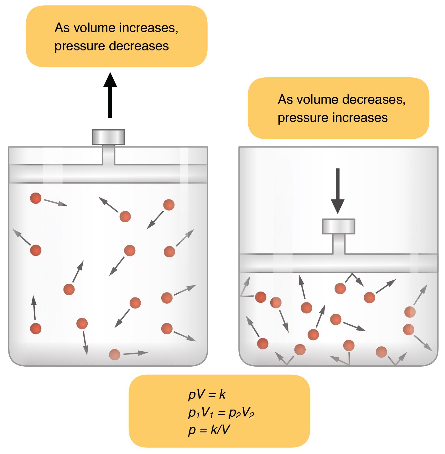
Boyle’s Law
Boyle’s law relates pressure and volume. It can be represented by:
P = 1/V
P = pressure
V = volume
Molecules of a gas will move at random within an enclosed space producing pressure on the walls of the space. The same amount of gas in a smaller space will exert a greater pressure than when in a larger space. So increasing the volume will lower the pressure for a given temperature and vice versa.
This is just what happens during inhalation. The diaphragm contracts and pulls downward increasing the volume of the thoracic cavity. The external intercostals also contract and expand the ribcage. The increased volume decreases the pressure inside of the lungs and air flows from higher pressure outside the lungs to lower pressure inside the lungs.
Expansion of the thoracic cavity causes the lungs to expand because of the pleural cavity. The pleural membranes secrete a fluid that forms a bond between the membranes. The force of this bond produces a small negative pressure.
During exhalation the diaphragm relaxes decreasing the volume of the thoracic cavity. The elastic fibers of the lungs work to move the lungs back to their original shape and the pressure increases moving air out of the lungs. Resting exhalation is considered a passive process.
Accessory Muscles of Respiration
Other muscles besides the diaphragm are involved in respiration when greater amounts of air need to be moved into the lungs. Muscles assisting in inhalation include the sternocleidomastoids, pectoralis minors and external intercostals. Muscles assisting in exhalation include the internal intercostals, and abdominals.
Examine Boyle’s Law below.
Partial Pressure
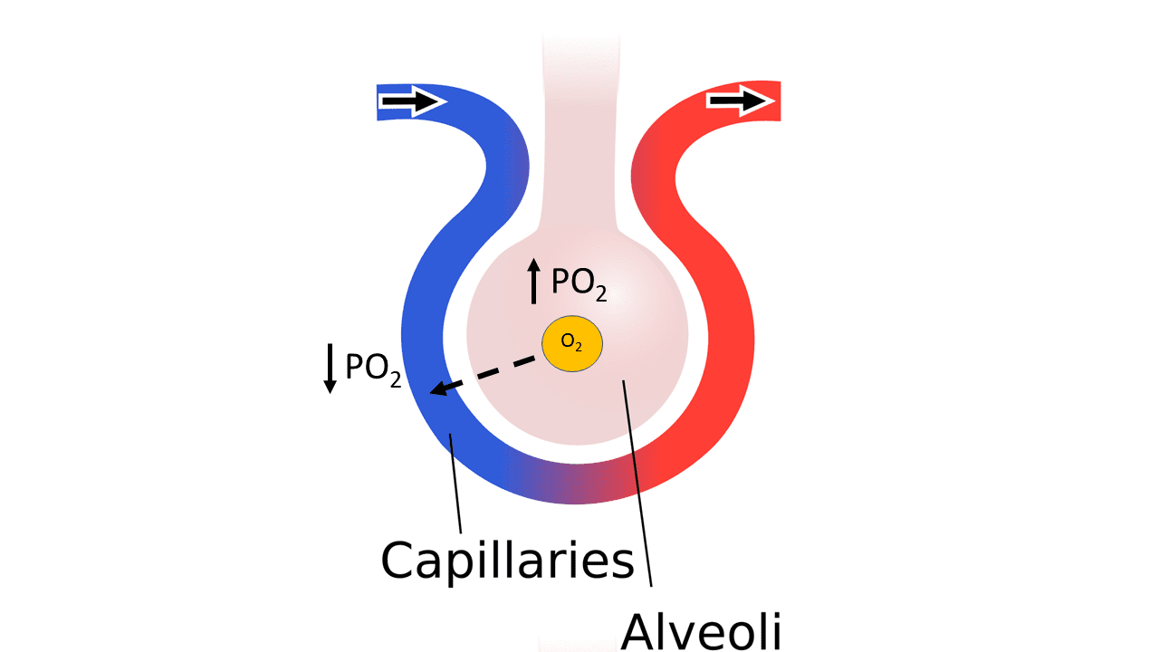
Once air enters the lungs, oxygen must move into the blood by diffusion. Oxygen will follow a partial pressure gradient by moving from the higher partial pressure in the lungs to the lower partial pressure in the blood.
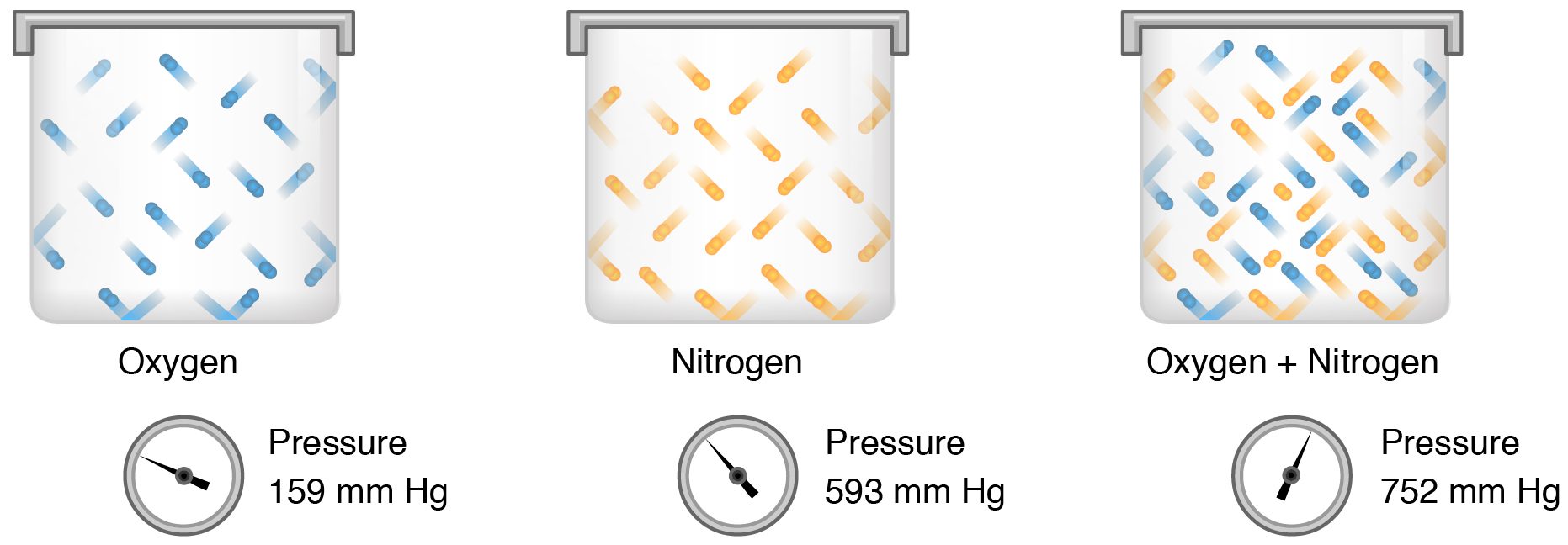
Since air is a mixture of gasses, each individual gas will exert its own pressure which contributes to the overall pressure of the gas.
The pressure each gas produces in the mixture of gases is known as the partial pressure of gas. We can represent the partial and total pressure of a gas such as air as follows:
P(nitrogen) + P (oxygen) + P (water vapor) + P (carbon dioxide) = P (air) = 760 mm Hg
For example if oxygen produces 20.9% of the total pressure of air then 20.9% of 760 mm Hg is about 159 mm Hg. So the partial pressure of oxygen is 159 mm Hg. We can denote partial pressure as PO2 or PCO2.
We can say that gas follows a partial pressure gradient. For example oxygen will move from a PO2 of 100 mm Hg to a PO2 of 80 mm Hg.
Examine the concept of partial pressure in the image below. Each gas exerts its own pressure on the container which then adds to the total pressure in the container on the right.
Movement of Oxygen and Carbon Dioxide
Once oxygen diffuses into the blood, it is carried by hemoglobin in the red blood cells (erythrocytes) to the cells where the partial pressure is lower. Oxygen can then diffuse into the cells.
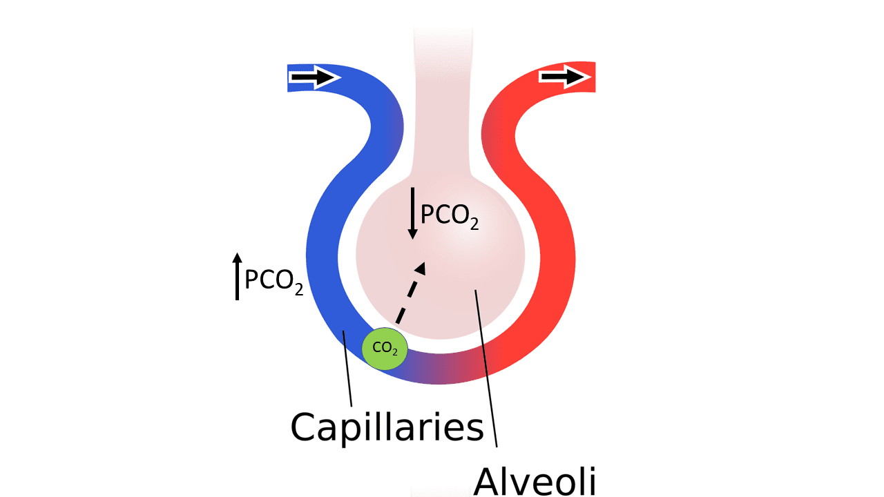
Carbon dioxide is produced by the cells, so the partial pressure of carbon dioxide is higher in the tissues surrounding the cells than in the oxygenated blood entering the cells. Carbon dioxide can then diffuse from the cells into the blood which transports the carbon dioxide to the lungs so then it can diffuse from the blood to the lower partial pressure in the lungs.
Examine how oxygen and carbon dioxide follow their partial pressure gradients in the images below.
Blood Vessels
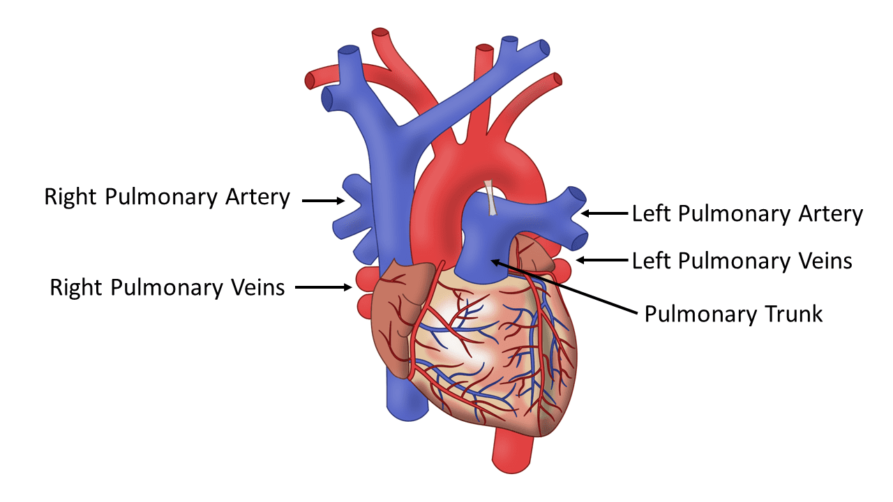
Deoxygenated blood is transported from the right side of the heart (right ventricle) to the pulmonary trunk that divides into right and left pulmonary arteries. The pulmonary arteries eventually become the capillaries surrounding the alveoli.
Once the blood becomes oxygenated by the lungs, it moves back to the left side of the heart by way of the pulmonary veins which carry blood to the left atrium.
Remember, the pulmonary arteries are the only arteries that carry deoxygenated blood and the pulmonary veins are the only veins carrying oxygenated blood.
Examine the blood vessels entering and exiting the lungs from the heart in the image below.
Respiratory Physiology Part 2
Vocabulary
Phrenic Nerve-nerve that controls the diaphragm.
Pons-middle portion of the brainstem.
Medulla Oblongata-lower portion of the brainstem.
Type I Alveolar Cells-mucous secreting cells lining the inside of the alveolus.
Type II Alveolar Cells-surfactant secreting cells lining the insise of the alveolus.
Surfactant-substance secreted by Type II Alveolar Cells that reduces surface tension.
Surface Tension-force created by weak bonds between water molecules.
Carbonic Acid-weak acid that forms from carbon dioxide and water.
Respiratory Acidosis-acidic condition in the blood caused by increased carbon dioxide.
Respiratry Alkalosis-alkaline condition in the blood caused by a decrease in carbon dioxide.
Introduction
Breathing is automatically controlled by the nervous system which monitors the blood for oxygen, carbon dioxide and pH. Once oxygen reaches the alveolus, a delicate balance of cellular secretions helps to maintain the integrity of the alveolus so that oxygen and carbon dioxide can freely move between the alveolus and capillaries. Besides delivering oxygen to the cells and eliminating carbon dioxide, the respiratory system plays a vital role in maintaining blood pH which must be maintained within a very narrow range. Since there is such a close relationship between the respiratory system and pH, problems in the respiratory system can cause imbalances in pH called respiratory acidosis and alkalosis.
Respiration and the Nervous System
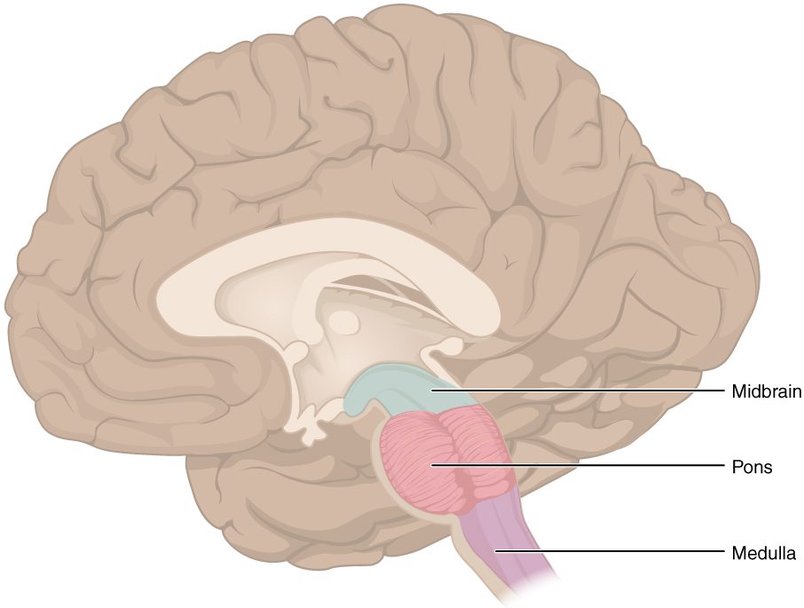
Neural control of respiration begins in the brainstem at the medulla oblongata and the pons. There are 2 paired groups of neurons in the medulla oblongata that work to control the rate and rhythm of breathing. Groups of neurons in the pons help to fine tune breathing and act as a backup system in case there is damage to the medulla oblongata.
The brainstem centers act unconsciously, but can be overriden by higher brain centers. For example, you can consciously increase or decrease your breathing.
The respiratory centers monitor the blood for oxygen, carbon dioxide and pH. There are also chemoreceptors located in other parts of the circulatory system such as in the carotid arteries and aorta.
The brainstem centers connect to the diaphragm by way of the right and left phrenic nerves.
Examine the parts of the brainstem involved in breathing.
Surface Tension and Surfactant
The alveoli contain 2 types of cells. The Type I cells secrete mucous on the inside of the alveolus. Since mucous contains mostly water, surface tension can develop which works to collapse the alveolus.
Surface tension is a force created by weak bonds between water molecules and creates an inward pulling force on the alveolus. The alveolar Type II cells secrete surfactant which works to break the weak bonds and reduce surface tension.
This concept is important in premature infants with underdeveloped lungs. In these cases, diminished secretion of surfactant can cause lung collapse or respiratory distress syndrome unless surfactant is administered.
Transport of Oxygen and Carbon Dioxide in the Blood
Most of the oxygen transported in blood is bound to hemoglobin to form oxyhemoglobin. A small amount of oxygen is dissolved in plasma. Each hemoglobin molecule can bind with four oxygen molecules. Hemoglobin can also release oxygen to form deoxyhemoglobin. There are almost 300 million hemoglobin molecules in one red blood cell. The functional characteristics of hemoglobin are also variable and respond to changes in PO2, pH, and temperature.
Carbon dioxide is transported in the blood by 3 mechanisms. These include carbon dioxide dissolved in plasma, carbon dioxide combining with hemoglobin and storage of carbon dioxide in the bicarbonate ion.
About 7% of the total carbon dioxide in blood is dissolved in plasma. Carbon dioxide also combines with hemoglobin to form a compound known as carbaminohemoglobin. About 23% of carbon dioxide is transported as carbaminohemoglobin. The majority of carbon dioxide (about 70%) is transported in the bicarbonate ion.
Carbon dioxide diffuses into red blood cells and encounters the enzyme carbonic anhydrase to form carbonic acid. Carbonic acid dissociates into bicarbonate and hydrogen ions. Bicarbonate ions diffuse out of the red blood cells into the plasma.
The reaction is reversible with either the storage or release of carbon dioxide depending on what is needed. For example in areas of low PCO2 such as in the alveoli the reaction will work in the direction to release CO2 for removal by the lungs. In areas of high PCO2 such as in the tissues the reaction will work in the direction to store CO2 in the bicarbonate ion.
Respiratory Acidosis and Alkalosis
Because most of the carbon dioxide is transported by the bicarbonate ion with subsequent release of hydrogen ions, a buildup of carbon dioxide in the blood will produce a lower pH. Carbon dioxide and water combine to form carbonic acid in the blood. Carbonic acid dissociates into bicarbonate and hydrogen ions. If the respiratory system cannot release enough carbon dioxide, the subsequent production of hydrogen ions makes the blood acidic. This is known as respiratory acidosis and can result from obstructive diseases such as emphysema or chronic bronchitis. You can generate a mild case of respiratory acidosis by simply holding your breath. The cells continue to produce carbon dioxide but the lungs are not removing it through exhalation. Carbon dioxide builds up in the lungs producing the hydrogen ion byproduct and the blood begins to become acidic.
Likewise you can produce a mild state of respiratory alkalosis by hyperventilating. In this case too much carbon dioxide is removed by the lungs and the hydrogen ion concentration subsequently decreases.
Respiratory Diseases
Vocabulary
Asthma-respiratory disease characterized by inflammation and narrowing of the airway.
Influenza-viral disorder that affects either the upper or lower respiratory systems or both.
Pneumonia-viral or bacterial infection characterized by fluid buildup in the lungs.
Tuberculosis-infectious bacterial disease of the lungs that can affect other parts of the body.
COPD-Chronic Obstructive Pulmonary Disease consisting of bronchitis and emphysema that causes decreased gas exchange in the lungs.
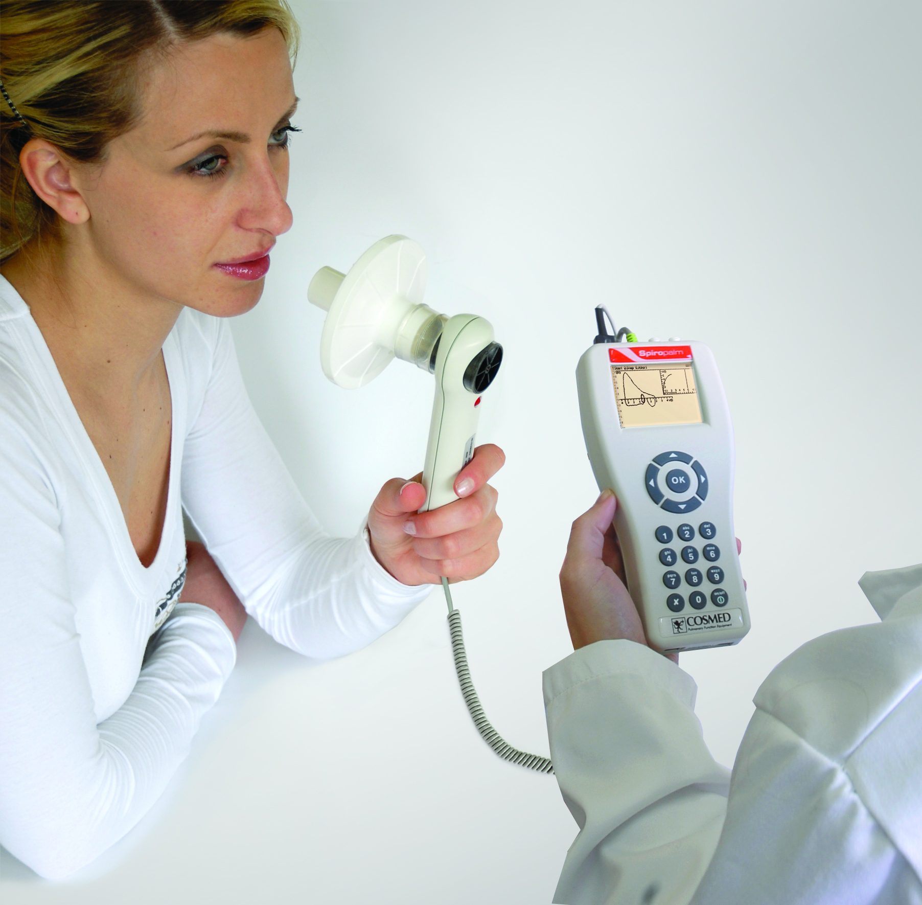
Spirometry-test for volumes of air entering and exiting the lungs that is conducted using a Spirometer that measures air flow.
Tidal Volume-the amount of air entering or exiting the lungs during resting breathing.
Residual Volume-the amount of air remaining in the lungs after a forceful exhalation.
Introduction
Certain diseases can affect the respiratory system in a variety of ways and levels of severity. It is important for health care practitioners to learn about diseases that affect the respiratory system so they can provide better care for their patients.
Asthma
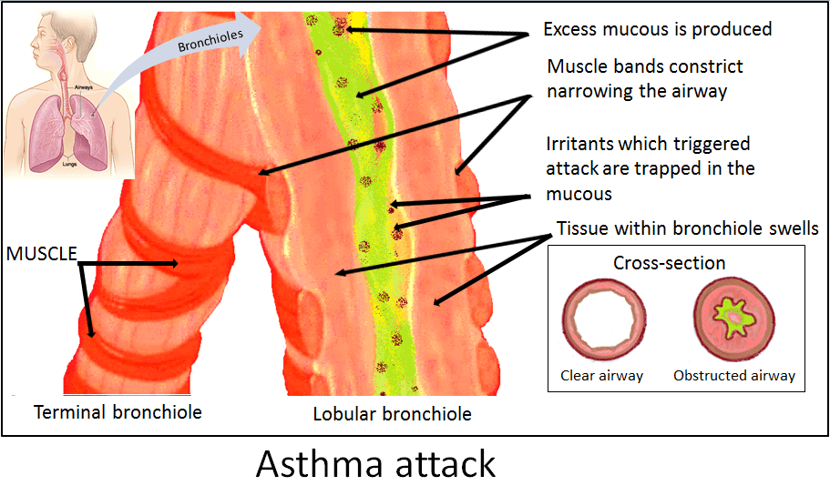
Asthma is a lung disease that is characterized by narrowing of the airway, inflammation and increased mucous production.
During an asthma attack inflammation in the airways causes excess mucous, swelling and narrowing. There is also wheezing, shortness of breath, and chest tightness.
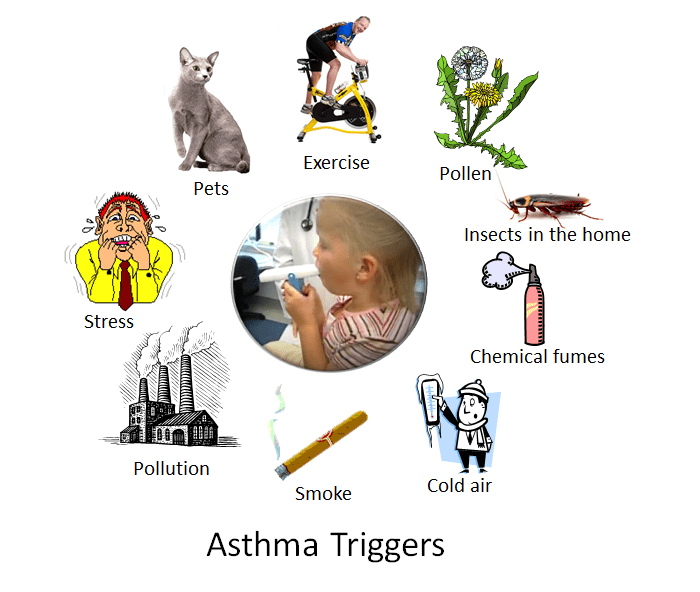
The cause of asthma is unknown, but some things in the environment can trigger an asthma attack. Some examples include cigarette smoke or other pollutants, things you may be allergic to like tree pollens, stress, infections, some medications, and exercise.
There is no cure for asthma, but there are treatments aimed at opening the airways (bronchodilators) and reducing inflammation.
Examine the following image regarding how asthma affects the airway (bronchi).
Examine the following image illustrating triggers for asthma.
Influenza
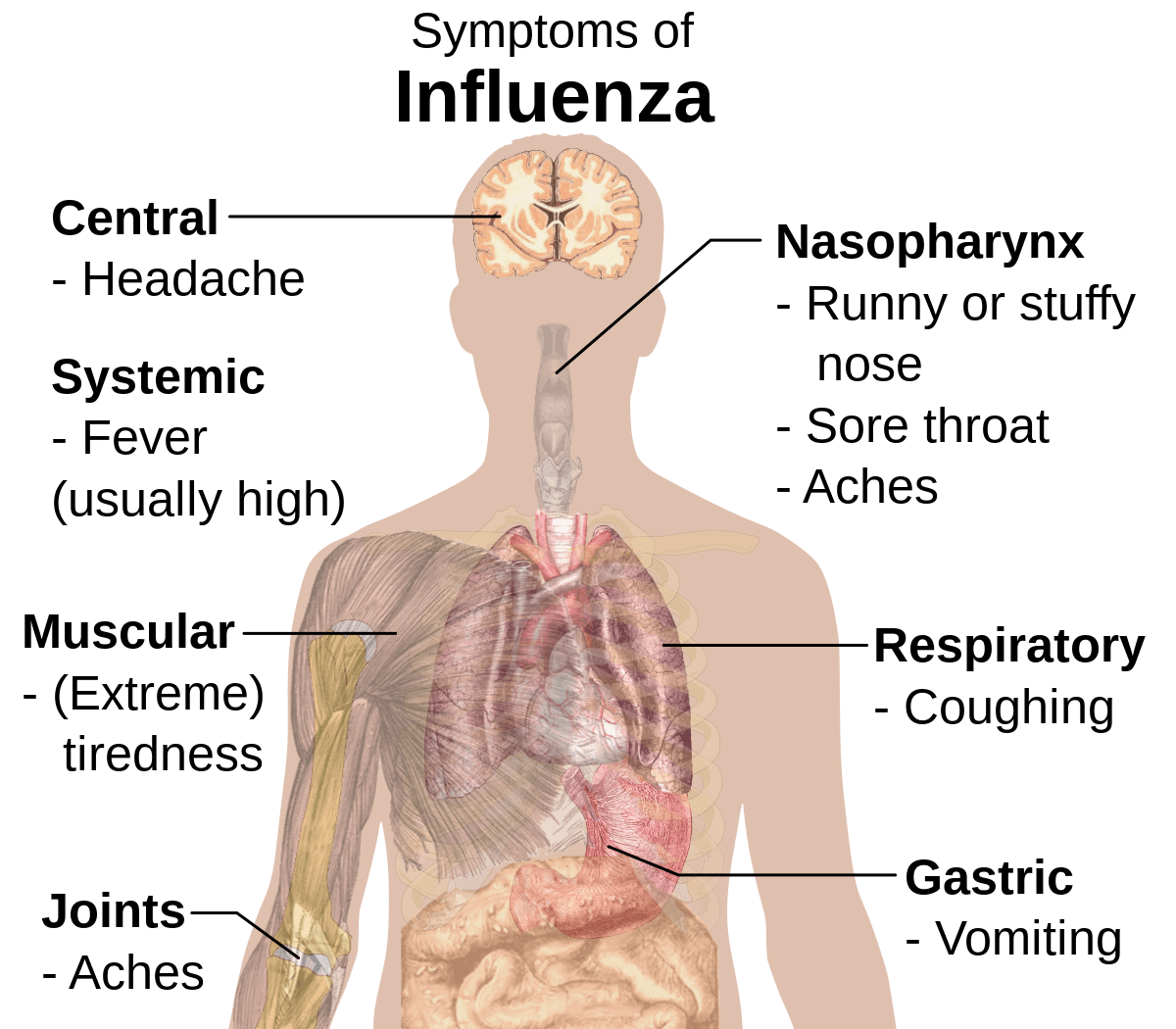
Influenza or flu can affect either the upper or lower respiratory systems or both. Influenza is carried by viruses and causes inflammation and mucous production. Influenza virus is transmitted by tiny droplets in the air from an infected person and incubates for a few days before developing symptoms. Influenza of the lower respiratory tract is usually more severe and can lead to pneumonia.
Examine the following image on how influenza affects the body.
Pneumonia
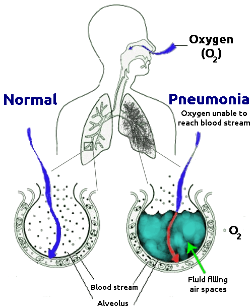
Pneumonia is an infection of the lungs caused by viruses or bacteria that result in fluid buildup in the lungs. Symptoms include fever, chest pain, cough, and difficulty breathing. Pneumonia is treated with either antibiotics, antivirals, or antifungal medications.
Examine the following image regarding how pneumonia affects the alveoli.
Tuberculosis
Tuberculosis (TB) is a disease caused by a bacterium known as Mycobacterium Tuberculosis that is spread through respiratory droplets by someone with active disease. TB causes chest pain, hemoptysis or coughing up blood, weight loss, and fever. TB can also affect other parts of the body such as the skeletal system, liver, adrenal glands, lymph nodes, and coverings of the brain (meninges). There are medications to treat TB that target the bacterium and symptoms.
Examine the following image regarding Tuberculosis.

Chronic Obstructive Pulmonary Disease (COPD)
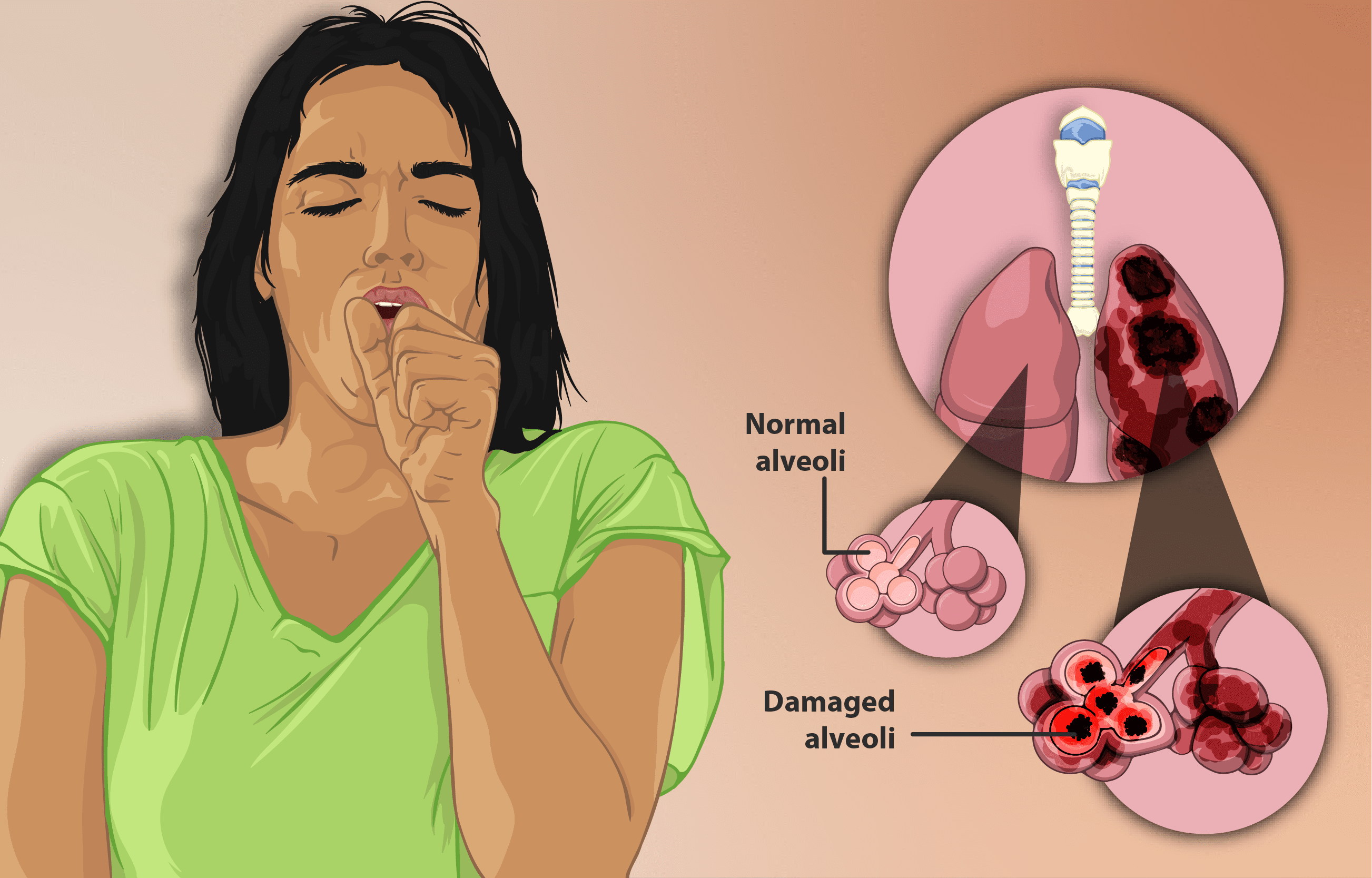
COPD, or chronic obstructive pulmonary disease is a combination of bronchitis and emphysema. Chronic bronchitis is a continuous inflammation of the bronchi with increased mucous production. Emphysema affects the alveoli by causing them to break apart which affects gas exchange. People suffering from COPD experience shortness of breath, wheezing, chronic cough, and mucous production. The symptoms can progress in severity and cause death.
COPD can be diagnosed with lung function tests using a spirometer to measure the amount of air entering and exiting the lungs.
Treatments for COPD begin with lifestyle changes and medications in early stages and can progress to the use of oxygen, and even surgery to remove damaged lung tissue.
Examine the following image about COPD.
Spirometry
Respiratory volumes can be measured with a device called a spirometer. Besides tidal volume other volumes can be measured including inspiratory reserve volume and expiratory reserve volume.
Inspiratory reserve volume (IRV) is the maximum amount of air that can be inhaled in addition to tidal volume. IRV is usually about 3300 ml in males and 1900 ml in females.
Expiratory reserve volume (ERV) is the maximum amount of air that can be exhaled in addition to tidal volume. ERV is about 1000 ml.
Residual volume (RV) is the amount of air remaining in the lungs after a maximal exhalation. RV is about 1200 ml in males and 1100 in females.
Combining respiratory volumes gives us respiratory capacities. These include vital capacity, inspiratory capacity, functional residual capacity and total lung capacity.
Vital capacity is the maximal amount of air that can move in and out of the lungs in a single breath. It is the sum of tidal volume, inspiratory reserve volume and expiratory reserve volume. It is about 4800 ml in males and 3400 ml in females.
Inspiratory capacity is the amount of air that can move into the lungs after resting inhalation and exhalation. Inspiratory capacity is the sum of tidal volume and inspiratory reserve volume.
Functional residual capacity is the air remaining in the lungs after a resting inhalation and exhalation. Functional residual capacity is the sum of expiratory reserve volume and residual volume.
Total lung capacity is the total volume of air in the lungs. It is the sum of vital capacity and residual volume. It is about 6000 ml in males and 4500 ml in females.