Introduction to Anatomical Terms
The Anatomical Position
- Definition: The standardized posture used in human anatomy to describe body structures, directional locations, and movements consistently.
- Key Standards:
- The individual stands upright (erect) facing the observer directly.
- Head, eyes, and toes face directly forward.
- Upper limbs are placed at the sides of the body.
- The palms face forward (anteriorly) with fingers extended.
- The thumbs point laterally (away from the body midline).
- The feet are parallel and point forward.

Anatomical Planes and Sections
- Definition of Plane: An imaginary flat surface that passes through the body to slice or section it for structural study.
- Frontal (Coronal) Plane:
- Divides the body or an organ into anterior (front) and posterior (back) portions.
- Transverse (Horizontal) Plane:
- Divides the body or an organ into superior (upper) and inferior (lower) portions.
- Sagittal Plane:
- Divides the body or an organ vertically into right and left sides.
- Midsagittal Plane: Passes directly along the midline, dividing the body into equal left and right halves.
- Parasagittal Plane: Divides the body into unequal left and right halves.

Directional Terms and Orientation
- Directional Terms Table:
- Ventral: Toward the front or belly. Example: The aorta is ventral to the vertebral column.
- Dorsal: Toward the back or spine. Example: The vertebral column is dorsal to the aorta.
- Anterior: Toward the ventral side. Example: The sternum is anterior to the heart.
- Posterior: Toward the dorsal side. Example: The esophagus is posterior to the trachea.
- Cephalic: Toward the head or superior end. Example: The cephalic end of the embryonic neural tube develops into the brain.
- Rostral: Toward the forehead or nose. Example: The forebrain is rostral to the brainstem.
- Caudal: Toward the tail or inferior end. Example: The spinal cord is caudal to the brain.
- Superior: Above. Example: The heart is superior to the diaphragm.
- Inferior: Below. Example: The liver is inferior to the diaphragm.
- Medial: Toward the median plane / midline of the body; on the inner side of. Example: The heart is medial to the lungs.
- Lateral: Away from the median plane / midline of the body; on the outer side of. Example: The eyes are lateral to the nose.
- Intermediate: Between a more medial and a more lateral structure. Example: The collarbone (clavicle) is intermediate between the breastbone (sternum) and shoulder.
- Proximal: Closer to the origin of a body part or point of attachment of a limb to the body trunk. Example: The elbow is proximal to the wrist.
- Distal: Farther from the origin of a body part or point of attachment of a limb to the body trunk. Example: The fingernails are at the distal ends of the fingers; the knee is distal to the thigh.
- Ipsilateral: On the same side of the body (right or left). Example: The liver is ipsilateral to the appendix.
- Contralateral: On opposite sides of the body (right and left). Example: The spleen is contralateral to the liver.
- Superficial (External): Toward or at the body surface. Example: The skin is superficial to the skeletal muscles.
- Deep (Internal): Away from the body surface; more internal. Example: The bones/lungs are deep to the muscles/skin.

- Directional Differences in Quadrupeds vs. Humans:
- In humans (bipeds), ventral and anterior are synonymous, as are dorsal and posterior.
- In four-legged animals (quadrupeds):
- Anterior refers to the leading portion of the body (the head).
- Ventral refers specifically to the belly or underside (which is the inferior surface in quadrupeds).
- Posterior refers to the tail/rear portion.
- Dorsal refers specifically to the back surface (which is the superior surface in quadrupeds).
Divisions of the Skeletal System
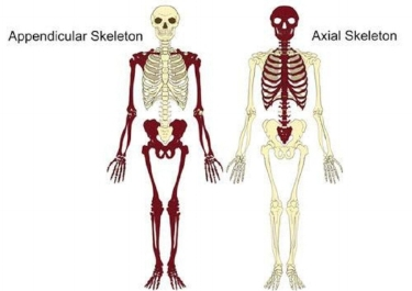
- Axial Skeleton:
- Forms the central vertical axis of the body.
- Composed of the skull (head), vertebral column, and thoracic cage (ribs and sternum).
- Appendicular Skeleton:
- Consists of the upper and lower limbs (appendages).
- Includes the attachment girdles:
- Pectoral (Shoulder) Girdle: Attaches upper limbs to the axial trunk.
- Pelvic (Hip) Girdle: Attaches lower limbs to the axial trunk.
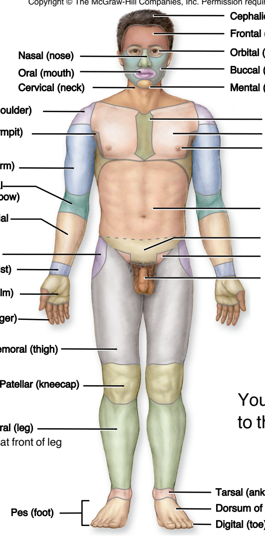
Body Regions
- Anterior Body Regions:
- Cephalic: Head region.
- Frontal: Forehead.
- Orbital: Eye region.
- Nasal: Nose region.
- Buccal: Cheek.
- Oral: Mouth.
- Mental: Chin.
- Cervical: Neck region.
- Acromial: Point of the shoulder.
- Axillary: Armpit region.
- Brachial: Arm (upper arm).
- Antecubital: Anterior surface of the elbow.
- Antebrachial: Forearm.
- Carpal: Wrist.
- Palmar: Palm of the hand.
- Digital: Fingers or toes.
- Sternal: Breastbone area.
- Pectoral / Thoracic: Chest area.
- Umbilical: Navel region.
- Coxal: Hip region.
- Inguinal: Groin region.
- Femoral: Thigh region.
- Patellar: Kneecap (anterior knee).
- Crural: Shin/anterior leg.
- Tarsal: Ankle region.
- Dorsum of Foot: Top surface of the foot.
- Pes: Foot.
- Posterior Body Regions:
- Cranial: Region surrounding the brain.
- Auricular / Otic: Ear region.
- Occipital: Base or back of the head.
- Vertebral: Spine / vertebral column area.
- Dorsal / Scapular: Shoulder blade / back region.
- Cubital / Olecranal: Posterior elbow.
- Lumbar: Lower back region between ribs and hips.
- Gluteal: Buttock region.
- Perineal: Region between the anus and external genitalia.
- Popliteal: Hollow behind the knee (posterior knee).
- Sural: Calf (posterior leg).
- Calcaneal: Heel of the foot.
- Plantar: Sole of the foot.

Body Cavities, Membranes, and Coeloms
- Cavities vs. Coeloms Distinction:
- There are 7 major body cavities in total, but only 3 true coeloms.
- All coeloms are cavities, but not all cavities are coeloms.
- Coelom Definition: A fluid-filled body space lined with serous membranes (mesothelium/epithelium) that encloses internal organs and provides clearance for movement without friction.
- Dorsal Cavities (Lined by meninges; not true coeloms):
- Cranial Cavity: Formed by cranial bones; contains the brain.
- Vertebral Canal: Formed by the vertebral column; contains the spinal cord.
- Ventral Cavities:
- Thoracic Cavity: Superior ventral cavity bounded by the chest wall; separated from the abdominal cavity by the diaphragm. Contains:
- Pleural Cavities (2 coeloms): Enclose the left and right lungs.
- Mediastinum: Central region between the lungs; contains the esophagus, trachea, thymus, and major blood vessels.
- Pericardial Cavity (1 coelom): Encloses the heart within the mediastinum.
- Abdominopelvic Cavity: Divided into:
- Abdominal Cavity: Superior portion containing the stomach, intestines, liver, gallbladder, spleen, and pancreas.
- Pelvic Cavity: Inferior portion containing the urinary bladder, internal reproductive organs, and rectum.
- Scrotal Cavity: Found exclusively in males, accommodating the testes.


- Serous Membranes (Example: Pericardium):
- Visceral Layer: Inner layer that adheres directly to the surface of the organ (e.g., visceral pericardium).
- Parietal Layer: Outer layer that lines the inner wall of the body cavity (e.g., parietal pericardium).
- Cavity Space: Fluid-filled potential space between the visceral and parietal layers.
- Balloon Analogy: Pushing a fist into a flexible balloon models heart tissue surrounded by serous layers:
- The fist represents the heart organ.
- The inner balloon wall touching the fist represents the visceral pericardium.
- The outer balloon wall represents the parietal pericardium.
- The air space inside the balloon represents the fluid-filled cavity.

Abdominopelvic Quadrants and Regions
- Four Abdominopelvic Quadrants:
- Formed by a vertical midline and horizontal transumbilical line crossing at the navel.
- Right Upper Quadrant (RUQ): Contains the right lobe of the liver, gallbladder, right kidney, and portions of the stomach and intestines.
- Left Upper Quadrant (LUQ): Contains the stomach, spleen, left lobe of the liver, pancreas, left kidney, and portions of the intestines.
- Right Lower Quadrant (RLQ): Contains the cecum, appendix, right ovary/spermatic cord, and right ureter.
- Left Lower Quadrant (LLQ): Contains the sigmoid colon, left ovary/spermatic cord, and left ureter.
- Nine Abdominopelvic Regions:
- Grid Boundaries:
- Vertical Lines: Midclavicular lines (drawn through the midpoint of each clavicle).
- Superior Horizontal Line: Subcostal line (passes across the lowest boundary of the 10th costal cartilage).
- Inferior Horizontal Line: Intertubercular line (passes horizontally between the tubercles of the iliac crests).
- Regional Breakdown:
- Epigastric Region: Upper central region above the stomach.
- Right Hypochondriac Region: Upper right region covered by the lower ribs.
- Left Hypochondriac Region: Upper left region covered by the lower ribs.
- Umbilical Region: Central region surrounding the umbilicus.
- Right Lumbar Region: Right middle region adjacent to the lumbar vertebrae.
- Left Lumbar Region: Left middle region adjacent to the lumbar vertebrae.
- Hypogastric (Pubic) Region: Lower central region below the umbilical area.
- Right Inguinal (Iliac) Region: Lower right region near the groin and iliac crest.
- Left Inguinal (Iliac) Region: Lower left region near the groin and iliac crest.

Human Organ Systems Overview
- Integumentary System:
- Major Organs: Skin, hair, nails, cutaneous glands.
- Functions: Protection, water retention, thermoregulation, vitamin D synthesis, cutaneous sensation.
- Skeletal System:
- Major Organs: Bones, cartilages, ligaments.
- Functions: Support, movement lever systems, protection of internal organs, blood formation, mineral storage, acid-base balance.
- Muscular System:
- Major Organs: Skeletal muscles.
- Functions: Movement, stability, posture, heat production, communication, control of body openings.
- Nervous System:
- Major Organs: Brain, spinal cord, nerves, ganglia.
- Functions: Rapid internal communication, coordination, motor control, sensation processing.
- Endocrine System:
- Major Organs: Pituitary, pineal, thyroid, parathyroid glands, thymus, adrenal glands, pancreas, ovaries, testes.
- Functions: Hormone production, long-term internal chemical coordination and metabolism control.
- Cardiovascular System:
- Major Organs: Heart, blood vessels.
- Functions: Distribution of oxygen, nutrients, hormones, metabolic wastes, immune cells, and heat.
- Lymphatic System:
- Major Organs: Lymph nodes, lymphatic vessels, thymus, spleen, tonsils.
- Functions: Fluid recovery from tissues, immune defense, pathogen detection, cell absorption.
- Respiratory System:
- Major Organs: Nose, pharynx, larynx, trachea, bronchi, lungs.
- Functions: Oxygen absorption, carbon dioxide discharge, speech, acid-base balance.
- Urinary System:
- Major Organs: Kidneys, ureters, urinary bladder, urethra.
- Functions: Waste elimination, regulation of blood pressure/volume, electrolyte and acid-base balance.
- Digestive System:
- Major Organs: Teeth, tongue, salivary glands, esophagus, stomach, small and large intestines, liver, gallbladder, pancreas.
- Principal Functions: Nutrient breakdown and absorption.
- Liver Specific Functions: Metabolism of carbohydrates, lipids, proteins, vitamins, and minerals; plasma protein synthesis; disposal of drugs, toxins, and hormones; blood cleansing.
- Male Reproductive System:
- Major Organs: Testes, epididymides, ductus deferentia, seminal vesicles, prostate, bulbourethral glands, penis.
- Principal Functions: Production and delivery of sperm; secretion of male sex hormones.
- Female Reproductive System:
- Major Organs: Ovaries, uterine tubes, uterus, vagina, vulva, mammary glands.
- Principal Functions: Production of oocytes, site of fertilization and fetal development, fetal nourishment, childbirth, lactation, secretion of female sex hormones.

