Fetal vs. Adult Circulation & How the Heart Beats
I - Fetal Circulation
Human development while inside the uterus has the following stages:
Zygote: the moment of fertilization until implantation into the uterus
Embryo: implantation until about 8 weeks of development
Fetus: 8 weeks until birth
Because fetuses are in a different environment before birth (no air), their lungs are undeveloped until right before birth, and are not used for gas exchange at all until birth.
There are 4 major differences in fetal circulation due to their non-functional lungs, in addition to the placenta, which is used for exchange instead.
Placenta: a network of intertwining but not continuous capillary beds that grow out of the uterus and out of the umbilical cord.
→ Separate blood streams: exchange between mother and child occurs by diffusion from 1 capillary through the walls into another.Ex: wastes, gases, nutrients, hormones, alcohol, drugs (include caffeine, Tylenol), and even some cells.

Ductus venosus (venous duct): passes through the liver, connecting the umbilical vein (oxygenated blood) with the vena cava.
Ductus arteriosus (arterial duct): between the aorta and pulmonary artery.
Redirects most “pulmonary loop blood” back into the body via the aorta.
→ Less blood goes through the lungs, where no gas exchange occurs in the fetus.
Formen ovale (oval opening): located between the atria.
Allows blood destined for the pulmonary loop to bypass the lungs and go to the systemic loop instead.
Umbilical arteries & veins: located between fetal and maternal circulatory loops.
Allows blood to travel from the placenta toward the vena cava (umbilical vein).
Allows blood to travel from the iliac arteries toward the placenta (umbilical arteries).
All 4 adaptations disappear before birth or shortly after.
Ex: An oval opening closes a few months before birth, usually, but in 25% of people, incomplete closure occurs. However, this is normally inconsequential because the hole closes during atrial contraction.
Ex: The ducts disintegrate about 8 days after birth.
II - Regulation of Heartbeat

The heartbeat is initiated within the heart tissue itself, not by the brain.
Sinoatrial node (aka “natural pacemaker”) is a group of specialized cells that depolarize like neural tissue.
Located in the posterior wall of the right atrium, near the opening of the anterior vena cava.
Its resting potential is higher than the threshold voltage (- 55 mV), so it depolarizes on its own (“automatic self-excitation”) at a certain rate.
Cardiac cycle: one complete round of the contraction and relaxation of the atria and the ventricles.
Steps of the cardiac cycle:
The sinoatrial (SA) node initiates the impulse.
The impulse travels through special branching fibres (muscle tissue) to both atria.
This causes atrial systole (aka “atrial contraction”)
The two atria contract simultaneously.
This will pump the blood to the ventricles through the AV valve.
The impulse will also travel through fibres to the atrioventricular (AV) node.
The AV node is located in the right atrium, near the lowest part of the septum.
The AV node continues the impulse through special muscle fibres called the Purkinje fibres to both ventricles.
This causes the ventricles to contract together to send blood out of the heart through the semilunar valves.
→ This is called ventricular systole (aka “ventricular contraction”)
Shortly after the ventricular systole begins, the atria start to relax.
This is called atrial diastole.
Once the ventricles complete systole, they relax and go into ventricular diastole.
Both the atria and ventricles will be in diastole at the same time.
During diastole, the atria fill with blood, and the ventricles partially fill with blood.
The cycle then starts again.
The atria and the ventricles need to contract at separate times to ensure that the blood flows in one direction.
What would happen if the atria and ventricles were to contract at the same time?
Heart sounds: “lubb dupp.”
“lubb”: the sound of the AV valve closure (at the beginning of the ventricular systole).
“dupp”: the sound of the semilunar valves closing (at the start of the ventricular diastole).
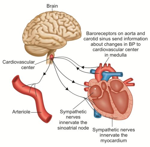
The heart rate is controlled by the sympathetic and parasympathetic nerves.
The cardiac centre in the brain sends signals to the SA node to initiate the impulses slower or faster.
The SA node also has receptors for adrenaline to increase the strength and frequency of the nerve impulses.