DNA Structure, Profiling, and Gel Electrophoresis Laboratory Study Guide
Historical Context and Application of DNA Profiling
The Case of Colin Pitchfork: DNA profiling first gained prominence following crimes committed against two young women in England during the mid-1980s.
Initially, a lead suspect was tried, but a DNA profiling method demonstrated that his DNA did not match the biological evidence found on the victims.
Authorities subsequently requested blood or saliva samples from local men to perform mass DNA sampling.
After six months of testing, no initial match was found.
It was later discovered that an individual named Colin Pitchfork had paid a friend to provide a DNA sample in his place.
Once a genuine sample was obtained from Pitchfork, his DNA was confirmed as a match to the samples collected from the victims, leading to his conviction.
Fundamental Definition and Function of DNA
Definition: DNA stands for deoxyribonucleic acid.
Biological Importance: DNA is the fundamental molecule that encodes all instructions specifying the plans for a cell's makeup.
The Blueprint Metaphor: DNA serves as the biological "blueprint." Just as a construction crew requires a master plan or blueprint to build a house, a cell utilizes the instructions encoded in DNA to build proteins and organize its structure.
The Molecular Structure of DNA
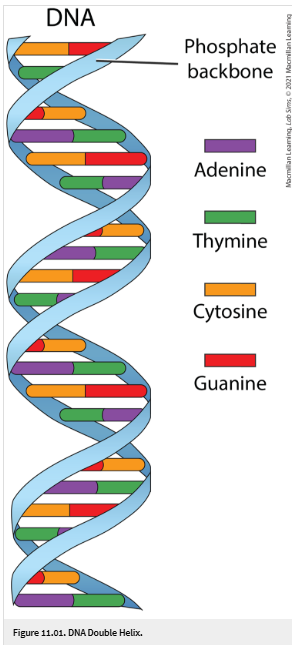
Double Helix Structure: DNA consists of two strands of nucleotides wound together in a spiral. It is often described as a "twisted ladder" or a rope ladder that has been rotated.
Nucleotides: The basic building blocks of DNA. A single nucleotide is composed of three components:
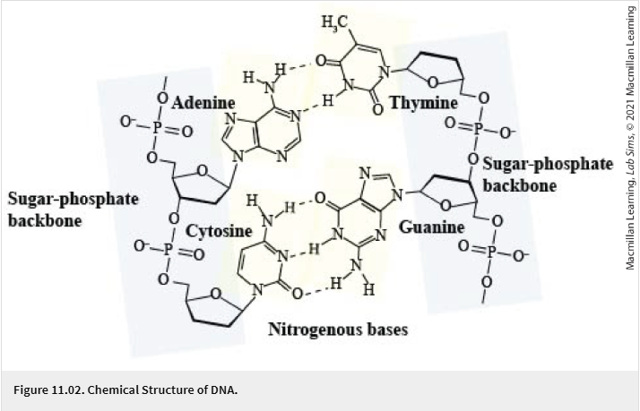
Pentose Sugar: Specifically, a five-carbon sugar known as deoxyribose.
Phosphate Group: Forms the structural framework along with the sugar.
Nitrogenous Bases: There are four types of bases in DNA:
Adenine ()
Thymine ()
Cytosine ()
Guanine ()
The Sugar-Phosphate Backbone: The "sides" of the ladder are made of alternating sugar and phosphate groups.
Chemical Bonding:
Covalent Bonds: These strong bonds connect the sugar and phosphate molecules along the backbone.
Hydrogen Bonds: These relatively weak bonds connect the nitrogenous bases in the center of the helix.
The instructor notes that Adenine and Thymine share hydrogen bonds, while Cytosine and Guanine share (Note: In standard molecular biology, these values are typically reversed, but the transcript states has and has ).
The weakness of hydrogen bonds is essential for biological processes, allowing the two strands to be separated easily during replication.
DNA Replication and the Complementary Base Pairing Rule
Complementary Base Pairing: Bases always pair in a specific manner:
Adenine () always pairs with Thymine ().
Cytosine () always pairs with Guanine ().
Replication Process (S-phase of Interphase):
The double strand is "unzipped" or split apart by breaking the hydrogen bonds between the bases.
Each "old" strand serves as a template for a "new" strand.
New nucleotides are brought in according to the complementary rules (e.g., if the old strand has an , the new strand will incorporate a ).
This results in two identical copies of the DNA molecule.
Genomic Characteristics and VNTRs
Human Genome Composition:
The human DNA sequence contains over (three billion) base pairs.
Despite this massive size, there are only approximately protein-coding genes.
Human DNA is identical between any two individuals.
VNTRs (Variable Number Tandem Repeats):
The difference between individuals is found in non-coding regions, specifically the VNTRs.
VNTRs are repeating base pair sequences (e.g., ) that occur throughout the DNA molecule.
The length of these sequences varies significantly between individuals, making them ideal for DNA testing and forensic identification.
Principles of Paternity Testing
Inheritance Patterns: A child inherits half of their DNA fragments (VNTRs) from their biological mother and the other half from their biological father.
Analysis Logic:
When looking at DNA bands on a gel, any band present in the child that does not match a band in the mother must be present in the biological father.
By comparing the child's remaining bands to potential fathers, the biological father can be identified with high certainty.
Even identical twins, while starting with the same genome, may develop minor mutations in VNTR sequences over time due to different environmental exposures and diets.
The DNA Profiling Process: Step-by-Step
Isolation: Cells (often from a cheek swab) are broken apart to release the DNA. Alcohol is typically used in the final step to precipitate and isolate the DNA from the cellular debris.
Restriction Enzyme Digest:
Restriction Enzymes act as "molecular scissors."
They are programmed to recognize and cut the DNA at specific sequences immediately before and after the targeted VNTR regions.
Polymerase Chain Reaction (PCR):
Metaphor: PCR is the "copy machine" of the laboratory.
A single isolated fragment is amplified into thousands or millions of copies so it can be visualized.
Three Major Steps of PCR Cycle:
Denaturation: High temperatures are used to separate the two DNA strands.
Annealing: Specific DNA primers attach to the starting points of the separated strands.
Elongation: New DNA strands are synthesized, extending from the primers. This process leads to exponential growth of the target sequence.
Gel Electrophoresis: The process used to separate the amplified DNA fragments by size for visualization.
Mechanics of Gel Electrophoresis
The Apparatus: A tank containing a power supply, a buffer solution, and a gel (which acts like a sponge with pores).
The Gel: Usually a blue-tinted slab with "wells" (rectangular holes) at the top where the DNA samples are loaded.
Charge and Migration:
DNA molecules are negatively charged.
During electrophoresis, the negative electrode is placed near the wells, and the positive electrode is placed at the far end.
When the current is turned on, the DNA is attracted to the positive electrode and migrates through the gel.
Fractionation by Size:
The migration rate is inversely proportional to the fragment size.
Smaller fragments move faster and further through the pores of the gel toward the bottom (positive electrode).
Larger fragments (high molecular weight) move more slowly and remain closer to the top (the wells).
The Buffer Solution:
Provides the correct for the experiment.
Protects DNA from being degraded or digested.
Conducts the electric current necessary for migration.
Loading Agents:
Glycerol: Added to DNA samples because it is heavy; it ensures the DNA sinks into and stays in the well rather than floating away in the top buffer.
Tracking Dye: A simple colored dye is added to help the researcher see where the sample is as it is loaded and as it moves during the run.
Laboratory Protocol and Procedure
Preparation:
Equip personal protective equipment (PPE).
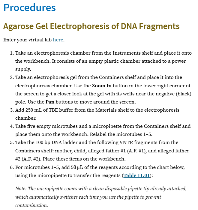
Place the electrophoresis chamber on the workbench and insert the gel.
Add of BTE buffer to the tank.
Sample Setup:
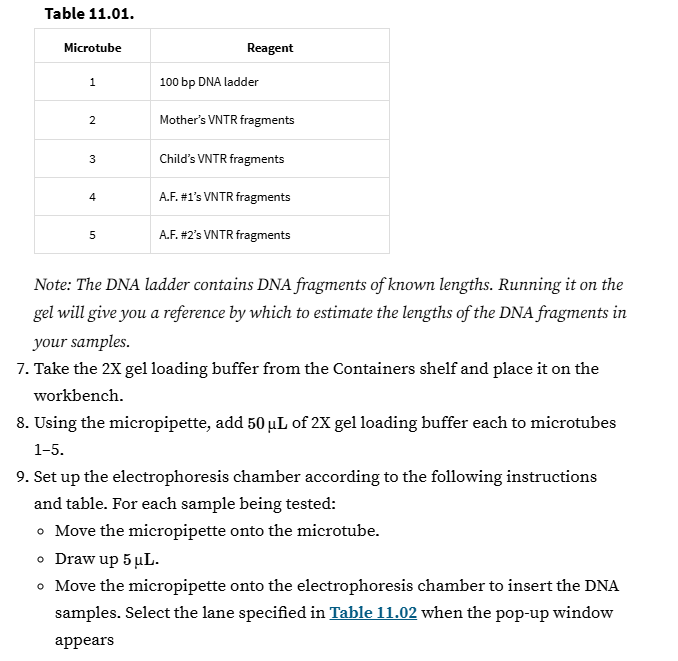
Label five microcentrifuge tubes ().
Add of the following to the respective tubes:
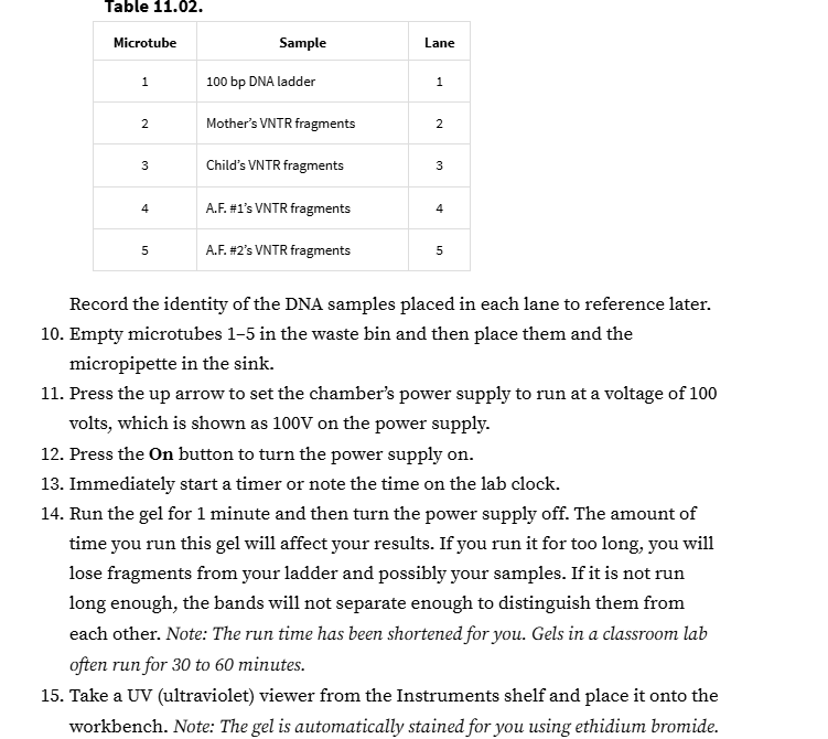
Tube 1: -base pair (bp) DNA ladder (marker).
Tube 2: Mother's VNTR fragments.
Tube 3: Child's VNTR fragments.
Tube 4: Potential Father 1's VNTR fragments.
Tube 5: Potential Father 2's VNTR fragments.
Add of Gel Loading Buffer to each tube.
Loading the Gel:
Use a micropipette to draw up from each tube and dispense into the corresponding wells ( through ) in the gel.
Running the Gel:
Set the voltage to .
Critical Timing: Turn the power supply on for exactly one minute. Do not exceed this time, or the bands may run off the end of the gel.
Visualization and Analysis
UV Light Exposure:
Because DNA is naturally transparent, the gel must be exposed to a second dye that binds to DNA.
Under UV light, this dye fluoresces, allowing the DNA bands to become visible.
Using a DNA Ladder (Marker):
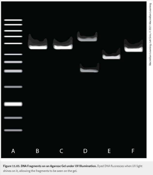
The ladder contains fragments of known sizes used for comparison.
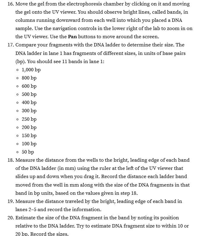
The -bp ladder used in this lab should produce distinct bands.
The top band is , the next is , and they decrease in size toward the bottom.
Determining Paternity:
Observe the bands in the child's column.
Cross-reference with the mother's bands.
Identify the child's bands that do not exist in the mother.
The man whose DNA fragments match these remaining bands is the biological father.
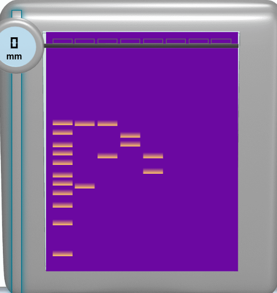
Around 35mm:
lane 2: 990bp
lane 3: 1000bp
40mm:
lane 4: 780bp
45mm:
lane 4: 600bp
50mm:
lane 3: 480bp
lane 5: 480bp
57mm
lane 5: 320bp
63bp
lane 2: 230-240bp