Chapter 12: Parturition and Postpartum Period
Study Notes
Parturition and Lactation
Gestation length
Specie
Average Length (days)
Swine 114
Sheep 148
Cattle 280
Mare 340
Factors influencing gestation length:
Maternal age
younger dams have shorter
Sex of fetus
males 1-2 days longer than females
Twins are carried 3-6 day than singles.
Genotype of fetus
breed differences exist
Cattle fetuses from breeds with shorter gestations have decreased gestation length when transferred to recipients with longer gestations.
Mule foals have longer gestations than horse foals.
Some of these differences may be due to different growth rates.
Species and breed differences in fetal size are largely due to genetic differences in the rate of cell division.
Most of the increase in weight occurs in the last two months of gestation.
Nutrition at this time is critical.
Undernutrition early in gestation has little effect on the fetus.
Late in gestation, poor nutrition decreases fetal growth and chances for survival after birth.
Age, Parity, Size of the mother affect birth weight.
PARTURITION: Labor & Delivery
Signs of approaching parturition:
Enlargement of mammary glands: may include milk or colostrum dripping from teats
Nest building behavior can be seen in swine if given the chance.
Mucous may be seen stringing from the vulva.
Stages of Labor
I. Dilation of Cervix:
regular uterine contractions caused by estrogen and PGF2a
Lasts from 2-12 hours
Marked by maternal restlessness
2. Expulsion of fetus:
stronger uterine contractions oxytocin is involved now. Abdominal contractions begin.
Rupture of chorioallantoic and amnion. Lasts from 30 min. to 3 hours.
Oxytocin secretion is stimulated by a neuroendocrine reflex.
3. Expulsion of the placenta:
uterine contractions decrease in strength. It cannot be separated from the second stage in sows or sheep with twins.
Of short duration in sows and mares.
Takes longer in ruminants because of the type of placenta.
Cows: the expulsion of placenta normally 6-12 hours If >24 hours, it is considered a retained placenta.
Retained placentas lead to increased risk of infections and can delay rebreeding.
Metritis: infection of the uterus
Antibiotic infusions to the uterus or antibiotic boluses will often lead to expulsion.
Oxytocin and PGF2a have been used to attempt to cause expulsion of fetus.
Manual removal is not recommended.
Vitamin A or E or Selenium deficiencies can increase the incidence of retained placentas.
Dystocia: difficult birth
Dystocia increases the incidence of retained placentas.
Two main causes of dystocia: large birth weights and abnormal presentation of the fetus
Large birth weights more of a problem in females giving birth
for the first time.
Parity: number of parturitions a female has experienced pregnancy
Nulliparous: none
Primiparous: 1
Multiparous: >1
Labor usually takes longer in primiparous females.
The normal presentation of the fetus is forward with the head between the front legs.
Other types of presentations can lead to dystocia.
Abnormal presentations more common in multiple births in monotocous species.
Some changes required for survival of neonate outside of the uterus.
Ductus Arteriosus: open in the fetus, shunts blood away from the lungs. This must close quickly after the umbilical cord is broken.
Foramen Ovale: an opening that allows blood to go from right atrium to left atrium, Closes shortly after birth
POSTPARTUM
Postpartum Period: repair of the reproductive tract to prepare for another pregnancy
Uterine Involution: restoration of the uterus to nonpregnant size and function will never return to the exact size as before pregnancy. Always slightly larger
Timing
Mares: rapid involution, about 2-3 weeks
Sows: rapid involution, 2-3 weeks
Ewes: return to nonpregnant size in about two weeks, Another two weeks required for regeneration of the endometrium
Cows: return to nonpregnant size 25 -30 days. full regeneration of endometrium takes another 2-3 weeks
Dystocia, retained placentas, metritis: all extend the time needed for uterine involution
One uterine horn may stay larger than the other.
Lochia
uterine discharge -- consists of fetal membranes, maternal tissue, blood
Lasts 2-7 days postpartum
Discharge due to the continued release of PGF2a from uterus. PGF2 secretion also promotes more rapid involution.
Release of oxytocin by suckling also promotes uterine involution
Postpartum Anestrous Period
For most species, reproductive activity does not resume immediately after parturition.
There is a period of time before estrous cycles start again.
Mares are an exception to this.
Most mares exhibit FOAL HEAT: 8-15 days postpartum.
They can be bred at this time if the reproductive tract is recovered from pregnancy.
Fertility is lower than at later estrus.
The other farm species exhibit a period of anestrus and anovulation postpartum.
Factors affecting the length of Postpartum Anestrus
Suckling of progeny
Nutrition of dam:prepartum and postpartum
Age of dam:primiparous females have longer anestrous periods
Season of year: sows weaned in summer are more likely to remain anestrus
Breed: Brahma based breeds have longer anestrous periods than Bos taurus breeds
Sows: Remain anestrus for the typical 3-4 week lactation period
Estrus is seen 4-7 days after weaning.
Poor nutrition can delay the return to estrus.
Split weaning: weaning the larger piglets 24- 48 hours before smaller ones can improve return to estrus
Ewes: Postpartum Period is complicated by seasonal anestrus.
Ewes lambing in spring do not cycle until the next fall because of long days of spring and summer.
Ewes lambing in the fall will resume estrous cycles in 5-6 weeks.
Cows: Dairy cows cycle 20-30 days postpartum
Usually bred on the second estrus.
Inadequate dietary energy in high producing cows can delay return to estrus.
Beef cows have longer anestrus periods than dairy because of the suckling of calves.
Calf removal after birth causes beef cows to return to estrus, similar to dairy cows.
48-hour calf removal shortly before the breeding season can increase the number of cows cycling.
Low energy diets before or after calving will lengthen the postpartum anestrus.
In many cows, the first ovulation postpartum is silent: without estrus.
This is followed by a short-lived CL and then a normal estrus and ovulation.
The CL is short-lived because of the early release of PGF2 alpha.
Suckling lengthens the postpartum interval by suppressing GnRH and LH secretion.
The psychological bonding of the dam to calf also delays rebreeding.
Low dietary energy also suppresses GnRH and LH secretion.
LACTATION
Essential for the survival of young
Anatomy of Mammary System
Cow: udder contains four glands, each with a single teat.
Each gland or quarter is a separate unit.
Ewe and doe: 2 glands with one teat each
Mare: 4 secretory regions fused into two glands
Each gland has a single teat which drains 2 of the secretory regions
Sow: 4-9 pairs of glands located along both sides of the midline.
Each gland has a single teat.
Colostrum
The first milk produced immediately after parturition. It is higher in protein and vitamin A than milk.
It contains immunoglobulins to provide passive immunity to neonate until its own immune system begins to function.
Other hormones involved in Lactation
Placental Lactogen: ruminants
Stimulates duct and alveoli growth
Some hormones synergize (make more effective)
Cortisol, thyroid hormones, insulin, GH synergize with estrogen, and progesterone during pregnancy to increase mammary development.
Prolactin is necessary for the start of lactation.
In cows, after lactation starts, prolactin is no longer necessary.
GH can increase milk production in cows by directing nutrient transfer to the mammary gland.
Prolactin will not cause this effect.
This effect of GH reduces the amount of nutrients available for other physiological systems.
Suckling
Suckling and or removal of milk is necessary for continued lactation.
The presence of milk in the cisterns prevents further milk synthesis.
Suckling causes the release of several hormones.
Oxytocin is essential for milk release, not milk synthesis.
Oxytocin is released by a Neuroendocrine Reflex.
Milk let-down in dairy cows is stimulated by washing and massaging of the udder in parlor.
Milk let-down can become a conditioned reflex so that sounds or sights can trigger oxytocin release.
Other hormones released by suckling include Prolactin, GH, and cortisol.
Stress can inhibit milk let-down through the release of Epinephrine from the Adrenal Medulla.
Section 1. Birth and Its Importance to the Producer
the creation, growth, and development of a potential new individual as they developed from zygote to embryo, to fetus, to mature fetus. The natural conclusion of this growth is the process of birth, which is more properly termed Parturition
In mammals, parturition represents the true separation of the new animal as an independent functional life form. Up until this point, it has been totally dependent on its mother’s physiology for survival. Now, while it will still be dependent on its mother for a time for nourishment, it will survive only if its own physiology is up to the task of maintaining its existence.
Look again at the figure of the three phases of development (Figure 1). An animal (or human) born before the completion of all these tasks has little hope of surviving on its own and will most likely die just after birth. While this is a tragedy for the individual, it also affects the producer. While the exact loss to the producer would depend on the species and the production scheme, there is no denying, death of an animal is a loss. But it goes further than just the loss of the animal. There are also the lost production days of its mother, the feed costs associated with those days, and the potential that the death may lead to issues with the mother that will cause her to be culled from the herd.
1st - 3rd | 2nd - 3rd | 3rd - 3rd |
Maternal Recognition | Completion of Differentiation | Most Rapid Increase in Weight (%) |
Implantation | ||
Embryo to Fetus | Sexual Differentiation | Maturation of Lungs |
Most Rapid increase in Cell number (%) | Organ Maturation | Parturition |
Section 2. Length of Gestation
So how long is gestation, and what factors can affect its length? Normal gestational lengths will vary from species to species (Table 1). However, there are a number of factors which can influence the length of gestation. One of the larger influences across species is maternal age. Young dames deliver sooner than older animals. There are two potential reasons. One, the animal is still growing herself and does not have the body frame nor the nutrients to allow the fetus to grow to full size. The other potential reason is that the uterus cannot expand sufficiently to accommodate the developing fetus.
Species | average length (days) | |
Swine | 114 | |
Sheep | 148 | |
Cattle | 280 | |
Mare | 340 |
Other factors that are known to affect gestational length include male fetuses, which on average will remain in utero an additional 1–2 days longer than females of the same species, and twins, which on average remain 3–6 days less than singleton pregnancies (in non-litter bearers). The genotype also plays a major role. When embryos from shorter-length gestation cattle are transferred into animals that normally have a longer gestational period, the fetuses still deliver according to their genetics, not those of the carrier. These differences appear to be due to genetic programming of the rate of cellular divisions, which leads to larger fetuses
Section 3. Parturition
While the exact mechanisms leading to the onset of parturition are not understood, we do know a number of things about the process. Parturition can be divided into three separate phases: (1) labor, (2) delivery of the fetus, and (3) delivery of the placenta. We also know that there are signs of approaching parturition we can use to tell an animal is near to delivery. The first is strictly how close the animal is getting to her normal gestation length. This works well in cattle and is an effective tool for hiring outside help at calving time. Other signs of approaching parturition include (1) enlargement of the mammary glands as they become organized and filled with milk, this may culminate in milk or colostrum dripping from the teats and (2) animals, especially swine, exhibiting nest-building behavior and/or mucous becoming liquidy and seen stringing from the vulva.
The first phase of parturition would be labor and would include dilation of the cervix. First contractions would be started by estrogen, which would trigger prostaglandin 2α (PGF2α) contractions as the placenta pulls away from the uterine wall. The combination of estrogen and PGF2α will lead to regular rhythmic contractions, which may last for hours. In general, this would be the longest phase of labor and would be marked by periods of restless.
As labor progresses, the uterus will contract around the fetus. The membranes of the placenta will separate, and in general, the amniotic fluid will leak out what is commonly called “water breaking". If labor is progressing normally, the cervix should be fully dilated open by this time, and the collapsing of the membranes and the uterus around the fetus will cause it to struggle. Its struggling will lead to the arc-reflex reaction, which causes oxytocin to be released from the posterior pituitary and stronger, abdominal contractions. Under normal conditions, the contractions will move the fetus from the uterus, through the open birth canal, to the outside world. However, in order for the fetus to smoothly pass through the birth canal, it must be both the right size and in the right orientation, or there will be delays (Figure 2).
the malpositioning of a limb in the uterus will cause it to “hang up” as it is being pushed down the birth canal. Further, if the fetus is larger than the opening in the pelvic bone, it would be difficult or impossible for the fetus to pass out of the reproductive tract. Any delay in delivery could have dire consequences for the fetus and is termed Dystocia, as once the membrane separates, the fetus will become starved for oxygen.
Dystocia: difficult delivery
Under normal conditions, this phase of labor will last between 30 minutes and 3 hours.
As the fetus passes through the birth canal, two very important anatomical/physiological changes must take place in its system if it is to survive. First, the ductus arteriosus, a blood vessel that has shunted blood away from the lungs and through the umbilical cord, must close. Second, the foramen ovale, an opening that has allowed blood to move directly from the right to left atria (which until now has been moving oxygenated blood from the placenta, through the umbilical cord, to the fetus), must close, as the lung begins to function and supply oxygen to the body.
Once the fetus delivers, the animal must still pass the placenta before the birth canal closes. However, once the fetus is on the ground as a neonate, contractions will decrease as oxytocin secretions stop.
Slower contractions generally mean this phase of labor will last longer than the delivery of the fetus. Although in some species, generally litter bearers, the fetus and the placenta deliver together. In singleton species, such as the cow and horse, this phase of labor will normally last between 6 and 12 hours. It is essential this phase of labor be completed because as long as the placenta is retained in the uterus, the birth canal will remain open. If we have an animal who does not pass her placenta after a prescribed period of time (in cattle 24 hours), we would say she has a retained placenta. What’s the big deal? The birth canal is open, she is bleeding, it’s warm and moist and full of nutrients. In short, it is a bacteria’s dream, and the animal is ripe for an infection, or what is properly termed Metritis
Metritis: uterine infection often associated with delivery
So, if retained placentas are an issue, what can we as producers do? First, we know that deficiencies of the vitamins A or E, or the element selenium, are associated with retained placentas. Therefore, it is critical to ensure the animal has access to these chemical agents during pregnancy. There are also a series of steps that can be done to treat retained placenta. The first should be obvious. If oxytocin and PGF2α naturally cause contraction of the uterus, then the administration of either hormone should stimulate placental delivery. This method can work and work well, but a word of caution. Never be standing behind the animal when administering these drugs. Because they are triggering strong contractions, it is much like causing severe abdominal cramps. The animal may kick in response. The second option is to place antibiotics directly into the uterus. This method has two advantages. It will help fight the infection, but the chemical nature of the antibiotic (usually acidic) will irritate the endometrium and causes it to release its own PGF2α, causing contractions leading to delivery. A third option is to combine the two techniques. One method that is discouraged is to remove the placenta through what is termed manual removal. There are many reasons manual removal might be an issue, but the primary concerns are retained pieces of the placenta, which will both slow the healing process and set the animal up to develop metritis as the tissue dies.
Section 4. The Postpartum Period
The postpartum period is characterized by two major physiological events. For most species, the first process is an anestrus period for the repair of the uterus—what is referred to as postpartum anestrus. The second event, which is absolutely necessary for mammals if the neonate is to survive, is the synthesis and release of milk products. Both require a large amount of energy, meaning, as a producer, this is a period we need to ensure proper nutrition if we want to maximize our animals’ production potentials.
Section 4.1 Uterine Repair
Think about the uterus and the events that have just concluded with parturition. We are left with a large, damaged and bleeding, cavity that must be repaired. The very first step in this repair is the Involution
Involution: shrinking of the uterus after parturition to close to its prepregnancy size
Involution represents a rapid return of the uterus to its prepregnancy size and shape. Just after parturition, this shrinking will cause a flow of blood and membranes termed Lochia
Lochia: Uterine discharge of fetal membranes, maternal membranes, and blood after parturition
Under normal conditions, lochia will last 2–7 days and should progress from liquidy bright red fluids to a darkening brown color over time. Extended bleeding after a week might indicate an issue needing veterinarian attention.
The second part of uterine repair is repair and regrowth of the endometrium. It should be obvious that parturition results in damage to the endometrium, as the placenta is torn away during labor. The exact amount of damage will be dependent on several factors (see below), but is mainly dependent on how a species’ placenta attaches to the endometrium. Table 2 shows the average time for uterine repair in most common domestic species.
species | timing of involution (in weeks) | Additional time for endometrial repair (in weeks) | Total (in weeks) |
Cows | 4 | 2-3 | 6-7 |
Ewes | 2 | 2 | 4 |
Mares | 2-3 | None | 2-3 |
Sows | 2-3 | None | 2-3 |
As suggested above, a number of things might delay the animal returning to cycle after parturition. Functionally, think of anything that leads to a difficult birth (dystocia) as causing issues that will lead to a delay in the next estrus. In general, difficult births will lead to more damage to the endometrium, which will require longer periods for repair. Other factors that have been shown to influence the length of the anestrus period are postpartum associated metritis, retained placentas, suckling (species-specific), poor nutrient of the dam, age of the dam, season of the year (especially in seasonal breeders), and even differences in breeds—where larger breeds tend to spend slightly longer for uterine involution and endometrial repair. As a producer, anything that leads to delay in return to estrus represents an additional production cost and a delay to future income.
Section 4.2 The horse, the usual exception
Unlike other species, the horse does not go through a postpartum anestrus. Instead, the horse exhibits a postpartum estrus or what is also called foal heat. In general, horses will return to estrus in 5–15 days post delivery. As described in Chapter 6, these are not always fertile cycles as the mare may not ovulate on this cycle. Further, these cycles must be properly managed to ensure the best outcomes for the mare, her new foal, and the potential new embryo growing inside her.
Section 4.3 Lactation
Lactation, the secretion of milk from mammary glands, is the culmination of a series of physiological events in the mammary tissue, which allow for the synthesis and release of milk providing the essential nutrients necessary for the neonate’s survival
Lactation: the secretion of milk from the mammary glands
It begins in early pregnancy where progesterone and estrogen from the placenta cause the mammary tissue to organize the glands that will synthesize the milk and the ducts that will then deliver the milk to the cistern. Prolactin, which will start being produced as early as week 8 of pregnancy, will stimulate the synthesis of milk in the glands with the aid of placental lactogen, cortisol, growth hormone, and thyroid hormone (all of which are synergistic with prolactin). While milk synthesis will start early in pregnancy, no milk will be expressed from the mammary until just before parturition, when a small amount of milk may be expressed due to delivery contracts, or at least a waxy build up on the teats composed of proteins and lipids.
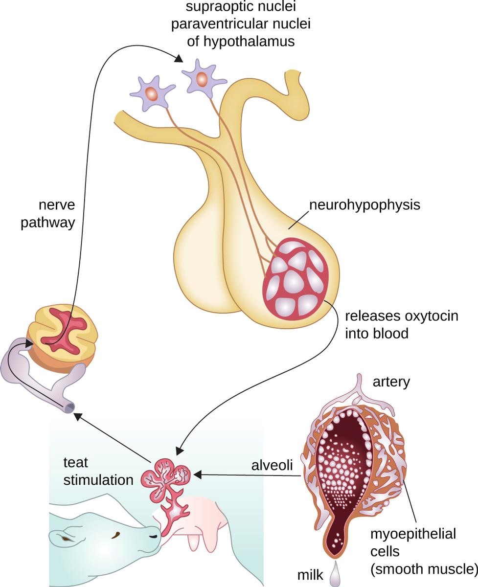
There are significant differences in the anatomy of the mammary glands of each species (Table 3). However, the expression of milk works the same in all species. This process was discussed in Chapter 3. To review, the process begins with the young animal suckling at its mother’s teat. The suckling sets off nerves in the teat, which send a nerve impulse up the spine to the thalamus in the brain. The impulse is routed to the hypothalamus where it stimulates the super optic and paraventricular nerves (the nerves that made the majority of oxytocin and served as its pathway to the posterior pituitary for storage). The nerve impulse follows the path down the axon to the posterior pituitary triggering the release of oxytocin into the bloodstream. Oxytocin travels to the mammary tissue where it binds to the glands triggering them to contract, forcing milk into the cistern, and the young animal receives dinner
Species | No. of Glands | |
Cows | 4 | |
Ewes | 2 | |
Mares | 2* | |
Sows | 4-9 pairs |
*The mare has four seperate secretory regions which fuse into two glands.
So a question, have you ever had the veterinarian give an animal an oxytocin shot to induce milk letdown? Did it work? If you have and it worked, all of the physiology to synthesis worked, but for whatever reason, the animal was not allowing the release of milk from the glands (this is often the result of some form of stress). Because the shot contained several times more oxytocin than normal, it overcame whatever was blocking milk letdown. However, if you have used the shot and it didn’t work, there was little to no milk in the glands, that is, it was never made. If it’s never made, it cannot be released so a second or third oxytocin shot will have no more effect than the first shot the veterinarian gave; you are just spending money.
Now let’s talk about what is in milk that the neonate needs. The first milk will be what is termed colostrum
colostrum: the first secretions from the mammary glands after parturition, which is high in antibodies
Colostrum is higher in protein, sugar, and vitamin A than milk later produced. Colostrum also contains higher levels of antibodies. Unlike adult animals, neonates have the ability to move intact immunoglobins across the gut to provide passive immunity until the young animal’s own immune system kicks into gear. Watch the following video for a more complete explanation of this process 1-Passive Immunity.
After the first few days, the milk will thin; having few if any immunoglobulins, and fewer nutrients in the same volume of fluid. As the neonate feeds more on grass and other solids, it will drink less and less milk. The mother’s milk production will slow as the glands stay full of milk for longer and longer periods until it finally stops (dries up).