Human Movement
Levels of Structural Organization of the Human Body
Introduction
Levels of Structural Organization
Definition: The orderly way scientists divide the complex human body, from the smallest units to the entire organism.
Important details — the six levels, from simplest to most complex:
Chemical Level
Cellular Level
Tissue Level
Organ Level
Organ System Level
Organismal Level
Chemical Level
Chemical Level
Definition: The simplest level of organization, consisting of atoms and molecules.
Examples of Atoms | Examples of Molecules |
|---|---|
Carbon (C) | Water (H₂O) |
Hydrogen (H) | Glucose (C₆H₁₂O₆) |
Oxygen (O) | DNA |
Nitrogen (N) | Proteins |
Calcium (Ca) |
Example: Hydrogen + Oxygen → Water (H + H + O = H₂O) shows atoms combining to form a molecule.
Importance: Chemical substances provide energy, build body structures, and regulate body processes.
Element | Symbol | Function |
|---|---|---|
Oxygen | O | Cellular respiration |
Carbon | C | Forms organic molecules |
Hydrogen | H | Component of water |
Nitrogen | N | Makes proteins and DNA |
Calcium | Ca | Bone formation |
Example: Drinking a glass of milk provides calcium ions that will eventually be used to strengthen bones — starting at the chemical level.
Cellular Level
Cellular Level
Definition: Cells are the smallest living units of the body, formed when atoms and molecules combine.
Important details: Cells obtain nutrients, produce energy, remove wastes, reproduce, and respond to stimuli.
Cell Type | Function |
|---|---|
Red Blood Cell | Carries oxygen |
Neuron | Conducts impulses |
Muscle Cell | Produces movement |
White Blood Cell | Fights infection |
Example: A single muscle cell contracting is what ultimately allows an entire muscle, like the biceps, to bend the elbow.
Tissue Level
Tissue
Definition: A group of similar cells performing a common function.
Tissue Type | Function |
|---|---|
Epithelial | Covers and lines surfaces |
Connective | Supports and binds |
Muscle | Produces movement |
Nervous | Conducts electrical signals |
Epithelial Tissue — covers and lines body surfaces, such as the outer layer of skin.
Connective Tissue — supports and binds structures together, such as tendons connecting muscle to bone.
Muscle Tissue — produces movement through contraction, such as the tissue making up the biceps.
Nervous Tissue — conducts electrical signals, such as the tissue making up nerves and the brain.
Example: The lining inside your cheek, made of epithelial tissue, protects the deeper layers of your mouth from friction while chewing.
Organ Level
Organ
Definition: A structure composed of two or more tissue types working together.
Example: The Heart
Tissue | Function |
|---|---|
Muscle tissue | Pumps blood |
Nervous tissue | Controls heartbeat |
Connective tissue | Supports heart |
Epithelial tissue | Lines chambers |
Example: The stomach is another organ made of muscle tissue (to churn food), epithelial tissue (to line its inner surface), connective tissue (to support its structure), and nervous tissue (to control digestive signals) — all working together for digestion.
Organ System Level
Organ System
Definition: A group of organs working together to perform a major body function.
The Eleven Organ Systems:
Organ System | Main Function |
|---|---|
Integumentary | Protection |
Skeletal | Support |
Muscular | Movement |
Nervous | Control |
Endocrine | Hormone regulation |
Cardiovascular | Transport |
Lymphatic | Immunity |
Respiratory | Gas exchange |
Digestive | Digestion |
Urinary | Waste removal |
Reproductive | Reproduction |
Example 1 — Digestive System: Mouth → Esophagus → Stomach → Small Intestine → Large Intestine (organs working together to digest food).
Example 2 — Cardiovascular System: Heart → Blood → Vessels → Blood (organs working together to circulate blood throughout the body).
Organismal Level
Organismal Level
Definition: The highest level of organization; all organ systems work together to form one living individual.
Example: A student who is breathing (respiratory system), digesting lunch (digestive system), and running to class (muscular and skeletal systems) all at the same time represents the organismal level — every system working together as one living person.
Summary of the Six Levels
Level | Description | Example |
|---|---|---|
Chemical | Atoms and molecules | Water, glucose, DNA |
Cellular | Smallest living units | Red blood cell, neuron |
Tissue | Groups of similar cells | Muscle tissue, nervous tissue |
Organ | Two or more tissues working together | Heart, stomach |
Organ System | Group of organs working together | Cardiovascular system |
Organismal | All systems working together | A whole living person |
The Language of Anatomy and Foundations of Gross Structure
Why Do We Need Anatomical Terminology?
Anatomical Terminology
Definition: A universal language that allows healthcare professionals and scientists to communicate accurately about body structures.
Example: Instead of saying "the pain is on the back of my lower leg," a nurse would chart it as "pain on the posterior aspect of the crus."
Anatomical Position
Anatomical Position
Definition: The standard reference position assumed for all anatomical descriptions.
Important details:
Standing upright
Head facing forward
Feet flat and slightly apart
Arms at the sides
Palms facing forward
Example: A textbook diagram showing a person facing the reader, arms slightly away from the body with palms out, is drawn in anatomical position so directions can be described consistently.
Directional Terms
Term | Meaning | Example |
|---|---|---|
Superior | Above | Nose is superior to the mouth |
Inferior | Below | Stomach is inferior to the heart |
Anterior | Front | Sternum is anterior to the heart |
Posterior | Back | Vertebral column is posterior to the heart |
Medial | Toward midline | Heart is medial to the lungs |
Lateral | Away from midline | Ears are lateral to the nose |
Proximal | Closer to attachment | Elbow is proximal to the wrist |
Distal | Farther from attachment | Fingers are distal to the elbow |
Superficial | Toward surface | Skin is superficial to muscles |
Deep | Away from surface | Bones are deep to muscles |
Example (applying several terms together): A scrape on the outer, upper part of the forearm, close to the skin, could be described as being on the "lateral, proximal, and superficial" aspect of the forearm.
Body Planes
Body Planes
Definition: Imaginary flat surfaces used to divide the body for descriptive purposes.
Sagittal Plane
Definition: Divides the body into right and left portions.
Important details: The Midsagittal Plane specifically divides the body into equal right and left halves.
Example: An X-ray taken from the side of the body, showing the front-to-back profile, is viewed along the sagittal plane.
Frontal (Coronal) Plane
Definition: Divides the body into front (anterior) and back (posterior) portions.
Example: A yearbook photo taken from the front, showing the body split into a front half and a back half, illustrates the frontal plane.
Transverse Plane
Definition: Divides the body into upper and lower portions.
Example: A CT scan "slice" of the abdomen, showing a cross-section from above, is taken along the transverse plane.
Major Body Cavities and Membranes
Body Cavities
Definition: Protective, fluid-filled compartments that hold and protect the internal organs so they are not loose inside the body.
Dorsal Cavity (Back)
Definition: The cavity located along the back of the body.
Important details:
Cranial cavity — houses the brain.
Vertebral/Spinal cavity — houses the spinal cord.
Example: A helmet protects the cranial cavity's contents (the brain) the same way the skull naturally does.
Ventral Cavity (Front)
Definition: The cavity located along the front of the body, divided by the diaphragm muscle.
Important details:
Thoracic cavity — chest area containing the lungs and heart.
Abdominopelvic cavity — contains digestive organs like the stomach and liver, and reproductive/urinary organs in the pelvis.
Example: A seatbelt across the chest and hips is designed to protect the thoracic and abdominopelvic cavities during a car accident.
Body Cavities and Organs (Table)
Cavity | Major Organs |
|---|---|
Cranial | Brain |
Vertebral | Spinal cord |
Thoracic | Heart, lungs |
Abdominal | Stomach, liver |
Pelvic | Urinary bladder, reproductive organs |
Approaches to Studying Gross Anatomy
Systemic Anatomy
Definition: Studying the body system by system (e.g., all the bones, then all the muscles, then the entire circulatory system).
Important details: This is the most common textbook approach.
Example: A textbook chapter devoted entirely to "The Skeletal System," covering bones throughout the whole body, uses the systemic approach.
Regional Anatomy
Definition: Studying all structures in a specific area of the body at once.
Important details: This is how medical students typically study via dissection.
Example: A cadaver lab session focused only on the structures of the neck — muscles, bones, blood vessels, and nerves all together — uses the regional approach.
Surface Anatomy
Definition: Studying internal structures as they relate to the overlying skin.
Example: A nurse locating a patient's pulse by feeling for the radial artery just under the skin of the wrist is applying surface anatomy.
Foundations of Gross Structure
1. The Integumentary System
Integumentary System
Definition: The body's outer protective layer, comprising the skin, hair, nails, and glands.
Important details: Acts as a primary defense against pathogens, regulates body temperature, prevents fluid loss, and synthesizes vitamin D.
Functions: Protection, Temperature regulation, Sensation, Vitamin D synthesis.
Example: Goosebumps forming on the skin when a person feels cold demonstrate the integumentary system's role in temperature regulation.
Epidermis
Definition: The thin, outermost layer of the skin.
Important details: Acts as a primary barrier against the environment, protecting the body from harmful pathogens, UV radiation, and dehydration, while also producing new skin cells and determining skin color.
Example: A minor scrape that only removes the very top layer of skin and heals within a day or two usually only affects the epidermis.
Dermis
Definition: The thick, supportive middle layer of skin located beneath the epidermis and above the subcutaneous fat.
Important details: Primarily composed of connective tissue, collagen, and elastin, which provide structural strength, elasticity, and resilience.
Example: A stretch mark forms when the dermis is stretched beyond its elastic capacity, such as during a growth spurt.
Hypodermis
Definition: The deepest layer of skin, primarily composed of fat cells and connective tissue.
Important details: Acts as a shock absorber, insulates the body to regulate temperature, and anchors the skin to underlying muscles and bones.
Example: The extra cushioning felt when sitting for a long time comes largely from the hypodermis.
Skin Layers (Table)
Layer | Function |
|---|---|
Epidermis | Protection |
Dermis | Blood vessels and nerves |
Hypodermis | Fat storage |
2. The Skeletal System
Skeletal System
Definition: The body's central support structure, comprising bones, cartilage, ligaments, and tendons.
Important details:
An adult human has 206 bones.
Shapes the body, protects vital organs, allows for movement via muscle attachment, produces blood cells, and stores minerals like calcium.
Functions: Support, Protection, Movement, Mineral storage, Blood cell production.
Example: The ribcage acting like a protective cage around the heart and lungs shows the skeletal system's protective function.
Classification of Bones
Long Bones
Definition: Cylindrical bones that are longer than they are wide.
Important details: Act as levers to facilitate movement. Examples: femur (thigh bone), humerus (upper arm), phalanges (finger bones).
Example: The femur acting as a lever when kicking a ball demonstrates the function of long bones.
Short Bones
Definition: Cube-shaped bones that provide stability and limited movement.
Important details: Examples: carpals (wrist) and tarsals (ankle).
Example: The small, blocky bones in the wrist that allow controlled, limited movement while typing are short bones.
Flat Bones
Definition: Thin, flat, and often curved bones.
Important details: Protect vital organs and offer broad surfaces for muscle attachment. Examples: sternum, ribs, scapulae, cranial skull bones.
Example: The skull protecting the brain from a minor bump to the head is an example of a flat bone doing its protective job.
Irregular Bones
Definition: Bones with complex shapes that don't fit into other categories.
Important details: Examples: vertebrae (spine) and the hip bones.
Example: The uniquely shaped vertebrae that stack to form the spine's curve are irregular bones.
Sesamoid Bones
Definition: Small, rounded bones embedded within tendons to protect them from compressive forces.
Important details: Examples: patella (kneecap) and the pisiform (a small wrist bone).
Example: The kneecap protecting the tendon that crosses the front of the knee while jumping is a sesamoid bone.
Sutural (Wormian) Bones
Definition: Tiny, extra bones that occur within the jagged, sutural joints between the skull bones.
Example: Some people have small extra bone fragments along their skull sutures that show up incidentally on an X-ray — these are sutural bones.
Major Bone Markings
Projections (Attachment Sites) — raised areas that allow muscles, tendons, and ligaments to anchor to bone.
Tuberosity: A large, rounded, often roughened projection (e.g., tibial tuberosity).
Tubercle: A small, rounded projection (e.g., greater tubercle of the humerus).
Trochanter: A very large, irregularly shaped process found only on the femur (e.g., greater trochanter).
Epicondyle: A raised area located on or above a condyle (e.g., medial epicondyle of the femur).
Crest: A prominent, narrow ridge of bone (e.g., iliac crest of the hip).
Spine: A sharp, slender, or pointed projection (e.g., ischial spine).
Line: A narrow ridge of bone, less prominent than a crest (e.g., superior nuchal line).
Example: A basketball player who develops soreness at the tibial tuberosity from repeated jumping is experiencing strain at a bone projection.
Articular Surfaces (Joint Formers) — smooth surfaces that help form joints where bones meet.
Head: The expanded, rounded end of a bone, often supported by a neck (e.g., head of the femur).
Condyle: A smooth, rounded articular projection (e.g., occipital condyles).
Facet: A nearly flat, smooth articular surface (e.g., articular facets of vertebrae).
Depressions (Basins and Grooves) — valleys in bone that guide joint motion or provide pathways for soft tissue.
Fossa: A shallow, basin-like depression (e.g., mandibular fossa).
Notch: An indentation at the edge of a bone (e.g., greater sciatic notch).
Groove (Sulcus): A furrow or elongated depression that houses vessels and nerves (e.g., intertubercular sulcus).
Openings (Passageways) — holes or slits that allow blood vessels and nerves to pass through or enter the bone.
Foramen: A round or oval opening through a bone (e.g., foramen magnum in the skull).
Fissure: A narrow, slit-like opening (e.g., superior orbital fissure).
Meatus: A canal-like passageway (e.g., external acoustic meatus/ear canal).
Sinus: A cavity within a bone, typically air-filled and lined with mucus (e.g., frontal sinus).
Example: The spinal cord passing safely through the foramen magnum at the base of the skull shows how an "opening" bone marking works.
3. The Muscular System
Muscular System
Definition: An intricate network of over 600 muscles that make up roughly half of total body weight, driven by the nervous system.
Important details: Its primary function is to contract and relax to facilitate physical movement, maintain posture, pump blood, and generate body heat.
Functions: Movement, Posture, Heat production.
Example: Shivering when cold is the muscular system rapidly contracting to generate heat.
Muscle
Definition: Specialized soft tissue made of elastic fibers that contracts and relaxes to produce movement, maintain posture, support organs, and pump blood.
Example: The calf muscle contracting to help you rise onto your toes.
Fascicle
Definition: A bundle of skeletal muscle fibers (or nerve fibers) held together by connective tissue.
Example: A bundle of uncooked spaghetti strands tied together resembles how fascicles bundle individual muscle fibers.
Muscle Fiber
Definition: An individual, elongated muscle cell that makes up muscle tissue and generates movement by contracting.
Example: Millions of these individual "threads" working together let the biceps contract when lifting a bag.
Myofibril
Definition: A basic, rod-like organelle of a muscle cell; groups of these make up muscle fibers and are primarily responsible for contraction and relaxation.
Myofilament
Definition: Thread-like protein structures that make up myofibrils; by interacting with one another they generate the force and movement required for muscle contractions.
Major Skeletal Muscles
Deltoid, Pectoralis major, Biceps brachii, Triceps brachii, Rectus abdominis
Gluteus maximus, Quadriceps, Gastrocnemius
Muscle | Function |
|---|---|
Deltoid | Arm abduction |
Biceps | Flexion |
Triceps | Extension |
Quadriceps | Knee extension |
Gastrocnemius | Plantar flexion |
Gross Motor Mechanics
Movement | Example |
|---|---|
Flexion | Bending elbow |
Extension | Straightening knee |
Abduction | Raising arm sideways |
Adduction | Returning arm |
Rotation | Turning head |
INTERNAL ANATOMY (from Foundations of Gross Structure)
4. Cardiovascular System (Overview)
Cardiovascular (Circulatory) System
Definition: A complex network responsible for transporting oxygen, nutrients, hormones, and waste products throughout the body.
Important details: Consists of three primary components — the heart, blood vessels, and blood — which maintain homeostasis and support cellular function.
Heart: A fist-sized, muscular organ that acts as the primary pump of the circulatory system, continuously circulating blood to deliver oxygen and nutrients while removing carbon dioxide.
Arteries: Muscular, elastic blood vessels that transport oxygen-rich blood and nutrients away from the heart to all cells and tissues.
Veins: Blood vessels that carry blood toward the heart; unlike arteries, veins primarily carry deoxygenated blood back to the heart and lungs to be re-oxygenated.
Capillaries: The body's smallest blood vessels (5–10 micrometers in diameter), forming a network that bridges arteries and veins; their primary function is cellular exchange.
(A full, detailed breakdown of the Cardiovascular System is covered separately in Section 5 below.)
5. Respiratory System (Overview)
Respiratory System
Definition: The network of organs and tissues that allows breathing, delivering oxygen to cells and removing waste carbon dioxide; also regulates blood pH and produces sound for speech.
Nasal cavity: The large, air-filled space above and behind the nose; divided into two passages by the nasal septum, it conditions and filters inhaled air and houses smell receptors.
Pharynx: The throat; a muscular passage connecting the nasal/oral cavities to the larynx and esophagus, directing air and food to their proper paths.
Larynx: The voice box; connects the throat to the windpipe, protects the airway during swallowing, regulates breathing, and houses the vocal cords.
Trachea: The windpipe; connects the larynx to the lungs, filtering, warming, and moistening air.
Bronchi: The main large airways that branch from the trachea into the lungs, distributing air and filtering particles.
Lungs: The primary respiratory organs; facilitate gas exchange between air and the bloodstream.
(A full, detailed breakdown of the Respiratory System is covered separately in Section 4 below.)
6. Digestive System
Digestive System
Definition: A continuous tract that processes food, absorbs vital nutrients, and expels waste, relying on hollow organs (the GI tract) and accessory organs (liver, pancreas).
Mouth (Oral Cavity): The body's primary opening for the digestive and respiratory systems; entry point for food and air, initiates digestion through chewing and saliva.
Esophagus: A hollow, muscular tube (about 25 cm long) connecting the throat to the stomach; uses peristalsis to propel food downward.
Stomach: A muscular, J-shaped organ in the upper abdomen; mixes food with gastric acids and enzymes to form chyme.
Small Intestine: A long, narrow tube connecting the stomach to the large intestine; completes chemical digestion and absorbs more than 90% of nutrients and water.
Large Intestine: The final segment of the digestive tract (about 5 feet long); absorbs water and electrolytes, converts waste into solid feces, and stores it for elimination.
Liver: The body's largest internal organ (about 3 pounds); performs over 500 vital functions including filtering toxins, regulating blood sugar, producing bile, and storing energy.
Pancreas: A tadpole-shaped gland behind the stomach; produces digestive enzymes and blood sugar-regulating hormones.
Example: Eating a heavy meal and feeling the food "move" from the stomach into the intestines over the next few hours illustrates the digestive system's sequential processing.
7. Urinary System
Urinary System
Definition: The body's filtration network; removes toxins, filters blood to produce urine, and maintains fluid and chemical balance.
Kidneys: A pair of fist-sized, bean-shaped organs in the lower back; filter blood, remove waste, regulate blood pressure, maintain electrolyte balance, and produce hormones.
Ureters: Muscular tubes (25–30 cm long) connecting the kidneys to the bladder, transporting urine via peristalsis.
Urinary Bladder: A hollow, muscular organ that temporarily stores urine (holds 400–600 mL), using the detrusor muscle to release fluid through the urethra.
Urethra: The tube that transports urine from the bladder out of the body; in males, also a passageway for semen during ejaculation.
Example: Drinking a large amount of water and needing to urinate soon after shows the kidneys and bladder working together to filter and store the extra fluid.
8. Nervous System (Overview)
Nervous System
Definition: The body's command center; composed of the brain, spinal cord, and a vast network of nerves that transmit electrical and chemical signals.
Important details: Regulates thought, movement, breathing, and sensory perception.
Brain: A complex, 3-pound organ that processes sensory information, regulates automatic bodily functions, and generates thoughts, emotions, and memories.
Spinal Cord: A long, fragile column of nerve tissue extending from the brainstem to the lower back; acts as the communication highway between the brain and the rest of the body.
Peripheral Nerves: The "cables" of the peripheral nervous system, sending sensory information to the brain and motor commands back to muscles and organs.
(A full, detailed breakdown of the Nervous System is covered separately in Section 3 below.)
9. Lymphatic System
Lymphatic System
Definition: A network of vessels, tissues, and organs that acts as the body's drainage, waste management, and defense network.
Important details: Maintains fluid balance, absorbs dietary fats, and plays a primary role in immune function.
Core Functions:
Fluid Balance: Collects excess fluid/proteins that leak from blood vessels into tissues and returns them to the bloodstream, preventing swelling (edema).
Immune Defense: Contains lymphocytes that identify and destroy bacteria, viruses, and abnormal cells.
Nutrient Absorption: Absorbs fats and fat-soluble vitamins from the digestive tract and delivers them to the bloodstream.
Key Components:
Lymph: The clear, watery fluid that circulates through the lymphatic system, carrying waste products and immune cells.
Lymphatic Vessels: A one-way network of thin tubes that transport lymph away from tissues and toward the heart.
Lymph Nodes: Small, bean-shaped structures that filter lymph, trapping harmful substances and producing immune cells (found in the neck, armpits, groin).
Spleen: An organ that filters the blood, removing damaged red blood cells and capturing bacteria.
Thymus: A small gland in the chest crucial for the maturation of T-cells.
Bone Marrow: Tissue inside bones that produces all blood cells, including lymphocytes.
Tonsils: Tissue masses in the throat that trap pathogens entering through the nose and mouth.
Example: Swollen lymph nodes in the neck during a cold are the lymph nodes actively trapping and fighting off the infection.
10. Endocrine System
Endocrine System
Definition: A network of glands and organs that produce and release hormones directly into the bloodstream to regulate metabolism, growth, tissue function, sleep, mood, and reproduction.
Hypothalamus: The control center linking the nervous system to the endocrine system; regulates body temperature, thirst, hunger, and sleep, and directs the pituitary gland.
Pituitary Gland: Known as the "master gland"; produces hormones that control several other endocrine glands.
Pineal Gland: Regulates the body's internal clock and sleep-wake cycle by producing melatonin.
Thyroid Gland: Controls metabolism, energy use, and sensitivity to other hormones.
Parathyroid Glands: Four tiny glands that precisely control calcium in the blood and bones.
Adrenal Glands: Located atop the kidneys; manage the "fight or flight" response, blood pressure, and stress hormones like cortisol.
Pancreas: Functions as both a digestive organ and an endocrine gland; produces insulin and glucagon to regulate blood sugar.
Ovaries (females): Produce eggs and secrete estrogen and progesterone.
Testes (males): Produce sperm and secrete androgens, primarily testosterone.
Example: Feeling your heart race and palms sweat right before an oral exam is the adrenal glands releasing stress hormones.
11. Reproductive System
Reproductive System
Definition: A network of organs, tissues, and hormones essential for producing offspring, relying on gonads (testes in males, ovaries in females) to produce sex cells and hormones.
Male:
Testes: The primary male gonads, located in the scrotum; produce sperm and synthesize testosterone.
Penis: The external sex organ through which males ejaculate and urinate.
Vas Deferens: A pair of muscular tubes connecting the epididymis to the ejaculatory duct, transporting mature sperm to the urethra.
Female:
Ovaries: The primary female reproductive glands; produce and release eggs and secrete estrogen and progesterone.
Uterus: A hollow, muscular, pear-shaped organ in the pelvis; plays vital roles in menstruation, fertility, pregnancy, and childbirth.
Vagina: The elastic, muscular reproductive organ through which mammals copulate and give birth.
Summary Table of Internal Anatomy
System | Main Function |
|---|---|
Cardiovascular | Transport |
Respiratory | Gas exchange |
Digestive | Digestion |
Urinary | Waste removal |
Nervous | Control |
Reproductive | Reproduction |
Endocrine | Metabolism, Growth and Development, Sleep, Mood, and Reproduction |
Lymphatic | Fluid Balance, Absorbs Dietary Fats, and Immune Function |
Anatomy vs. Physiology in Sports Medicine
Introduction
Human Anatomy
Definition: The study of the structures and physical relationships of body parts — the body's "hardware."
Example: Learning the names and locations of the shoulder muscles is studying anatomy.
Human Physiology
Definition: The study of how body structures function and work together to sustain life and motion — the body's "software."
Example: Learning how those shoulder muscles actually coordinate to lift an arm overhead is studying physiology.
The Smartphone Analogy
Anatomy is the phone's hardware — the screen, circuit boards, camera lens, battery, and wiring.
Physiology is the operating system — how the processor talks to the battery, how touches become actions, and how software runs apps.
The Principle of Complementarity
Principle of Complementarity
Definition: Function always reflects structure — a structure's physical form dictates what it can do.
Everyday Analogy: A spoon is shaped like a smooth, hollow bowl because its function is to hold liquid; a fork has sharp tines because its function is to pierce solid food — you can't easily eat soup with a fork.
Anatomical Examples of Complementarity
Body Part | Structure (Form) | Function (Job) | How They Complement Each Other |
|---|---|---|---|
Heart | Thick, involuntary cardiac muscle with hollow chambers and one-way valves | Pumps blood continuously throughout the body | Thick muscle provides force to squeeze blood out; valves ensure blood flows only forward |
Lungs (Alveoli) | Microscopic air sacs with extremely thin, single-layered cellular walls | Rapidly exchanges oxygen and carbon dioxide with the blood | Ultra-thin walls let gases pass through instantly; thick walls would block gas exchange |
Teeth | Sharp, pointed incisors in front; flat, heavy molars in back | Cutting and grinding food | Sharp front teeth act like scissors to bite; flat back teeth act like millstones to grind |
The Sports Medicine View
Structural Anatomy — The Deltoid's "Form"
Definition: The shoulder muscle (deltoid) is pennate (specifically multipennate), meaning its fibers are shaped like feathers attaching diagonally to a central tendon.
Important details: Because of this design, fibers run in multiple directions — anterior, lateral, and posterior.
Example: A badminton player who can smash overhead, swing sideways, and reach backward all with the same shoulder relies on this multi-directional fiber arrangement.
Exercise Physiology — Multi-Angle Force Vectors ("Function")
Definition: Because the deltoid's fibers pull from different angles, the muscle can move the arm in almost any direction.
Important details: A "force vector" is the direction of a pull. An athlete cannot effectively train the whole shoulder with just one exercise; multi-angle movements are needed.
Example: A gym-goer who only does front raises but never side or rear raises will develop an imbalanced shoulder because they're only training one angle of the deltoid's pull.
Achilles Tendon Example
Anatomical Observation: The Achilles tendon is thick, fibrous, and composed of dense regular connective tissue.
Physiological Application: During a sprint, this structural design allows the tendon to withstand and store massive mechanical elastic energy, acting like a spring to propel a runner forward.
Medical Context: Understanding this loading threshold allows sports physical therapists to design rehabilitation protocols for Achilles tendinitis, balancing tissue remodeling with functional load.
Example: A recovering runner slowly increasing their sprint distance week by week, rather than jumping straight back to full speed, respects the Achilles tendon's structural loading threshold.
Homeostasis: The Central Theme of Performance
What is Homeostasis?
Homeostasis
Definition: The process by which the body maintains a relatively stable internal environment despite massive external fluctuations.
Important details: This is a dynamic equilibrium — a continuous series of fine adjustments within a narrow, healthy physiological window.
Analogy: The body is like a "smart house" — outside conditions may be freezing, blazing, or stormy, but inside, temperature, lights, and water pressure stay steady.
Example: Sweating during a hot outdoor PE class, and then shivering slightly when walking into an air-conditioned room afterward, both show the body working to maintain homeostasis.
"Massive external fluctuations": Huge changes in surroundings (e.g., running in extreme heat, or being at high altitude with thin air).
"Dynamic equilibrium": It looks still, but is constantly moving — like a cyclist making dozens of tiny steering adjustments per second to stay balanced.
"A narrow, healthy physiological window": The strict safety zone the body must stay in to survive (e.g., normal body temperature must stay near 98.6°F/37°C).
The Feedback Loop Architecture
Feedback Loop
Definition: A three-part circuit used by the nervous and endocrine systems to regulate homeostatic equilibrium.
Important details:
Receptor (Sensor): Detects environmental or internal deviations (e.g., baroreceptors sensing a blood pressure spike during a heavy lift).
Control Center (Integrator): Compares inputs against a predetermined set point (e.g., the medulla oblongata calculating heart rate adjustments).
Effector: Carries out physical adjustments to hit the target state.
Negative vs. Positive Feedback Loops
Feature | Negative Feedback Loop (Stabilizing) | Positive Feedback Loop (Amplifying) |
|---|---|---|
Primary Mechanism | Reverses a stimulus to bring the system back to its set point | Amplifies a stimulus to accelerate a process toward an endpoint |
Role in Exercise | Prevents cellular damage, heat stroke, metabolic collapse | Coordinates emergency clotting or rapid system shifts during trauma |
Sports Examples | Thermoregulation (sweating), blood pressure regulation | Platelet aggregation cascade after a ligament tear or wound |
Thermoregulation (Negative Feedback) — Detailed Example
Definition: The process by which the body cools itself down when metabolic heat rises during exercise.
Important details:
Receptor: Central thermoreceptors in the hypothalamus register a blood temperature spike.
Control Center: The preoptic area of the hypothalamus determines the temperature exceeds 37°C.
Effectors: Eccrine sweat glands trigger sweat secretion (cooling via evaporation); cutaneous blood vessels vasodilate, routing hot blood to the surface to radiate heat outward.
Result: Temperature drops back to the set point, removing the stimulus and concluding the loop.
Example: A student's face turning red and sweaty while running laps during PE class shows vasodilation and sweating working together to cool the body.
Hemostasis / Blood Clotting (Positive Feedback) — Detailed Example
Definition: The process by which the body clots blood and prevents blood loss through a self-amplifying loop.
Important details:
When a wound occurs, the vessel wall releases substances that start clotting.
Platelets adhere to the wound and release substances that attract more platelets.
As platelets accumulate, more chemicals are released, drawing in even more platelets — accelerating the process until the clot is large enough to stop the bleeding.
Receptor: Damaged tissue exposes collagen fibers, catching passing platelets.
Chemical Signal: Trapped platelets release ADP and thromboxane into the bloodstream.
Effector: These chemicals attract and activate neighboring platelets.
Result: The cycle accelerates rapidly until a fibrin clot fully seals the wound.
Example: A student who scrapes their knee during a relay race notices the bleeding gradually slows and stops within a few minutes — this is the positive feedback clotting cascade at work.
Cellular Physiology of Motion
Membrane Transport Mechanisms
Passive Transport
Simple Diffusion: Driven by kinetic energy down a concentration gradient.
Example: Oxygen moves passively from high partial pressure in blood capillaries into muscle tissue cells with low partial pressure.
Facilitated Diffusion: Large or polar molecules cross via membrane proteins.
Example: During recovery, insulin and muscle contractions cause GLUT4 transport proteins to fuse with the cell membrane, allowing glucose to enter down its concentration gradient to replenish glycogen reserves.
Active Transport
Primary Active Transport: Uses ATP directly.
Example: The Sodium-Potassium Pump (Na+/K+ ATPase) pumps Na+ ions out of the cell and K+ ions in against their steep concentration gradients, maintaining the resting membrane potential required for muscle firing.
Secondary Active Transport: Uses an established electrochemical gradient.
Example: The Sodium-Calcium Exchanger (NCX) harnesses the downhill influx of Na+ into the cell to pump excess calcium (Ca²⁺) out against its gradient, helping reset muscle fiber resting states.
Cell Signaling: Sympathetic Activation
Cell Signaling (Fight-or-Flight Example)
Definition: The process by which a chemical message (like adrenaline) travels from outside a cell to change its behavior inside.
Scenario: Imagine you unexpectedly see a snake on a hiking trail. Your brain perceives danger and releases a rush of epinephrine (adrenaline) into your bloodstream.
The 3 Steps of Cell Signaling
Reception: Adrenaline molecules travel through the blood like messages sent to the entire body. When adrenaline reaches a heart muscle cell, it binds to a Beta-1 receptor — like a doorbell that only rings when adrenaline presses it.
Transduction: The receptor activates internal G-proteins, which activate an enzyme to create a second messenger molecule called cAMP — an internal alarm system that amplifies the signal.
Response: The flood of cAMP forces heart cell proteins to contract harder and faster — thousands of heart cells contract rapidly, and you feel your heart pounding as it pumps oxygen to your legs so you can run.
Formal Version (Epinephrine and Skeletal Muscle):
Reception: Epinephrine acts as a primary ligand, binding to β2-adrenergic receptors on skeletal muscle and liver cells.
Transduction: Binding alters the receptor shape, activating a G-protein that triggers adenylyl cyclase to generate cAMP, amplifying the initial signal.
Response: Muscle cells rapidly break down stored glycogen into glucose for immediate ATP production, preparing the athlete for explosive movement.
Action Potentials in Skeletal Muscle
Action Potential
Definition: An electrical impulse that propagates down an excitable cell membrane to trigger mechanical contraction — a rapid, temporary shift in a cell's membrane voltage that allows neurons and muscles to transmit electrical signals.
Important details — the process unfolds in four phases:
Resting State: At rest, a neuron's internal voltage is around -70 mV, maintained by the sodium-potassium pump (3 Na+ out for every 2 K+ in).
Depolarization: When an adequate stimulus hits threshold (around -55 mV), voltage-gated sodium channels burst open, Na+ rushes in, and the membrane potential shoots up to about +40 mV.
Repolarization: Sodium channels close and voltage-gated potassium channels open; K+ rushes outward, returning the cell's interior to a negative charge.
Hyperpolarization & Recovery: Potassium channels are slow to close, briefly dipping the voltage even lower than resting potential; during this "refractory period," the neuron resets before firing again.
Example: A sprinter's leg muscle firing in a fraction of a second at the sound of the starting gun involves thousands of these action potentials cascading almost instantly along nerve and muscle membranes.
Systemic Function: The 9 Vital Life-Sustaining Processes
The 9 Vital Processes
Definition: Nine distinct, interconnected operations that all organ systems must coordinate to support physical movement and sustain life.
Movement
Definition: The coordinated interaction of the skeletal, muscular, and nervous systems to shift physical locations, move body parts, or transport internal contents.
System Mechanism: Nerve impulses reach the neuromuscular junction, releasing acetylcholine (ACh), which binds to receptors on the muscle motor end plate, initiating an action potential that releases intracellular calcium; calcium uncovers binding sites on actin, allowing myosin heads to perform a power stroke.
Exercise Application: Modifying motor unit recruitment via strength training increases the volume of actin-myosin cross-bridges that can fire simultaneously, maximizing power output.
Responsiveness (Irritability)
Definition: The ability to sense environmental changes and execute targeted physiological adaptations.
Exercise Application: During an agility drill, photoreceptors in the retina register a change in a teammate's direction; this is processed in the occipital lobe and cerebellum, updating motor pathways to adjust foot placement within milliseconds.
Growth
Definition: An increase in body size, achieved by scaling either the number of cells or the physical dimensions of existing cells.
System Mechanism: Resistance exercise induces microscopic muscle damage, stimulating pituitary secretion of growth hormone and local release of insulin-like growth factor (IGF-1), accelerating protein synthesis.
Clinical Application: Underpins muscular hypertrophy during strength training and governs bone remodeling patterns via Wolff's Law.
Reproduction (Cellular)
Definition: The formation of new cells for tissue growth, maintenance, and repair, or the production of a new organism.
System Mechanism: Cellular division (mitosis) replaces degraded or damaged structures with fresh, functional tissue cells.
Sports Medicine Context: When an athlete experiences a grade II muscle strain, satellite cells adjacent to the damaged fibers activate, proliferate via mitosis, and fuse with the damaged area to rebuild muscle architecture.
Respiration
Definition: The exchange of gases between the atmosphere, the blood, and active metabolic cells.
Exercise Integration: Exercising muscles consume O₂ and produce CO₂, creating a steep pressure gradient across the alveolar-capillary membrane that accelerates diffusion rates, matching tissue metabolic demands.
Digestion
Definition: The mechanical and chemical breakdown of ingested food into simple molecular components that can pass into the bloodstream.
Exercise Application: During intense exercise, the sympathetic nervous system downregulates gastrointestinal blood flow to prioritize active muscles — this can sometimes cause cramping if an athlete eats too close to competition.
Absorption
Definition: The physical transport of digested nutrients, water, and electrolytes across the gastrointestinal epithelial barrier into blood or lymph vessels.
Sports Medicine Context: Performance rehydration drinks combine sodium with glucose, using the SGLT-1 cotransporter — sodium moving down its gradient pulls glucose into the cell with it, drawing water along via osmosis and accelerating rehydration.
Circulation
Definition: The transportation of nutrients, respiratory gases, hormones, and metabolic wastes through fluid pathways to all sectors of the body.
System Mechanism: Driven by alternating phases of heart contraction (Systole) and relaxation (Diastole).
The Exercise Equation: Cardiac Output = Heart Rate (HR) × Stroke Volume (SV).
Excretion
Definition: The removal of metabolic wastes, excess ions, and cellular byproducts from the body's internal fluids.
System Mechanism: The kidney's nephrons filter plasma at the glomerulus via high hydrostatic pressure (ultrafiltration); essential items are reabsorbed as filtrate travels down the tubule, while unwanted byproducts are retained for removal.
Exercise Application: Heavy sweating lowers blood volume; the brain secretes Antidiuretic Hormone (ADH), which upregulates water channels (aquaporins) in the nephron's collecting ducts to reabsorb water, yielding dark, concentrated urine.
Summary Matrix: Systemic Integration in Sports Medicine
Life Process | Key System Involved | Action During Exercise | Clinical/Medical Connection |
|---|---|---|---|
Movement | Muscular / Skeletal | Actin-myosin cross-bridge cycling drives locomotion | Rehabilitation of strains/tears via progressive structural loading |
Responsiveness | Nervous / Sensory | Proprioceptors sense tendon strain and activate spinal reflexes | Assessing neural function and reaction time via reflex testing |
Growth | Endocrine / Muscular | Resistance training upregulates local protein synthesis | Hypertrophy pathways; treating muscle atrophy after injury |
Reproduction | Cellular / Integumentary | Satellite cells replicate to rebuild damaged muscle architecture | Tissue regeneration timelines for returning an athlete to play |
Respiration | Respiratory / Cardiovascular | Acceleration of simple diffusion rates across alveolar walls | Managing exercise-induced bronchospasm or high-altitude training |
Digestion | Gastrointestinal | Downregulated by sympathetic shunting to favor muscle blood flow | Managing exercise-induced GI distress through meal timing |
Absorption | Gastrointestinal | Na+/Glucose cotransporters draw water into capillaries | Formulating sports beverages to maximize gastric clearance |
Circulation | Cardiovascular | Cardiac output scales to deliver nutrients and oxygen | Monitoring athletic bradycardia and structural heart changes |
Excretion | Urinary / Renal | ADH preserves water volume at the nephron tubules | Using specific gravity urine tests to evaluate dehydration |
The Function and Interaction of the Nervous System in Fitness and Exercise
Main Functions of the Nervous System
Nervous System
Definition: A network of specialized cells and organs that work together to receive, process, and respond to information; the body's control and communication center.
1. Receives Sensory Information
Definition: Gathering information from the environment and from within the body through sensory receptors.
Important details: Eyes detect movement and light; ears detect sounds; skin senses touch, pressure, and temperature.
Example: Feeling the texture of a basketball right before dribbling is sensory information being received through the skin.
2. Processes and Interprets Information
Definition: The brain and spinal cord analyze incoming information and determine the appropriate response.
Example: Seeing a teammate open on the court and deciding whether to pass or shoot.
3. Sends Motor Commands
Definition: The nervous system sends instructions to muscles and glands.
Example: The brain signaling the leg muscles to jump for a rebound.
4. Coordinates Body Activities
Definition: The nervous system coordinates both voluntary and involuntary actions.
Important details: Voluntary — running, jumping, lifting weights. Involuntary — breathing, heartbeat, digestion.
Example: While sprinting (voluntary), your heart rate also automatically rises (involuntary) — both are coordinated by the nervous system at the same time.
Major Parts of the Nervous System
A. Central Nervous System (CNS)
Brain: The command center; interprets sensory information, controls movement, maintains balance and coordination, stores memories and supports learning.
Spinal Cord: The communication pathway between the brain and the rest of the body; transmits messages to and from the brain, and controls reflex actions.
Example: Quickly pulling your hand away from a hot pan before you even consciously register the pain is a spinal cord reflex.
B. Peripheral Nervous System (PNS)
Definition: Nerves that connect the CNS to the rest of the body.
Sensory Nerves: Carry information from sensory receptors to the brain and spinal cord.
Example: Detecting the buzzer sound signaling the end of a basketball quarter.
Motor Nerves: Carry instructions from the brain and spinal cord to muscles.
Example: Moving your legs the instant a race begins.
How the Nervous System Produces Movement
Example — Catching a Frisbee:
Eyes see the approaching frisbee.
Sensory nerves send information to the brain.
The brain processes the information.
The brain decides how to respond.
Motor nerves send signals to the arm and hand muscles.
Muscles contract.
The frisbee is caught.
This process happens within fractions of a second.
Interaction of the Nervous System with Other Body Systems During Exercise
A. Nervous System and Musculoskeletal System
The nervous system controls and coordinates movement, sending signals to muscles.
The musculoskeletal system produces movement through muscle contractions and bone movement.
Example: During a lunge, the brain sends signals through motor nerves; the quadriceps and gluteal muscles contract; the hip and knee joints move as the body lowers and rises. Without nerve signals, muscles cannot contract properly.
B. Nervous System and Cardiovascular System
During exercise, the brain detects increased activity and signals the heart to beat faster, delivering more blood, oxygen, and nutrients to working muscles.
Example: A student's heart rate climbing as soon as a PE fitness test begins, even before real fatigue sets in, shows the nervous system anticipating and adjusting cardiovascular output.
C. Nervous System and Respiratory System
During exercise, muscles require more oxygen; the nervous system signals the respiratory muscles to work harder, making breathing faster and deeper.
Example: Gasping for breath immediately after a 100-meter dash shows the nervous system driving faster, deeper breathing to restore oxygen levels.
Importance of the Nervous System in Fitness and Exercise
A healthy nervous system improves:
Reaction Time — the ability to respond quickly to stimuli (e.g., a goalkeeper reacting to a penalty kick).
Coordination — the ability to use different body parts together smoothly (e.g., juggling a soccer ball with both feet).
Balance — the ability to maintain body position and stability (e.g., balancing on a slackline).
Agility — the ability to change direction quickly and efficiently (e.g., dodging a defender in football).
Muscle Control — the ability to perform precise and accurate movements (e.g., threading a needle-thin pass in badminton).
Benefits of Regular Exercise on the Nervous System
Regular physical activity helps maintain nervous system health by:
Improving communication between nerves and muscles
Enhancing coordination and balance
Increasing reaction time
Supporting brain function and memory
Reducing stress and anxiety
Promoting overall well-being
Activities that support nervous system health: Walking, Running, Dancing, Swimming, Cycling, Sports and recreational activities.
Caring for the Nervous System
Get enough sleep.
Exercise regularly.
Eat nutritious foods.
Stay hydrated.
Manage stress effectively.
Avoid harmful substances such as tobacco, alcohol, and illegal drugs.
Wear protective equipment during sports and physical activities.
The Respiratory System (Detailed)
Respiratory System
Definition: The system responsible for supplying the body with oxygen and removing carbon dioxide, a waste product of cellular respiration.
Important details:
Oxygen is essential because body cells use it to produce energy needed for movement, growth, repair, and other life processes.
Carbon dioxide must be removed to maintain the body's normal internal environment.
During exercise, the demand for oxygen increases significantly, requiring the respiratory system to work more efficiently.
Example: A swimmer noticing they need to take bigger breaths between laps as the workout intensifies shows the respiratory system responding to increased oxygen demand.
Upper Respiratory Tract
Nose and Nasal Cavity
Definition: The primary entrance for air into the body.
Important details: Tiny hairs (cilia) and mucus trap dust, microorganisms, and airborne particles; the nasal cavity also warms and moistens incoming air and houses smell receptors.
Pharynx
Definition: The throat; a muscular passageway shared by the respiratory and digestive systems.
Important details: Carries air from the nasal cavity to the larynx, and directs food from the mouth to the esophagus during swallowing.
Larynx
Definition: The voice box; connects the pharynx to the trachea.
Important details: Contains the vocal cords that produce sound during speech; protects the airway through the epiglottis, a flap of cartilage that closes over the larynx during swallowing to prevent food/liquid from entering the lungs.
Example: Accidentally swallowing water "the wrong way" and coughing forcefully happens because the epiglottis didn't fully seal the larynx in time.
Lower Respiratory Tract
Trachea (Windpipe)
Definition: A tube that carries air from the larynx to the bronchi.
Important details: Supported by C-shaped rings of cartilage that keep the airway open while allowing flexibility during swallowing; lined with cilia and mucus that trap particles and move them upward for removal.
Bronchi
Definition: The major air passages that conduct air into the lungs.
Important details: The trachea divides into right and left primary bronchi, which divide into secondary (lobar) bronchi supplying each lobe, then tertiary (segmental) bronchi supplying specific segments, continuing into progressively smaller bronchioles.
The Lungs
Definition: The primary organs of respiration, containing millions of tiny air sacs called alveoli where gas exchange occurs.
Important details: The right lung has three lobes; the left lung has two lobes to accommodate the heart's position. During inhalation, lungs expand; during exhalation, they recoil. During exercise, breathing rate and depth increase to provide more oxygen and remove more CO₂.
The Alveoli
Definition: Microscopic air sacs at the ends of the bronchioles where gas exchange occurs.
Important details: Extremely thin walls and an extensive capillary network create an ideal gas exchange surface. Oxygen diffuses into blood and binds to hemoglobin; CO₂ diffuses from blood into the alveoli to be exhaled. Hundreds of millions of alveoli provide a very large surface area for rapid, efficient exchange.
Example: A student running the 800-meter dash relies on their alveoli rapidly exchanging more oxygen and CO₂ than they would while sitting in class.
The Diaphragm
Definition: A dome-shaped skeletal muscle beneath the lungs that forms the floor of the thoracic cavity; the primary muscle responsible for breathing.
Important details: During inhalation, it contracts and flattens, increasing thoracic volume and lowering lung pressure so air flows in. During exhalation, it relaxes and returns to a dome shape, decreasing thoracic volume and helping push air out. During exercise, it contracts more rapidly and forcefully.
How the Components Work Together
When a person inhales, air enters through the nose, is filtered/warmed/moistened, passes through the pharynx and larynx, travels down the trachea into the bronchi, bronchioles, and finally reaches the alveoli.
Within the alveoli, oxygen diffuses into surrounding capillaries while CO₂ moves from blood into the alveoli.
Oxygen-rich blood is transported by the cardiovascular system to muscles and organs, where it's used for cellular respiration.
Carbon dioxide produced by working muscles is carried back to the lungs through the bloodstream and exhaled.
Example: After sprinting up a flight of stairs, a student's rapid, deep breaths for the next minute represent this entire cycle working overtime to restore normal oxygen and CO₂ levels.
Interaction with Other Body Systems
The respiratory system supplies oxygen to the blood; the cardiovascular system transports oxygen-rich blood to working muscles; the muscular system uses this oxygen to produce ATP.
As muscles become more active, they consume more oxygen and produce more CO₂. In response, the brain's respiratory center increases breathing rate and depth, while the cardiovascular system increases heart rate and circulation.
Importance in Fitness and Exercise
A healthy respiratory system improves the body's ability to deliver oxygen to working muscles and remove CO₂ efficiently, enhancing endurance, improving oxygen utilization, delaying fatigue, and supporting faster recovery.
Activities such as running, swimming, cycling, and dancing strengthen the lungs and respiratory muscles.
Caring for the Respiratory System
Avoid smoking, vaping, and exposure to secondhand smoke.
Practice good hygiene (regular handwashing, covering mouth/nose when coughing or sneezing).
Engage in regular aerobic exercise and deep-breathing exercises.
Maintain good indoor air quality by reducing dust, smoke, and pollutants.
The Cardiovascular System (Detailed)
Cardiovascular System
Definition: The body's transportation network; delivers oxygen, nutrients, hormones, and other essential substances to body cells while removing carbon dioxide and other wastes.
Important details: Consists of three major components — the heart, blood, and blood vessels — which work together to maintain homeostasis. During exercise, it becomes more active because working muscles require more oxygen and nutrients.
The Heart
Heart
Definition: A strong, muscular organ located slightly left of the chest's center, about the size of a clenched fist; functions as the body's pump.
Important details: Beats rhythmically without conscious effort; during exercise, it increases both rate and force of contraction to pump more blood per minute.
Chambers:
The right atrium receives oxygen-poor blood from the body via the superior and inferior vena cava.
This flows into the right ventricle, which pumps it to the lungs via the pulmonary arteries for oxygenation.
Oxygen-rich blood returns via the pulmonary veins into the left atrium, then passes into the left ventricle.
The left ventricle has the thickest muscular wall because it pumps oxygen-rich blood throughout the entire body through the aorta.
Example: Feeling your heartbeat get noticeably stronger (not just faster) during an intense cardio workout reflects the increased force of ventricular contraction.
Blood
Blood
Definition: A specialized connective tissue that circulates throughout the body.
Important details: An average adult has about 4.5–5.5 liters of blood, roughly 7–8% of total body weight. Functions include transporting oxygen, delivering nutrients, carrying hormones, removing waste, regulating body temperature, defending against infection, and clotting to prevent blood loss.
Composed of four major components: red blood cells, white blood cells, platelets, and plasma.
Red Blood Cells (Erythrocytes)
Definition: The most abundant blood cells, responsible for transporting respiratory gases.
Important details: Their biconcave shape increases surface area and flexibility for passing through capillaries. Contain hemoglobin, which binds oxygen in the lungs and releases it to tissues, and carry some CO₂ back to the lungs.
Example: An athlete training at high altitude, where the air has less oxygen, may develop more red blood cells over time to compensate.
White Blood Cells (Leukocytes)
Definition: Essential parts of the immune system that protect against bacteria, viruses, fungi, parasites, and other pathogens.
Important details: Different types perform specialized functions including destroying pathogens, producing antibodies, and coordinating immune responses.
Platelets (Thrombocytes)
Definition: Tiny cell fragments that play a vital role in blood clotting.
Important details: Gather at injury sites to form a temporary plug and release chemicals to activate clotting, preventing excessive blood loss and promoting healing.
Plasma
Definition: The straw-colored liquid portion of blood, making up about 55% of total blood volume.
Important details: Consists mainly of water plus proteins, nutrients, hormones, electrolytes, waste products, and dissolved gases; helps regulate temperature, blood pressure, and fluid/electrolyte balance.
Blood Vessels
Arteries
Definition: Blood vessels that carry blood away from the heart.
Important details: Thick, muscular, elastic walls withstand high pressure. Most carry oxygen-rich blood (exception: pulmonary arteries carry oxygen-poor blood to the lungs). During exercise, arteries supplying active muscles dilate for greater blood flow. The aorta is the largest artery in the body.
Veins
Definition: Blood vessels that return blood to the heart.
Important details: Thinner walls, lower pressure than arteries. Many contain one-way valves to prevent backward flow, especially in the limbs. Most carry oxygen-poor blood (exception: pulmonary veins carry oxygen-rich blood). The superior and inferior vena cava are the body's largest veins.
Capillaries
Definition: The smallest blood vessels; the primary sites for exchange between blood and body tissues.
Important details: Walls are only one cell thick, allowing oxygen, nutrients, and hormones to diffuse into cells while CO₂ and waste move into the bloodstream; connect arteries and veins.
Example: A bruise forming after a hard bump shows small capillaries breaking and leaking blood into the surrounding tissue.
How the Components Work Together
Oxygen enters the lungs and diffuses into the blood, binding to hemoglobin. The heart pumps this blood through arteries to working muscles. In the capillaries, oxygen and nutrients move into muscle cells to support ATP production. As muscles work, they produce CO₂ and waste, which diffuse into capillaries; veins carry this blood back to the heart, which pumps it to the lungs for gas exchange.
Interaction with Other Body Systems
The respiratory system supplies oxygen and removes CO₂ via the lungs; the cardiovascular system transports these gases to and from tissues; the muscular system uses oxygen and nutrients for contraction. As exercise intensity increases, the nervous system stimulates the heart to beat faster and more forcefully.
Importance in Fitness and Exercise
A healthy cardiovascular system improves oxygen/nutrient delivery, enhances endurance, delays fatigue, speeds recovery, and supports athletic performance. Regular aerobic activities strengthen the heart, improve circulation, lower blood pressure, and promote healthy cholesterol levels.
Caring for the Cardiovascular System
Engage in regular physical activity.
Eat a balanced diet rich in fruits, vegetables, whole grains, lean proteins, and healthy fats.
Limit foods high in saturated fat, trans fat, sodium, and added sugars.
Maintain a healthy body weight, get adequate sleep, manage stress.
Avoid tobacco and excessive alcohol; have regular medical check-ups.
The Musculoskeletal System (Detailed)
Musculoskeletal System
Definition: The body's framework, allowing people to stand upright, maintain posture, protect internal organs, and perform movements.
Important details: Composed of bones, muscles, joints, tendons, ligaments, and cartilage working together as an integrated system.
Major Components
Bones
Definition: Living organs of connective tissue and minerals that provide structural support for the body.
Important details: Maintain body shape, protect organs, store minerals (calcium, phosphorus), produce blood cells in bone marrow, and function as levers during movement.
Important bones for movement: humerus, radius, ulna, femur, patella, tibia, fibula.
Skeletal Muscles
Definition: Voluntary muscles attached to bones through tendons, producing movement by contracting and pulling on bones across joints.
Important details: Because muscles can only pull (not push), they usually work in opposing pairs called antagonistic muscles — when one contracts, the opposing muscle relaxes.
Major muscles: biceps brachii (flexes elbow), triceps brachii (extends elbow), deltoid (moves arm in multiple directions), quadriceps femoris (extends knee), hamstrings (flex knee, extend hip), gastrocnemius (plantar flexion), tibialis anterior (dorsiflexion).
Example: When doing a bicep curl, the biceps brachii contracts (agonist) while the triceps brachii relaxes (antagonist) to allow smooth bending of the elbow.
Joints
Definition: The locations where two or more bones meet, providing stability while allowing movement.
Important details: Synovial joints are the most common type and are responsible for nearly all voluntary body movements. They contain a fluid-filled joint cavity lined by a synovial membrane that produces synovial fluid, which lubricates the joint, reduces friction, and nourishes the articular cartilage.
Six Major Types of Synovial Joints:
Ball-and-socket joints (shoulder, hip) — movement in almost every direction.
Hinge joints (elbow, knee) — bending and straightening.
Condyloid joints (wrist) — movement in two planes, no complete rotation.
Pivot joints (first two cervical vertebrae) — enable head rotation side to side.
Planar joints (carpal bones, some vertebrae) — sliding/gliding movements.
Saddle joint (thumb) — wide range of motion for grasping and manipulating objects.
Tendons
Definition: Strong bands of fibrous connective tissue that connect muscles to bones.
Important details: Transmit the force generated by muscle contraction to the skeleton, producing movement; strong yet slightly elastic. The Achilles tendon (connecting calf muscles to the heel) enables walking, running, and jumping.
Ligaments
Definition: Tough, flexible bands of connective tissue that connect bones to other bones.
Important details: Stabilize joints by holding bones together while limiting excessive movement; especially important in highly mobile joints (knee, shoulder, ankle). Sprains occur when ligaments are stretched or torn.
Cartilage
Definition: A smooth, resilient connective tissue found at the ends of bones and other structures.
Important details: Cushions joints by absorbing shock and reducing friction, allowing smooth movement even during high-impact activities.
How the Musculoskeletal System Produces Movement
The brain sends electrical signals through motor neurons to skeletal muscles.
Muscles contract and pull on tendons attached to bones.
Bones act as rigid levers and joints serve as pivot points, so the force generated produces movement.
Ligaments stabilize the joints throughout the movement; cartilage minimizes friction and absorbs shock.
Interaction with Other Body Systems
The nervous system initiates and coordinates muscle contractions.
The cardiovascular system supplies oxygen, nutrients, and hormones while removing CO₂ and waste.
The respiratory system provides oxygen for energy production and removes CO₂.
Importance in Fitness and Exercise
Strong bones resist fractures; well-conditioned muscles generate force for strength, speed, power, and endurance; flexible joints improve agility and reduce injury risk. Regular physical activity strengthens bones, increases muscle mass, improves flexibility, and enhances coordination and balance.
Caring for the Musculoskeletal System
Regular weight-bearing and resistance exercises.
A balanced diet rich in calcium, vitamin D, protein, and other nutrients.
Proper posture; warm up before exercise, cool down afterward; use correct body mechanics when lifting.
Adequate sleep and rest for recovery.
Wear appropriate protective equipment during sports.
Human Movement 1: Linear and Angular Movements
What is Human Movement?
Human Movement
Definition: Any change in the position of the body or its body parts, occurring through the interaction of several body systems.
Components Involved in Human Movement
Skeletal System — provides support and structure; serves as attachment points for muscles.
Muscular System — produces force through contraction; creates movement by pulling on bones.
Nervous System — controls and coordinates movement; sends signals from the brain and spinal cord to muscles.
Joints — allow bones to move relative to one another; determine the types of movement possible.
Example: Without the coordinated action of all four components, a patient recovering from a stroke may be unable to lift their affected arm, even if the bones and muscles themselves are structurally intact.
Why Study Human Movement?
Clinical Applications: Assess patient mobility, identify movement limitations, monitor rehabilitation progress, evaluate musculoskeletal injuries, improve patient care and treatment planning.
Example: A physical therapist observing how far a patient recovering from ankle surgery can flex their foot uses movement analysis to plan the next phase of rehabilitation.
Linear Movement
Linear Movement
Definition: Movement that occurs when the entire body or a body segment moves in the same direction.
Important details:
All parts move in the same direction, at approximately the same speed, and travel the same distance.
Characteristics: follows a straight or nearly straight path; minimal change in body segment angles; often involves movement from one location to another; commonly observed during locomotion.
Activity | Linear Movement Observed |
|---|---|
Walking forward | Body moves forward |
Running in a straight line | Body moves forward |
Riding an elevator | Body moves upward or downward |
Wheelchair propulsion | Body moves forward |
Sliding across a floor | Body moves in one direction |
Healthcare Connection: Healthcare professionals evaluate linear movement when assessing walking ability (gait), balance, mobility, and functional independence.
Example: A patient using a walker to move steadily down a hospital corridor is demonstrating linear movement that a physical therapist would assess for gait quality.
Angular Movement
Angular Movement
Definition: Movement that occurs when a body segment moves around a joint, involving a change in the angle between body segments.
Important details: Occurs around a joint; changes the angle between body segments; the most common type of movement in the human body; essential for daily activities.
Activity | Angular Movement Observed |
|---|---|
Bending the elbow | Forearm moves around the elbow joint |
Straightening the knee | Lower leg moves around the knee joint |
Raising the arm | Arm moves around the shoulder joint |
Nodding the head | Head moves around the neck joints |
Kicking a ball | Leg moves around the hip and knee joints |
Healthcare Connection: Assessed to evaluate joint mobility, range of motion, muscle function, recovery after injury, and physical performance.
Example: A basketball player rehabbing a torn ACL slowly working on bending and straightening the knee joint is practicing controlled angular movement.
Comparing Linear and Angular Movement
Linear Movement | Angular Movement |
|---|---|
Body moves in one direction | Body segment moves around a joint |
Little or no change in angle | Angle changes |
Often involves whole-body movement | Usually involves body segments |
Example: Walking forward | Example: Bending the elbow |
Human Movement in Everyday Activities
Example 1: Walking
Linear Movement: The body moves forward.
Angular Movement: The hips move; the knees bend and straighten; the ankles move during each step.
Example 2: Reaching for a Book
Linear Movement: The body may move closer to the shelf.
Angular Movement: The shoulder joint moves; the elbow joint moves.
Example 3: Climbing Stairs
Linear Movement: The body moves upward.
Angular Movement: The hips move; the knees bend and straighten; the ankles adjust position.
Importance of Movement Analysis in Healthcare
Helps professionals identify movement impairments, monitor patient recovery, develop rehabilitation plans, improve mobility and independence, and enhance quality of life.
Real-Life Example: A patient recovering from a stroke may have difficulty moving one arm. By observing movement patterns, healthcare professionals can identify affected joints, evaluate function, and design rehabilitation exercises.
Key Terms to Remember
Term | Definition |
|---|---|
Human Movement | Any change in body position |
Linear Movement | Movement in one direction |
Angular Movement | Movement around a joint |
Joint | Point where two bones meet |
Movement Analysis | Study of how the body moves |
Human Movement 1: Flexion and Extension
Understanding Joints
Joint (Articulation)
Definition: The point where two or more bones meet.
Important details: Joints allow movement, provide support, maintain stability, connect bones together, and help the body perform everyday activities.
Major Joints of the Human Body
Joint | Location | Major Bones Involved |
|---|---|---|
Shoulder | Between the arm and trunk | Humerus and Scapula |
Elbow | Between upper arm and forearm | Humerus, Radius, Ulna |
Wrist | Between forearm and hand | Radius and Carpal Bones |
Hip | Between pelvis and thigh | Pelvis and Femur |
Knee | Between thigh and lower leg | Femur and Tibia |
Ankle | Between leg and foot | Tibia, Fibula, and Talus |
Neck (Cervical Spine) | Head and neck region | Cervical Vertebrae |
Trunk (Spine) | Vertebral column | Vertebrae |
Classification of Joints
Joint Type | Movement Allowed | Example |
|---|---|---|
Fibrous Joint | Little or no movement | Sutures of the skull |
Cartilaginous Joint | Limited movement | Intervertebral discs, pubic symphysis |
Synovial Joint | Freely movable | Shoulder, elbow, hip, knee |
Fibrous Joints
Definition: Joints connected by dense connective tissue that allow little to no movement.
Important details: Provide stability and protection. Examples: sutures of the skull, distal tibiofibular joint. Because they are largely immovable, they don't typically perform flexion/extension.
Cartilaginous Joints
Definition: Joints connected by cartilage that allow limited movement.
Important details: Provide support, flexibility, and shock absorption. Examples: intervertebral discs, pubic symphysis.
Synovial Joints
Definition: The most movable joints in the human body.
Important details: Nearly all major movements (flexion, extension, abduction, adduction, rotation) occur here. Examples: shoulder, elbow, wrist, hip, knee, ankle.
Structures of a typical synovial joint:
Articular Cartilage: A smooth layer covering bone ends; reduces friction and absorbs shock.
Joint Capsule: A strong connective tissue structure surrounding the joint, holding bones together.
Synovial Membrane: Lines the inside of the joint capsule and produces synovial fluid.
Synovial Fluid: Lubricating fluid that reduces friction and allows smooth bone movement.
Ligaments: Connect bones to other bones and stabilize the joint.
Major Synovial Joint Types
Hinge Joints
Definition: Joints that function like a door hinge, primarily allowing movement in one plane.
Important details: Movements allowed — flexion, extension. Locations: elbow, knee, interphalangeal (finger) joints, toe joints.
Example: Bending and straightening the knee while going up and down stairs uses the knee's hinge joint.
Ball-and-Socket Joints
Definition: Joints consisting of a rounded bone head fitting into a cup-shaped socket, providing the greatest range of motion in the body.
Important details: Movements allowed — flexion, extension, abduction, adduction, internal/external rotation, circumduction. Locations: shoulder, hip.
Example: Winding up to throw a ball uses the shoulder's ball-and-socket range of motion.
Pivot Joints
Definition: Joints that allow one bone to rotate around another.
Important details: Movement allowed — rotation. Locations: atlas-axis joint (neck), proximal radioulnar joint (forearm).
Example: Shaking your head "no" uses the pivot joint at the top of the neck.
Condyloid (Ellipsoidal) Joints
Definition: Joints that allow movement in two planes, providing greater flexibility than hinge joints.
Important details: Movements allowed — flexion, extension, abduction, adduction, circumduction. Locations: wrist joint, knuckle joints.
Saddle Joints
Definition: Joints with surfaces shaped like a saddle, allowing movement in multiple directions.
Important details: Movements allowed — flexion, extension, abduction, adduction, circumduction. Location: carpometacarpal joint of the thumb.
Example: Texting quickly with your thumbs relies heavily on the saddle joint's wide range of motion.
Gliding (Plane) Joints
Definition: Joints with relatively flat surfaces that slide past one another.
Important details: Movement allowed — small sliding movements. Locations: intercarpal joints of the wrist, intertarsal joints of the foot, facet joints of the vertebral column.
What is Flexion?
Flexion
Definition: A movement that generally decreases the angle between two body segments.
Important details: Body parts move closer together; usually involves a bending movement.
Examples given in source: bending the elbow, bending the knee, looking downward, bending forward at the waist, bringing the thigh toward the abdomen.
Example: Curling a dumbbell up toward the shoulder is elbow flexion.
What is Extension?
Extension
Definition: A movement that generally increases the angle between two body segments.
Important details: Body parts move farther apart; usually involves a straightening movement.
Examples given in source: straightening the elbow, straightening the knee, standing upright from a bent position, looking upward, moving the thigh backward.
Example: Pushing a heavy door open with a straightening arm is elbow extension.
Comparing Flexion and Extension
Flexion | Extension |
|---|---|
Decreases angle between body segments | Increases angle between body segments |
Bending movement | Straightening movement |
Brings body segments closer together | Moves body segments farther apart |
Example: Bending the elbow | Example: Straightening the elbow |
Example: Sitting down | Example: Standing up |
Flexion and Extension at Major Joints
Shoulder: Flexion — arm moves forward/upward in front of the body (e.g., reaching forward, raising a hand in class). Extension — arm moves backward behind the body (e.g., swinging the arm backward while walking).
Elbow: Flexion — forearm moves toward the upper arm (e.g., eating, performing a biceps curl). Extension — forearm straightens away from the upper arm (e.g., reaching for an object, pushing open a door).
Hip: Flexion — thigh moves toward the trunk (e.g., sitting down, climbing stairs, marching). Extension — thigh moves backward or returns from flexion (e.g., standing upright, walking, running).
Knee: Flexion — lower leg moves toward the thigh (e.g., walking, climbing stairs, sitting). Extension — lower leg straightens (e.g., standing, kicking a ball, walking).
Neck: Flexion — chin moves toward the chest (e.g., looking down at a phone, reading). Extension — head moves backward (e.g., looking up at a sign, watching fireworks).
Trunk: Flexion — upper body bends forward (e.g., tying shoes, picking up objects). Extension — body straightens or bends backward (e.g., standing upright, stretching).
Wrist and Fingers: Wrist flexion — bending the hand toward the palm. Wrist extension — moving the hand backward. Finger flexion — closing the hand to form a fist. Finger extension — straightening the fingers. Important for writing, typing, holding objects, and healthcare procedures.
Range of Motion (ROM)
Range of Motion (ROM)
Definition: The amount of movement available at a joint.
Important details: Assessed to determine joint health, mobility, recovery after injury, and physical function.
Active Range of Motion (AROM): The patient moves the joint independently.
Example: A patient bending and straightening their own knee without assistance.
Passive Range of Motion (PROM): Another person moves the patient's joint.
Example: A physical therapist moving a relaxed patient's knee for them.
Importance of ROM Assessment: Detects movement limitations, monitors rehabilitation progress, evaluates pain during movement, helps develop treatment plans.
Flexion and Extension in Activities of Daily Living (ADLs)
Activity | Flexion Involved | Extension Involved |
|---|---|---|
Sitting down | Hip and Knee Flexion | — |
Standing up | — | Hip and Knee Extension |
Eating | Elbow Flexion | Elbow Extension |
Looking at a phone | Neck Flexion | — |
Looking upward | — | Neck Extension |
Walking | Knee Flexion | Knee Extension |
Clinical Applications
Nursing: Assessing mobility, monitoring recovery, evaluating fall risk, assisting with patient transfers.
Physical Therapy: Evaluating joint function, measuring rehabilitation progress, restoring movement.
Occupational Therapy: Assessing movement during dressing, eating, grooming, and self-care activities.
Sports Medicine: Evaluating performance, injury prevention, joint mobility.
Conditions Affecting Flexion and Extension
Arthritis — joint inflammation may reduce ROM and cause pain.
Fractures — immobilization may temporarily restrict movement.
Stroke — neurological damage may impair voluntary movement.
Joint Replacement Surgery — patients often require rehabilitation to regain flexion and extension.
Muscle Strains and Sprains — pain and tissue damage may limit movement.
Hyperextension
Hyperextension
Definition: Extension that continues beyond the normal anatomical position.
Important details: Examples include excessive backward bending of the neck, overextending the knee, and arching the lower back excessively. Can increase the risk of injury when excessive force is applied.
Example: A gymnast landing with a knee that bends slightly backward instead of stopping straight is at risk of a hyperextension injury.
Common Misconceptions
❌ Flexion always means moving forward. ✔ Flexion means decreasing the angle between body segments.
❌ Extension always means moving backward. ✔ Extension means increasing the angle between body segments.
❌ Flexion and extension only occur at the elbow and knee. ✔ These movements occur throughout the body.
❌ Flexion and extension are only important in sports. ✔ They are essential for healthcare assessment and everyday activities.
Key Terms to Remember
Term | Definition |
|---|---|
Joint | Point where two or more bones meet |
Flexion | Movement that decreases joint angle |
Extension | Movement that increases joint angle |
Hyperextension | Extension beyond anatomical position |
Angular Movement | Movement occurring around a joint |
Range of Motion (ROM) | Amount of movement available at a joint |
Active Range of Motion | Patient moves independently |
Passive Range of Motion | Joint moved by another person |
Activities of Daily Living (ADLs) | Everyday self-care activities |
Human Movement 1: Abduction, Adduction, Internal and External Rotation
Ball-and-Socket Joints: The Masters of These Movements
Definition: A ball-and-socket joint consists of a rounded bone head fitting into a cup-shaped socket, allowing movement in multiple directions.
Important details: These joints can perform flexion, extension, abduction, adduction, internal rotation, external rotation, and circumduction. Examples: shoulder joint (head of humerus + glenoid cavity of scapula) and hip joint (head of femur + acetabulum of pelvis).
Anatomical Position and the Body Midline
Body Midline
Definition: An imaginary vertical line that divides the body into right and left halves.
Important details: Many movement descriptors are based on the relationship of a body part to this midline.
Abduction
Abduction
Definition: The movement of a body part away from the body's midline.
Important details: Comes from the Latin word meaning "to lead away." The distance between the body segment and the midline increases; occurs primarily at ball-and-socket joints.
Shoulder Abduction
Definition: Occurs when the arm moves sideways away from the body.
Important details: Bones involved — humerus, scapula. Primary muscles — deltoid, supraspinatus.
Example: Spreading both arms out to the sides to keep balance while walking on a narrow curb uses shoulder abduction.
Hip Abduction
Definition: Occurs when the leg moves away from the body's midline.
Important details: Bones involved — femur, pelvis. Primary muscles — gluteus medius, gluteus minimus.
Example: Stepping sideways to guard an opponent in basketball uses hip abduction.
Finger and Toe Abduction
Definition: Occurs when the fingers or toes spread apart.
Important details: The reference point for finger abduction is the middle finger; for toe abduction, the second toe.
Clinical Importance: Assessed when evaluating shoulder/hip injuries, muscle weakness, joint mobility, and rehabilitation progress. Limited abduction may indicate rotator cuff injury, frozen shoulder, or hip pathology.
Adduction
Adduction
Definition: The movement of a body part toward the body's midline.
Important details: Comes from the Latin word meaning "to lead toward." The distance between the body segment and the midline decreases; opposite of abduction; commonly follows abduction to restore body position.
Shoulder Adduction
Definition: Occurs when the arm moves toward the body after being abducted.
Important details: Primary muscles — latissimus dorsi, pectoralis major, teres major.
Example: Bringing your arms back down to your sides after doing a jumping jack is shoulder adduction.
Hip Adduction
Definition: Occurs when the leg moves toward the body's midline.
Important details: Primary muscles — adductor longus, adductor brevis, adductor magnus.
Example: Squeezing a soccer ball between your knees uses hip adduction.
Comparing Abduction and Adduction
Abduction | Adduction |
|---|---|
Movement away from the midline | Movement toward the midline |
Increases distance from center of body | Decreases distance from center of body |
Example: Raising arm sideways | Example: Lowering arm to side |
Example: Side leg raise | Example: Bringing legs together |
Understanding Rotation
Rotation
Definition: Movement that occurs when a bone turns around its own longitudinal axis, unlike flexion/extension (joint angle changes) or abduction/adduction (movement relative to the midline).
Analogy: Similar to turning a screwdriver or rotating a doorknob around its central axis.
Internal Rotation (Medial Rotation)
Internal Rotation
Definition: Movement in which a bone rotates toward the body's midline; also called medial rotation.
Shoulder Internal Rotation
Definition: The humerus rotates inward toward the body.
Important details: Primary muscles — subscapularis, pectoralis major, latissimus dorsi.
Example: Reaching behind your back to tuck in a shirt uses shoulder internal rotation.
Hip Internal Rotation
Definition: The femur rotates inward.
Important details: Primary muscles — tensor fasciae latae, portions of gluteus medius.
Example: Turning your toes inward while standing pigeon-toed demonstrates hip internal rotation.
External Rotation (Lateral Rotation)
External Rotation
Definition: Movement in which a bone rotates away from the body's midline; also called lateral rotation.
Shoulder External Rotation
Definition: The humerus rotates outward.
Important details: Primary muscles — infraspinatus, teres minor.
Example: Winding your arm back before throwing a ball uses shoulder external rotation.
Hip External Rotation
Definition: The femur rotates outward.
Important details: Primary muscles — gluteus maximus, deep lateral rotators.
Example: Sitting cross-legged on the floor requires hip external rotation.
Comparing Internal and External Rotation
Internal Rotation | External Rotation |
|---|---|
Rotates toward midline | Rotates away from midline |
Medial Rotation | Lateral Rotation |
Turns inward | Turns outward |
Example: Toes inward | Example: Toes outward |
Muscles Responsible for These Movements
Movement | Primary Muscles |
|---|---|
Shoulder Abduction | Deltoid, Supraspinatus |
Shoulder Adduction | Latissimus Dorsi, Pectoralis Major |
Shoulder Internal Rotation | Subscapularis |
Shoulder External Rotation | Infraspinatus, Teres Minor |
Hip Abduction | Gluteus Medius, Gluteus Minimus |
Hip Adduction | Adductor Group |
Hip Internal Rotation | Tensor Fasciae Latae |
Hip External Rotation | Gluteus Maximus, Deep Lateral Rotators |
Clinical Applications
Nursing: Assisting patients, evaluating mobility, monitoring recovery.
Physical Therapy: Evaluating joint function, muscle strength, rehabilitation progress.
Occupational Therapy: Assessing dressing, grooming, self-care activities.
Sports Medicine: Evaluating performance, injury prevention, return-to-play readiness.
Conditions Affecting These Movements
Rotator Cuff Injury — may limit shoulder abduction and rotation.
Frozen Shoulder — may reduce abduction, adduction, internal rotation, external rotation.
Hip Arthritis — may reduce hip rotation and abduction.
Stroke — may impair voluntary movement and coordination.
Common Misconceptions
❌ Abduction means moving forward. ✔ Abduction means moving away from the body's midline.
❌ Rotation is the same as flexion. ✔ Rotation occurs around a longitudinal axis.
❌ All joints can perform rotation. ✔ Only certain joints, particularly ball-and-socket joints, can perform substantial rotational movements.
The Shoulder Complex
Why the Shoulder is the Most Mobile Joint
Shoulder Complex
Definition: A highly sophisticated system of bones, ligaments, and muscles that together give the shoulder the greatest range of motion of any joint in the body.
Important details: This extreme mobility is due to its structural design — a ball-and-socket configuration where the "ball" (head of the humerus) is remarkably large compared to the shallow "socket" (glenoid fossa), often compared to a golf ball sitting on a golf tee. This creates a biomechanical trade-off: extreme mobility sacrifices inherent structural stability.
Example: A swimmer's ability to rotate their arm through a full circle during freestyle strokes shows the shoulder's exceptional mobility, but also why swimmers are prone to shoulder overuse injuries.
Role in the Kinetic Chain
Definition: The shoulder complex functions as a crucial link in the kinetic chain — the pathway through which forces generated from the ground travel upward through the body.
Example: When a volleyball player spikes a ball, force begins in the legs, transfers through the core and torso, and is finally accelerated through the shoulder complex into the hand. A weak link anywhere in this chain forces the shoulder to work harder, increasing injury risk.
Bones of the Shoulder
Humerus: The upper arm bone; its proximal end features a smooth, spherical head that articulates with the scapula.
Scapula (Shoulder Blade): A flat, triangular bone on the posterior thoracic cage. Key landmarks: the glenoid fossa (socket), acromion process (top shelf), and coracoid process (front projection).
Clavicle (Collarbone): An S-shaped bone connecting the upper extremity to the axial skeleton via the sternum; acts as a mechanical strut propping the shoulder away from the chest.
Sternum (Breastbone): The flat bone at the center of the chest, providing the central anchor point for the shoulder complex via the clavicle.
Joints of the Shoulder
Glenohumeral (GH) Joint: The true ball-and-socket joint between the humerus and glenoid fossa.
Scapulothoracic (ST) Joint: A functional (not true anatomical) joint where the scapula's anterior surface glides across the posterior chest wall.
Acromioclavicular (AC) Joint: A plane synovial joint connecting the acromion process of the scapula to the lateral end of the clavicle.
Sternoclavicular (SC) Joint: The structural pivot point connecting the medial end of the clavicle to the sternum.
Ligaments and Special Structures
Glenohumeral Ligaments (Superior, Middle, Inferior): Thickenings of the joint capsule that prevent the humerus from slipping out of place.
Coracoclavicular Ligament (Conoid and Trapezoid): Anchors the clavicle to the coracoid process, keeping the shoulder blade attached to the collarbone.
Glenoid Labrum: A deep ring of fibrocartilage on the rim of the glenoid fossa; deepens the socket by roughly 50%, providing structural suction and stability without blocking movement.
Shoulder Capsule: A loose, fibrous sac wrapping around the GH joint, filled with synovial fluid; its redundant folds allow the arm to be raised fully overhead.
Example: A person who dislocates their shoulder repeatedly may have a torn glenoid labrum, reducing the socket's depth and stability.
Muscles of the Shoulder
The Rotator Cuff (SITS)
Definition: Four essential muscles whose primary task is to dynamically pull the head of the humerus firmly into the shallow glenoid fossa during movement.
Supraspinatus: Abducts the arm; initiates the first 15° of raising the arm sideways.
Infraspinatus: Externally (laterally) rotates the arm.
Teres Minor: Externally (laterally) rotates the arm.
Subscapularis: Internally (medially) rotates the arm (on the anterior underside of the scapula).
Other Major Muscle Groups
Deltoid: A three-headed muscle (Anterior, Lateral, Posterior) covering the shoulder like a cap; responsible for flexion, abduction, and extension of the humerus.
Pectoralis Major: Large chest muscle; powerful adductor and internal rotator of the arm.
Latissimus Dorsi: Large back muscle; extends, adducts, and internally rotates the arm ("the swimmer's muscle").
Trapezius: Large diamond-shaped back muscle; upper fibers elevate the scapula, middle fibers retract it, lower fibers depress it.
Serratus Anterior: Located on the side of the ribs; protracts the scapula and holds it flat against the rib cage (crucial for punching and reaching).
Rhomboids (Major/Minor): Located between the spine and scapula; retract and downwardly rotate the shoulder blade.
Levator Scapulae: Runs down the side of the neck to elevate the scapula.
Six Basic Shoulder Movements
The glenohumeral joint moves through three distinct geometric planes:
Flexion: Raising the arm straight forward and up toward the ceiling (Sagittal Plane).
Extension: Moving the arm backward behind the plane of the body (Sagittal Plane).
Abduction: Moving the arm away from the midline out to the side (Frontal Plane).
Adduction: Bringing the arm back down toward the side of the body (Frontal Plane).
Internal (Medial) Rotation: Rotating the arm inward toward the abdomen (Transverse Plane).
External (Lateral) Rotation: Rotating the arm outward away from the abdomen (Transverse Plane).
Movement Analysis & Biomechanics
Prime Movers (Agonists): The main muscles responsible for generating the force to create movement.
Synergists: Helper muscles that assist the prime mover or fine-tune the trajectory of the movement.
Stabilizers: Muscles that contract statically (isometrically) to hold one bone steady so another muscle can pull against it effectively.
Scapulohumeral Rhythm
Scapulohumeral Rhythm
Definition: The coordinated 2:1 ratio between the humerus and scapula needed to lift the arm fully overhead (180° of total abduction or flexion).
Important details: For every 2° of movement at the Glenohumeral joint, the Scapulothoracic joint must upwardly rotate by 1°. Out of 180° total overhead reach, roughly 120° occurs at the GH joint, and 60° occurs through upward rotation of the scapula.
Example: A volleyball player who cannot reach fully overhead to spike may have restricted scapulothoracic rotation disrupting this 2:1 rhythm.
The Kinetic Chain of the Upper Extremities
Upper Extremity Kinetic Chain
Definition: The concept that the joints and segments of the arm and shoulder complex are interconnected, so movement at one joint directly influences adjacent segments.
Key Segments:
The Shoulder Complex (Girdle): Composed of the SC, AC, and GH joints, plus the scapulothoracic functional joint; connects the trunk/core to the arm.
The Arm (Elbow): Acts primarily as a hinge joint to adjust the length of the lever arm.
The Forearm (Radioulnar Joints): Allows pronation and supination.
The Wrist and Hand: The final point of force application and fine motor control.
Proximal Stability vs. Distal Mobility
Proximal Stability: The ability of the core, thoracic spine, and scapula to lock down and form a stable foundation.
Distal Mobility: The freedom of the furthest segments (arm, elbow, hand) to move cleanly through space.
The Golden Rule: "You cannot fire a cannon from a canoe." If the scapula is loose and unstable, the arm cannot produce power safely.
The Catch-Up Phenomenon
Definition: If a proximal segment fails to do its job, distal joints must "catch up" or overcompensate.
Example: If the scapula fails to upwardly rotate when lifting an object overhead, the shoulder joint pinches; the body may then force the elbow to flare out or the lower back to over-arch. The weight may still get lifted, but the joints take unnecessary damage.
Push-Up Biomechanics
Definition: A closed-kinetic-chain movement highlighting horizontal adduction and scapular control.
Important details:
Descent Phase (Eccentric): Pectoralis major, anterior deltoid, and triceps lengthen under tension to control the descent; scapulae slowly retract.
Ascent Phase (Concentric): Pectoralis major and anterior deltoid contract to horizontally adduct the shoulder; the serratus anterior contracts hard to protract the scapulae at the top, pinning the shoulder blades flat against the ribs.
Overhead Shoulder Press Biomechanics
Definition: An open-kinetic-chain overhead movement demonstrating extreme vertical stabilization.
Important details:
Drive Phase (Concentric): Anterior/lateral deltoids and supraspinatus initiate abduction/flexion; as the bar clears the head, the upper trapezius and serratus anterior forcefully rotate the scapula upward.
Lockout Phase (Stabilization): Rotator cuff muscles lock the humeral head down, while the lower trapezius balances the upper trapezius to stabilize the scapula against the load.
Open Chain vs. Closed Chain Movements
In open chain movements, the limb swings freely; in closed chain movements, the hand or foot pushes against a stationary surface or object.
Example: A leg extension machine exercise is open chain (the foot moves freely in the air), while a squat is closed chain (the foot stays planted on the ground).
Common Movement Errors
Scapular Winging: The medial border of the scapula sticks out like a wing, pointing to a weak serratus anterior or long thoracic nerve dysfunction.
Excessive Shrugging during Lifting: Over-activating the upper trapezius, pulling the shoulder complex toward the ears, usually due to weak deltoids or poor rotator cuff strength.
Forward Shoulder Posture (Rounded Shoulders): Prolonged sitting or imbalanced training shortens/tightens the pectoralis major and minor, pulling the scapulae into protraction and internal rotation, closing down space inside the joint capsule.
Anatomy Spotlight: Subacromial Impingement Syndrome
Definition: A condition where the space beneath the acromion process (the subacromial space) becomes compressed, pinching the supraspinatus tendon and subacromial bursa.
Important details: Poor mechanics during overhead pressing or push-ups (shrugging, extreme internal rotation) causes the humeral head to glide upward and slam into the acromion, causing fraying, micro-tears, chronic inflammation, and pain over time.
Common Shoulder Injuries
Impingement Syndrome: Mechanical rubbing/trapping of the supraspinatus tendon and subacromial bursa beneath the acromion, usually from repetitive overhead activities.
Rotator Cuff Tear: A partial or complete tear of one or more SITS tendons (most commonly supraspinatus), caused by sudden high-velocity loading or chronic overuse; results in a marked inability to lift the arm sideways against resistance.
Shoulder Instability (Dislocation): Occurs when the humeral head is forced completely out of the glenoid fossa, most frequently anteriorly, during forced external rotation and abduction (e.g., a football tackle).
Frozen Shoulder (Adhesive Capsulitis): Extreme stiffness and progressive, painful loss of active and passive ROM; the joint capsule becomes inflamed, thickened, and forms scar tissue bands that physically restrict the joint.
The Elbow and Forearm Complex
Introduction
Elbow and Forearm Complex
Definition: The critical structural "bridge" within the upper extremity kinetic chain that balances structural length, alters reach, and determines the spatial orientation of the hand.
Important details: Functions via a dual-mechanism architecture that pairs sagittal stability under load with transverse rotational mobility.
Joint Classifications and Articulations
A. Humeroulnar Joint
Definition: Articulation between the spool-shaped trochlea of the humerus and the hook-like trochlear notch of the ulna.
Important details: A strict uniaxial hinge joint (1 degree of freedom). Permitted movements are restricted to the sagittal plane — flexion and extension.
B. Humeroradial Joint
Definition: Articulation between the spherical capitulum of the humerus and the shallow fovea on the head of the radius.
Important details: Structurally resembles a ball-and-socket, but soft-tissue geometry and its rigid parallel attachment to the ulna force it to act as a hinge/gliding joint, gliding during flexion/extension and spinning/pivoting during pronation/supination.
C. Radioulnar Joints (Proximal and Distal)
Definition: The proximal radioulnar joint is formed by the radial head pivoting within the radial notch of the ulna, held by the annular ligament; the distal radioulnar joint links the ulnar head with the ulnar notch of the radius.
Important details: A linked uniaxial pivot mechanism (1 degree of freedom) governing movements in the transverse plane.
Supination: Places the radius parallel to the ulna (palm forward/upward).
Pronation: Causes the radius to cross diagonally over the stationary ulna (palm backward/downward).
Example: Turning a doorknob or using a screwdriver relies on the radioulnar joints' pronation and supination.
Muscles of the Elbow and Forearm Complex
1. The Elbow Flexors ("The Pullers") — anterior side of the arm.
Biceps Brachii: A two-headed muscle crossing both the shoulder and elbow joints; a powerful elbow flexor, but only when the forearm is supinated (palms up); also the most powerful supinator in the body.
Brachialis: Lies deep to the biceps, crosses only the elbow joint, and inserts into the ulna; the "workhorse" of elbow flexion, working equally well regardless of forearm position.
Brachioradialis: Originates on the lower humerus and inserts near the wrist on the radial side; flexes the elbow most effectively in a neutral (thumbs-up) position, and helps return the forearm to neutral from extreme pronation or supination.
2. The Elbow Extensors ("The Pushers") — posterior side of the arm.
Triceps Brachii: A massive three-headed muscle making up the bulk of the upper arm's posterior compartment; the primary driver of elbow extension; the long head also assists in extending and adducting the shoulder.
Anconeus: A tiny, triangular muscle at the elbow joint; assists the triceps in extension and helps stabilize the joint.
3. The Forearm Rotators (Pronators & Supinators)
Pronators (Palms Down):
Pronator Teres: Located near the elbow; pulls the radius across the ulna to turn the palm downward.
Pronator Quadratus: A flat, square muscle near the wrist; the primary, deep driver of pronation.
Supinators (Palms Up):
Supinator: A deep muscle wrapping around the upper part of the radius; works during quiet, unresisted supination.
Biceps Brachii: Steps in as a high-power supinator when speed or resistance is required.
Functional Anatomy & Muscle Actions Table
Joint Action | Primary Agonist(s) | Key Synergist / Stabilizer |
|---|---|---|
Elbow Flexion | Brachialis (pure flexor), Biceps Brachii (load/supination), Brachioradialis (speed) | Pronator Teres |
Elbow Extension | Triceps Brachii (Long, Lateral, Medial Heads) | Anconeus |
Forearm Supination | Supinator (unloaded), Biceps Brachii (high load/speed) | Brachioradialis (from pronated start) |
Forearm Pronation | Pronator Quadratus (deep prime mover), Pronator Teres (high speed/force) | Brachioradialis (from supinated start) |
Biomechanical Analysis: The Dumbbell Bicep Curl
A. Concentric Phase (The Ascent)
Definition: The lifting portion of a bicep curl, combining elbow flexion with forearm supination.
Important details: The movement begins with the arm extended and the forearm in a neutral/pronated position. As the load ascends, the elbow flexes while the forearm rotates outward into full supination. The Biceps Brachii's distal tendon inserts into the radial tuberosity — as it contracts, it first unwinds the radius (generating supination torque) before achieving maximum leverage for elbow flexion at roughly 90°. The Brachialis acts as a fixed anchor pulling flatly on the ulna regardless of forearm rotation.
Example: A student curling a light dumbbell will notice their palm naturally rotates to face upward as the weight rises — this is the biceps' dual flexion-supination action.
B. Eccentric Phase (The Descent)
Definition: The lowering portion of a bicep curl, where muscles remain active to control the weight's descent.
Important details: The Biceps Brachii, Brachialis, and Brachioradialis remain active to counteract gravity and safely decelerate the weight. Allowing the weight to drop quickly removes muscle tension and shifts high peak shock directly onto the anterior joint capsule and structural tendons.
C. Kinetic Chain Compensation: Torso Swinging
Definition: A compensation pattern where the body recruits other muscles/joints when a load exceeds the elbow flexors' capacity.
Important details: Lifters commonly hyperextend the lumbar spine or flex the shoulder (recruiting the anterior deltoid) to generate momentum, reducing targeted tension on the elbow flexors and introducing unwanted shear stress to the lower back and shoulder.
Example: A gym-goer using a weight too heavy for strict curls and noticeably swinging their upper body to "help" lift it is demonstrating this compensation.
Kinetic Chain Interdependence & The Catch-Up Phenomenon
Definition: In high-velocity movements (tennis serve, baseball pitch, volleyball spike), energy is meant to cascade smoothly from the core through a mobile, stable shoulder girdle.
Important details: If the proximal anchor (shoulder joint or scapulothoracic rhythm) suffers from mobility restrictions or poor stability, energy flow stalls. Distal segments are forced to accelerate rapidly to "catch up," resulting in a sudden, high-velocity snap or whip at the elbow joint.
Clinical Application: Lateral Epicondylitis (Tennis Elbow)
Lateral Epicondylitis
Definition: A chronic overuse injury marked by tendinosis and microtearing at the common extensor tendon origin, primarily affecting the Extensor Carpi Radialis Brevis (ECRB).
Important details: When an athlete lacks proper shoulder external rotation or scapular control, they cannot cleanly position the arm during a stroke. To compensate during a backhand, they whip the wrist and forearm into rapid pronation or forced wrist extension. The small extensor muscles on the lateral forearm then experience intense, repetitive eccentric loads that exceed their structural tolerance.
Example: A tennis player who keeps getting elbow pain despite resting and icing may actually need to fix their shoulder mechanics first, since the elbow pain is really a symptom of a proximal kinetic chain problem.
The Terminal Link — The Wrist, Hand, and Fingers
Introduction: The Concept of the "Terminal Link"
Terminal Link
Definition: The final segment of a kinetic chain that directly interacts with the external environment, translating the raw power generated by the large, proximal muscles of the torso and shoulder into highly precise, functional actions.
Example: A surgeon's steady, controlled finger movements during a delicate procedure rely on the hand acting as the terminal link, translating proximal stability into fine motor precision.
Anatomical Foundations
Bones
Definition: The skeleton of the hand and wrist comprises 27 bones grouped into three regions: carpals (wrist), metacarpals (palm), and phalanges (fingers).
Ligaments
Definition: Strong bands of connective tissue that connect bone to bone to provide stability and control movement.
Important details: The hand contains over 100 ligaments. Key structures include:
Collateral Ligaments, Volar Plates, Deep Transverse Metacarpal Ligament, Retinacular Ligaments — stabilize the fingers.
Radiocarpal and Ulnocarpal Ligaments, Scapholunate Ligament, Collateral Ligaments — major wrist ligaments.
Major Joints of the Terminal Link
Joint Name | Anatomical Classification | Primary Movements Allowed | Functional Role / Notes |
|---|---|---|---|
Wrist Joint (Radiocarpal Joint) | Condyloid (Ellipsoid), biaxial | Flexion/Extension; Radial/Ulnar deviation (Abduction/Adduction); Circumduction | Connects forearm to hand; foundation for hand positioning |
Intercarpal Joints (IC) | Plane (Gliding), multiaxial | Small gliding movements | Between carpal bones; provide flexibility and shock absorption |
Carpometacarpal Joint of the Thumb (CMC 1) | Saddle (Sellar), biaxial | Flexion/Extension; Abduction/Adduction; Opposition/Reposition | Essential for grip strength and pinch mechanics; highly mobile |
Carpometacarpal Joints of Fingers 2–5 (CMC 2-5) | Plane (Gliding), multiaxial | Minimal gliding/sliding | 2nd/3rd CMC nearly immobile for stability; 4th/5th slightly mobile to cup the palm |
Metacarpophalangeal Joints (MCP / "Knuckles") | Condyloid (Ellipsoid), biaxial | Flexion/Extension; Abduction/Adduction; Circumduction | Connects fingers to palm; the thumb MCP behaves more like a hinge with very limited abduction/adduction |
Proximal Interphalangeal Joints (PIP) | Hinge (Ginglymus), uniaxial | Flexion/Extension | Between the closest and middle finger bones; crucial for making a tight fist |
Distal Interphalangeal Joints (DIP) | Hinge (Ginglymus), uniaxial | Flexion/Extension | Nearest the fingertips; allows fine manipulation and picking up small objects |
Neuromuscular Control: Major Muscle Groups
Extrinsic Muscles: Located in the forearm with long tendons extending into the hand; provide power.
Intrinsic Muscles: Located entirely within the hand; provide fine-motor control and precision.
Wrist Flexors and Extensors
Wrist Flexors (palm/anterior side, originate near the medial epicondyle of the humerus):
Flexor Carpi Radialis (FCR): Flexes and abducts the wrist (thumb side).
Flexor Carpi Ulnaris (FCU): Flexes and adducts the wrist (pinky side).
Palmaris Longus: A small, sometimes absent muscle assisting wrist flexion.
Flexor Digitorum Superficialis & Profundus: Deeper muscles that flex the wrist and curl the fingers.
Wrist Extensors (back/posterior side, originate near the lateral epicondyle of the humerus):
Extensor Carpi Radialis Longus (ECRL) & Brevis (ECRB): Extend the wrist and aid radial deviation (thumb side).
Extensor Carpi Ulnaris (ECU): Extends and adducts the wrist (pinky side).
Extensor Digitorum: Extends the wrist and straightens the fingers.
Finger Flexors and Extensors
Finger Flexors (anterior forearm compartment):
Flexor Digitorum Superficialis (FDS): Flexes the middle joints (PIP joints) of the four fingers.
Flexor Digitorum Profundus (FDP): Runs deeper; flexes the fingertip joints (DIP joints).
Flexor Pollicis Longus (FPL): Controls flexion of the thumb.
Finger Extensors (posterior forearm compartment):
Extensor Digitorum Communis (EDC): The main muscle for straightening the four fingers at the knuckles.
Extensor Indicis (EI): Assists with independent extension of the index finger (useful for pointing).
Extensor Digiti Minimi (EDM): Assists with independent extension of the pinky finger.
Extensors of the Thumb: Includes the extensor pollicis longus (EPL) and extensor pollicis brevis (EPB).
Thumb Flexors and Extensors
Thumb Flexors:
Flexor Pollicis Longus (FPL): An extrinsic muscle that bends the tip of the thumb.
Flexor Pollicis Brevis (FPB): An intrinsic thenar muscle that bends the base (MCP joint) of the thumb.
Thumb Extensors:
Extensor Pollicis Longus (EPL): Straightens the furthest joint of the thumb and pulls it backward.
Extensor Pollicis Brevis (EPB): Straightens the proximal joint at the base of the thumb.
Abductor Pollicis Longus (APL): Assists in extending and pulling the thumb away from the hand.
Kinesiology & Biomechanics: Movement Analysis
A. Wrist (Radiocarpal Joint) Movements
Flexion: Moving the palm toward the anterior aspect of the forearm.
Extension: Moving the back of the hand toward the posterior aspect of the forearm.
Radial Deviation (Abduction): Tilting the wrist laterally toward the thumb side.
Ulnar Deviation (Adduction): Tilting the wrist medially toward the pinky side.
B. Finger and Thumb Movements
Finger Flexion/Extension: Curling the fingers into a fist (flexion) and straightening them out (extension).
Finger Abduction/Adduction: Spreading the fingers apart (abduction) and bringing them back together (adduction); the reference axis is the midline of the middle finger.
Thumb Opposition: A complex, combined movement (circumduction, abduction, internal rotation) allowing the tip of the thumb to touch the tips of the other four fingers — a unique evolutionary adaptation for precise tool manipulation.
Example: Pinching a small bead between the thumb and index finger relies on thumb opposition.
Practical Lab: Grip Mechanics
Power Grip
Definition: Used when an object must be held forcefully against the palm; the fingers flex tightly around the object and the thumb applies counter-pressure.
Important details: Relies heavily on isometric contractions of the extrinsic flexors.
Example: Gripping a bat tightly while swinging.
Precision Grip
Definition: Used when an object must be manipulated with high control; the object is held between the fingertips and the thumb without touching the palm.
Important details: Relies heavily on the intrinsic hand muscles.
Example: Holding a pencil while writing.
Clinical Applications & Ergonomic Care
A. Carpal Tunnel Syndrome (CTS)
Pathology: The carpal tunnel is a narrow passageway on the palm side of the wrist, made of bones and the transverse carpal ligament; the median nerve and nine tendons pass through it. Repetitive wrist flexion or awkward postures compress the median nerve.
Symptoms: Tingling, numbness in the thumb, index, and middle fingers; weak grip strength; shooting pain that often worsens at night.
Prevention: Maintain a neutral wrist alignment, avoid resting wrists directly on hard edges while typing, use ergonomic wrist rests.
B. Trigger Finger (Stenosing Tenosynovitis)
Pathology: Inflammation of the sheath surrounding the flexor tendons of the fingers, causing a nodule that prevents the tendon from sliding smoothly.
Symptoms: The affected finger becomes painful, clicks, or locks in a bent (flexed) position, requiring manual straightening.
Prevention: Avoid prolonged, tight gripping of hand tools without padding; take regular rest breaks to stretch the fingers.
C. Grip Fatigue
Pathology: Metabolic exhaustion of the forearm flexor muscles due to prolonged, uninterrupted isometric contractions.
Symptoms: A rapid drop in grip strength, cramping, and a dull ache along the anterior forearm.
Prevention: Implement active rest, stretch the wrist extensors and flexors between work sets, and use auxiliary aids (like lifting straps) when grip is the limiting factor.
The Kinetic Synthesis (Whole-Chain Integration)
Core Concept
Open Kinetic Chain (OKC)
Definition: The concept describing how, like a cracking whip, energy travels sequentially through the body's linked segments and accelerates exponentially at the end.
Important details: When an athlete spikes a volleyball, hits a tennis serve, or throws a boxing hook, individual joints never operate in isolation — force is generated from the ground up, transferred through the core, and funneled sequentially through the upper extremity out to the fingertips. If there is a break anywhere in the chain, the entire system suffers.
The Three Phases of Force Flow
Phase 1: Proximal Acceleration (The Foundation)
Definition: Large, powerful proximal segments (core, hips, shoulder girdle) generate massive force and rotational torque.
Important details: Key movements — pelvis and trunk rotation, scapular retraction, deep glenohumeral (shoulder) external rotation. Muscle agonists — core musculature, rotator cuff (infraspinatus, teres minor), latissimus dorsi/pectoralis major stretching eccentrically to store elastic energy.
Phase 2: Linear Extension (The Conduit)
Definition: Stored energy from the trunk and shoulder is transferred down the arm, converting rotational force into directional velocity.
Important details: Key movements — rapid scapular protraction, glenohumeral internal rotation, rapid elbow extension. Muscle agonists — pectoralis major, anterior deltoid, serratus anterior, triceps brachii.
Phase 3: Terminal Snap & Deceleration (The Release & Protection)
Definition: Force reaches the final, smallest distal segments to be transferred to an object; immediately after release, the body must safely dissipate remaining energy to prevent injury.
Important details: Key movements — forearm pronation, wrist flexion/ulnar deviation, followed by rapid eccentric deceleration. Muscle agonists — forearm flexors, pronator teres; the posterior rotator cuff and periscapular muscles fire eccentrically as brakes to keep the shoulder joint from flying out of its socket.
The "Catch-Up" Phenomenon
Catch-Up Phenomenon
Definition: What happens when a proximal segment (e.g., core or shoulder) is restricted, weak, or misaligned during Phase 1, failing to transfer sufficient kinetic energy to the next segment.
Important details: To achieve the desired output velocity, the smaller, more distal segments (elbow and wrist) must work overtime, accelerating abnormally fast past safe physiological thresholds to "catch up" for the deficit created proximally.
Pathological Cross-Section Example: Restricted shoulder external rotation (proximal deficit, Phase 1) can lead to excessive, violent elbow valgus stress and wrist flexion (distal overcompensation, Phases 2 & 3), resulting in chronic tissue degradation such as medial epicondylitis ("golfer's/pitcher's elbow") or ligament tears (e.g., UCL degradation).
Golfer's/Pitcher's Elbow vs. Tennis Elbow
Golfer's Elbow (Medial Epicondylitis)
Definition: Pain on the bony bump on the inside of the elbow, caused by strain to the flexor muscles that bend the wrist downward.
Important details: Aggravated by gripping, throwing, or lifting with the palm up.
Tennis Elbow (Lateral Epicondylitis)
Definition: Pain on the bony bump on the outside of the elbow, caused by strain to the extensor muscles that bend the wrist backward.
Important details: Aggravated by gripping, racket sports, or lifting with the palm down.
Shared Symptoms & Treatments: Both conditions cause pain radiating down the forearm, weak grip, and stiffness. Common treatments include resting the arm, applying ice, using a counterforce brace, and performing targeted strengthening exercises.
Movement Patterns of the Hip Joint
The Hip Joint: The Foundation of Lower Limb Movement
Hip Joint
Definition: A ball-and-socket synovial joint that connects the trunk to the lower limbs while supporting the body's weight; the foundation of nearly every movement involving the lower limbs.
Important details: Considered one of the strongest joints because it withstands tremendous forces, yet one of the most mobile because it allows movement in multiple directions. It can perform flexion, extension, abduction, adduction, internal rotation, and external rotation.
Did You Know? Although the hip is highly mobile, it is much more stable than the shoulder because the acetabulum is deeper than the shoulder socket, allowing it to support body weight safely while still providing a wide range of movement.
Example: Climbing stairs requires the hip to flex to lift the leg onto the next step, then extend to push the body upward; even standing on one leg while climbing requires the hip muscles to stabilize the pelvis.
Structures of the Hip Joint
Head of the Femur: The rounded upper end of the thigh bone; forms the "ball" of the hip joint, allowing movement in many directions while remaining securely positioned inside the pelvis.
Acetabulum: A deep cup-shaped socket on the lateral side of the pelvis that receives the femoral head, providing excellent stability and reducing the likelihood of dislocation compared to the shoulder.
Articular Cartilage and Acetabular Labrum: Both the femoral head and acetabulum are covered with smooth hyaline cartilage, which reduces friction and distributes pressure. The acetabular labrum, a ring of fibrocartilage around the rim, deepens the socket by about 10%, improving stability.
Ligaments:
Iliofemoral ligament — prevents excessive hip extension.
Pubofemoral ligament — limits excessive abduction and lateral rotation.
Ischiofemoral ligament — limits excessive medial rotation.
Ligament of the head of the femur — contributes to joint integrity and carries a small blood supply.
Synovial Membrane and Synovial Fluid: Enclose the joint and produce fluid that lubricates the joint, nourishes the cartilage, and reduces friction.
Clinical Connection: When synovial fluid decreases or cartilage is damaged, movement becomes painful due to increased friction between bones.
Primary Movement Patterns of the Hip
Hip Flexion
Definition: The thigh moves toward the trunk.
Important details: Performed during walking, running, climbing stairs, marching, kicking, and stepping over obstacles. Primary muscles — iliopsoas, rectus femoris, sartorius. Greater hip flexion allows a longer stride, making running faster and more efficient.
Example: Lifting your leg to step over a fallen branch on a hiking trail requires hip flexion.
Hip Extension
Definition: The thigh moves backward relative to the pelvis.
Important details: Powers standing up from a chair, climbing stairs, jumping, sprinting, and pushing the body forward. Primary muscles — gluteus maximus, hamstrings.
Example: Pushing off the ground powerfully at the start of a sprint relies on hip extension.
Hip Abduction
Definition: The leg moves away from the body's midline.
Important details: One of the body's most important stabilizing actions — every time one foot leaves the ground during walking, the hip abductors keep the pelvis level. Weak abductors allow the pelvis to drop toward the unsupported side. Primary muscles — gluteus medius, gluteus minimus, tensor fasciae latae.
Example: A runner whose hips visibly "wobble" side to side may have weak hip abductors failing to stabilize the pelvis.
Hip Adduction
Definition: The leg moves toward the body's midline.
Important details: The adductor muscles stabilize the lower limb during walking and control side-to-side movement during cutting, pivoting, and changing direction.
Example: Quickly changing direction to dodge a defender in football requires strong hip adduction control.
Internal Rotation
Definition: Turns the femur toward the body's midline.
Important details: Though relatively small in range, this helps align the lower limb during walking and running and contributes to smooth turning movements.
External Rotation
Definition: Turns the femur away from the body's midline.
Important details: Enhances stability during weight-bearing activities, particularly important in sports involving rapid changes in direction or rotational movements.
Common Movement Errors
Efficient movement depends not only on how far the hip can move but also on how well it controls movement.
Common errors include limited hip flexion, hip drop during walking, excessive inward rotation of the thigh, and reduced hip extension.
Important details: These compensations often increase stress on the knees, ankles, and lower back, reducing movement efficiency and increasing injury risk.
Promoting Healthy Hip Movement
Regular stretching to preserve joint range of motion.
Strengthening muscles surrounding the hip to improve stability and force production.
Balance and coordination exercises to enhance neuromuscular control.
Proper posture and movement mechanics to reduce unnecessary joint stress.
Movement Patterns of the Knee Joint
The Knee Joint
Knee Joint
Definition: A hinge synovial joint connecting the femur to the tibia, with the patella protecting the front of the joint and improving the mechanical efficiency of the knee-straightening muscles.
Important details: Its main movements are flexion (bending) and extension (straightening); when flexed, it also permits a limited amount of medial and lateral rotation. The knee must constantly balance mobility with stability.
Structures Involved in Knee Movement
Femur: The longest and strongest bone in the body; its lower end forms the upper portion of the knee joint.
Tibia: The primary weight-bearing bone of the lower leg; its upper surface forms the lower portion of the knee joint.
Patella: A sesamoid bone embedded within the quadriceps tendon; protects the front of the knee and increases the mechanical advantage of the quadriceps during extension, acting as a pulley.
Menisci: Two crescent-shaped fibrocartilaginous structures (medial and lateral) between the femur and tibia; improve the fit between the bones, distribute body weight, absorb shock, and reduce friction.
Ligaments:
Anterior Cruciate Ligament (ACL) — prevents excessive forward movement of the tibia relative to the femur, contributes to rotational stability.
Posterior Cruciate Ligament (PCL) — prevents excessive backward movement of the tibia.
Medial Collateral Ligament (MCL) — stabilizes the inner aspect of the knee.
Lateral Collateral Ligament (LCL) — supports the outer side of the knee.
Articular Cartilage and Synovial Fluid: Cover the femur, tibia, and patella surfaces to reduce friction and distribute loads; synovial fluid lubricates the joint and nourishes the cartilage.
Clinical Connection: Damage to the menisci or ligaments often reduces knee stability and may alter normal movement patterns; ACL tears commonly occur during sudden changes in direction or awkward landings.
Primary Movement Patterns of the Knee
Knee Flexion
Definition: Decreases the angle between the thigh and lower leg — the bending of the knee.
Important details: Essential for lowering the body's center of mass during squatting, absorbing impact when landing from a jump, preparing for movement before take-off, shortening the limb to step over obstacles, and clearing the foot during walking/running. Primary muscles — hamstrings (biceps femoris, semitendinosus, semimembranosus); secondary muscles — gastrocnemius, sartorius, gracilis.
Example: Bending the knees when landing softly after a jump rope routine absorbs impact and protects the joint.
Knee Extension
Definition: Increases the angle between the thigh and lower leg — straightening the knee.
Important details: Supports body weight during standing, raises the body from a seated or squatting position, generates force during jumping and sprinting, stabilizes the lower limb during weight-bearing activities. Primary muscles — quadriceps femoris (rectus femoris, vastus lateralis, vastus medialis, vastus intermedius).
Example: Standing up quickly from a low bench requires powerful quadriceps contraction to extend the knees.
Knee Rotation
Definition: Limited medial and lateral rotation when the knee is flexed.
Important details: Medial rotation — the tibia rotates inward relative to the femur; lateral rotation — the tibia rotates outward. Important during pivoting, turning, and changing direction in sports such as basketball, football, and tennis.
Knee Movement in Common Activities
Walking: The knee alternates between flexion and extension; flexion shortens the limb during swing, extension prepares the leg for weight acceptance during stance.
Squatting: The knees flex as the body lowers; the quadriceps contract eccentrically to control the descent, then concentrically to extend the knees back up.
Jumping and Landing: Knee extension generates take-off force; controlled knee flexion upon landing dissipates impact forces.
Kicking: The knee flexes during preparation, then extends rapidly to accelerate the foot toward the ball; the hamstrings help control the motion before and after contact.
Knee Stability
Knee Stability
Definition: The result of combined muscular strength, ligament support, joint structure, neuromuscular coordination, proprioception, and proper alignment.
Important details: Proprioception — the body's awareness of joint position — allows rapid adjustments that maintain stability on uneven surfaces or during sudden direction changes. Proper alignment of hip, knee, and ankle ensures forces are distributed evenly.
Did You Know? Many non-contact knee injuries occur not because the knee is weak, but because the muscles activate too late or the lower limb is poorly aligned during landing and cutting movements.
Common Knee Movement Errors
Dynamic knee valgus (excessive inward collapse of the knees during squatting or landing), limited knee flexion, and incomplete knee extension.
Important details: These may result from muscle weakness, reduced mobility, poor neuromuscular control, or improper alignment, potentially increasing stress on the ligaments, cartilage, and surrounding joints.
Promoting Healthy Knee Movement
Regular flexibility exercises to preserve joint mobility.
Resistance training to strengthen the quadriceps, hamstrings, and surrounding muscles.
Balance and proprioceptive exercises to improve neuromuscular control.
Proper warm-up, movement technique, and good alignment during walking, running, squatting, and jumping.
Movement Patterns of the Ankle and Foot
The Ankle and Foot
Ankle and Foot
Definition: An integrated unit that supports the body's weight and allows movement across varied surfaces; the ankle primarily allows the foot to move up and down, while the foot adapts to surfaces, distributes weight, absorbs shock, and provides balance.
Important details: The ankle is primarily a hinge-type synovial joint formed by the tibia, fibula, and talus. Its primary movements are dorsiflexion and plantarflexion; the foot complements these through inversion and eversion.
Example: Walking on a rocky beach requires the ankle and foot to continuously adjust position to maintain balance on the uneven surface.
Structures Involved in Ankle Movement
Tibia: The larger, stronger bone of the lower leg; its distal end forms the upper portion of the ankle joint and transmits body weight from the leg to the foot.
Fibula: Located laterally; contributes little to weight-bearing but provides important mechanical stability and serves as an attachment site for stabilizing muscles/ligaments.
Talus: The ankle bone connecting the leg to the foot; receives body weight from the tibia and distributes it throughout the foot. Unlike most bones, it has no muscular attachments and depends on surrounding ligaments for stability.
Ligaments, Articular Cartilage, Synovial Membrane and Fluid: Stabilize the joint and reduce friction, allowing smooth, pain-free movement.
Clinical Connection: Damage to the ankle ligaments (e.g., during an ankle sprain) may reduce joint stability and impair normal movement; rehabilitation focuses on restoring both mobility and balance.
Structures Involved in Foot Movement
Bones of the Foot: The foot contains 26 bones — 7 tarsal bones, 5 metatarsals, and 14 phalanges — organized into three regions:
Hindfoot (talus and calcaneus) — receives body weight from the leg.
Midfoot (navicular, cuboid, cuneiform bones) — contributes to the arches of the foot.
Forefoot (metatarsals and phalanges) — provides support during standing and propulsion during walking/running.
Ligaments, Articular Cartilage, Synovial Membrane, and Synovial Fluid: Stabilize the foot's joints, maintain arch integrity, reduce friction, and facilitate smooth movement.
Primary Movement Patterns of the Ankle and Foot
Dorsiflexion
Definition: Brings the top of the foot toward the shin.
Important details: Essential during the swing phase of walking, allowing the toes to clear the ground and preventing tripping; also enables the body to move forward over the foot, improves squat depth, and contributes to balance. Primary muscles — tibialis anterior, extensor hallucis longus, extensor digitorum longus, fibularis tertius.
Example: Lifting the front of your foot up before your heel strikes the ground while walking demonstrates dorsiflexion.
Plantarflexion
Definition: Points the foot downward.
Important details: Generates the force needed to propel the body forward during walking and running, push the body upward during jumping, and improve running speed. Primary muscles — gastrocnemius, soleus, tibialis posterior, flexor hallucis longus, flexor digitorum longus, fibularis longus.
Example: Rising up onto your toes to reach a high shelf uses plantarflexion.
Eversion
Definition: Turns the sole of the foot outward.
Important details: Allows the foot to adapt to uneven terrain, improves lateral stability, and distributes body weight more evenly. Primary muscles — fibularis longus, fibularis brevis, fibularis tertius.
Inversion
Definition: Turns the sole of the foot inward.
Important details: Contributes to balance, allows adjustment while walking on uneven surfaces, and assists in changing direction during sports. Primary muscles — tibialis posterior, tibialis anterior.
Did You Know? Most ankle sprains occur when the foot suddenly moves into excessive inversion, overstretching or tearing the ligaments on the outside of the ankle.
Ankle and Foot Movement During Daily Activities
During walking, dorsiflexion clears the foot during swing while plantarflexion propels the body forward during push-off.
During running, these movements occur more rapidly with greater force.
When landing from a jump, the ankle dorsiflexes to absorb impact while muscles contract eccentrically to control movement.
On uneven ground, inversion and eversion allow the foot to adapt, helping maintain balance and reducing stress on the knee and hip.
Common Ankle and Foot Movement Errors
Limited dorsiflexion, excessive pronation, excessive supination, poor shock absorption during landing, and inadequate push-off.
Important details: These may result from muscle weakness, tightness, reduced joint mobility, poor balance, or improper movement mechanics; the body often compensates by placing greater stress on the knees, hips, or lower back.
Promoting Healthy Ankle and Foot Movement
Regular stretching to maintain flexibility of the calf muscles, Achilles tendon, and plantar fascia.
Strengthening the muscles of the lower leg and intrinsic foot muscles.
Balance and proprioceptive exercises for neuromuscular control.
Proper footwear, good movement mechanics, warming up, and gradual training progression.
Walking and the Gait Cycle
Introduction
Walking
Definition: A fundamental form of human locomotion in which the body moves forward through the coordinated movement of the lower limbs while maintaining continuous contact with the ground.
Important details: Requires the hips, knees, ankles, and feet to work together to support body weight, maintain balance, absorb forces, and propel the body forward. While one limb supports the body and accepts its weight, the opposite limb moves forward to prepare for the next step.
Example: Crossing a balance beam requires this same coordinated weight-shifting sequence between the hips, knees, ankles, and feet.
The Gait Cycle
Gait Cycle
Definition: The complete sequence of movements performed by one lower limb during walking; begins when a foot contacts the ground and ends when that same foot contacts the ground again.
Important details: Has two major phases — Stance Phase and Swing Phase.
1. Stance Phase
Definition: Begins when the foot makes contact with the ground and ends when that same foot leaves the ground.
Important details: During stance, the limb is responsible for accepting body weight, supporting the body, absorbing impact, maintaining balance and stability, progressing the body forward, and generating propulsion before the foot leaves the ground. The stance phase occupies approximately 60% of a normal walking gait cycle.
2. Swing Phase
Definition: Begins when the foot leaves the ground and ends when that same foot contacts the ground again.
Important details: During swing, the leg moves forward, repositions itself for the next step, shortens sufficiently to allow the foot to clear the ground, and prepares the limb for the next ground contact. The knee flexes during swing to shorten the limb, while ankle dorsiflexion keeps the toes from dragging. Makes up the remaining approximately 40% of the walking gait cycle.
The Other Leg Is Doing the Opposite Job: When the right leg is in stance, the left leg is generally in swing, then the roles reverse. There are also brief periods of double-limb support, when both feet are simultaneously in contact with the ground, contributing to the stability of walking.
How the Joints Contribute
Hip: Alternates between flexion (advancing the limb during swing) and extension (positioning the limb and contributing to progression during stance). Muscle pair: Hip flexors (iliopsoas) ↔ Hip extensors (gluteus maximus, hamstrings) — an agonist-antagonist relationship.
Knee: Alternates between flexion (shortens the limb and helps provide foot clearance during swing; contributes to shock absorption during stance) and extension (prepares the limb for weight acceptance). Muscle pair: Knee flexors (hamstrings) ↔ Knee extensors (quadriceps femoris).
Ankle and Foot: Contribute primarily through dorsiflexion (helps clear the foot during swing) and plantarflexion (contributes to propulsion during push-off). Muscle pair: Dorsiflexors (tibialis anterior) ↔ Plantarflexors (gastrocnemius + soleus). The foot also adapts to the ground through inversion and eversion for balance.
During Stance vs. During Swing
During Stance: Hip moves toward controlled extension → Knee undergoes controlled flexion followed by extension → Ankle/foot moves through controlled dorsiflexion followed by plantarflexion for propulsion. The muscles are not just producing these movements — they are controlling the rate and magnitude of movement (e.g., controlled knee flexion absorbs impact, then extension prepares the limb for continued support).
During Swing: The objective shifts from support to advancing the limb: Hip flexion advances the thigh → Knee flexion shortens the limb → Ankle dorsiflexion clears the toes → Knee/hip reposition the limb → Foot prepares for the next ground contact. The knee and ankle do not work independently — hip advancement, knee flexion, and ankle dorsiflexion together allow the leg to move forward without the toes dragging.
Abnormal Gait
Abnormal Gait
Definition: A walking pattern in which the normal movement, alignment, coordination, or control of the lower limbs is altered.
Important details: A person may still be able to walk, but their movement may contain compensatory patterns or inefficient mechanics. A limitation in one joint can cause another joint or body segment to change its movement to complete the task, which is why gait should be viewed as a coordinated movement pattern rather than isolated joint actions.
Antalgic Gait
Definition: A gait pattern primarily caused by pain, usually in the lower limb.
Important details: Because weight-bearing increases discomfort, the person unconsciously reduces the time spent supporting the body on the painful limb, producing an uneven walking pattern — a protective strategy to reduce pain during walking.
Example: A student with a sprained ankle who quickly shifts weight off that foot with every step is displaying an antalgic gait.
Trendelenburg Gait
Definition: A gait pattern that occurs when the muscles stabilizing the pelvis during single-leg support (commonly the gluteus medius) are too weak to adequately perform their function.
Important details: When one leg supports the body, inadequate hip abductor control allows the pelvis to drop toward the unsupported side; the person may also shift the trunk toward the supporting side to maintain balance.
Foot Drop
Definition: A condition where a person has difficulty maintaining adequate ankle dorsiflexion, particularly while the leg is advanced during swing.
Important details: Because the foot may remain pointed downward, the toes can come too close to the ground or drag during walking, resulting from weakness or impaired control of the dorsiflexor muscles.
Steppage Gait
Definition: A compensatory walking pattern that may develop when adequate foot clearance cannot be achieved normally, such as with foot drop.
Important details: The person lifts the knee and hip higher than usual during swing to create additional clearance for the foot, demonstrating how the body can modify movement at one joint to compensate for a problem elsewhere in the kinetic chain.
Shuffling Gait
Definition: A gait pattern characterized by short, dragging steps in which the feet do not clear the ground normally.
Important details: The person may have difficulty lifting the feet adequately during swing, resulting in reduced foot clearance and a shortened step length; can be associated with problems affecting movement initiation, muscle control, balance, or neurological function.
Running and Jumping
Running
Running
Definition: A form of human locomotion in which the body is propelled forward through a coordinated sequence of lower-limb movements, relying on the hips, knees, ankles, and feet to support the body, absorb forces, generate propulsion, and advance the body forward.
Important details: Requires greater force production, faster limb movement, and greater demands on balance and coordination than walking.
Flight Phase
Definition: A period during running when neither foot is in contact with the ground.
Important details: In walking, at least one foot remains in contact with the ground throughout, whereas running temporarily suspends the body in the air between ground contacts. This changes the demands placed on the lower limbs because the body must repeatedly transition between ground contact, propulsion, flight, and landing.
Running Stance Phase: Relatively brief compared to walking because the body moves at higher speed and the limb must rapidly transition from absorbing forces to producing propulsion; the hip, knee, ankle, and foot movements are coordinated to manage the forces generated at foot contact.
Running Swing Phase: Begins when the foot leaves the ground; the hip and knee change position to advance the limb while the ankle and foot help clear the ground. Repositioning must be quicker than in walking because running provides less time between ground contacts.
Joint Contributions in Running:
Hip: Hip flexion (iliopsoas) brings the thigh forward during swing; hip extension (gluteus maximus) contributes to positioning the limb and supporting forward progression.
Knee: Controlled knee flexion during ground contact manages landing forces, followed by extension for propulsion; during swing, knee flexion shortens the limb before extending again for the next contact. Muscles — hamstrings (flexion), quadriceps (extension).
Ankle and Foot: Dorsiflexion positions the foot during swing; plantarflexion contributes to propulsion during ground contact. Muscles — tibialis anterior (dorsiflexors), gastrocnemius and soleus (plantarflexors).
Example: A sprinter's exaggerated high-knee action during the acceleration phase of a 100-meter dash reflects greater hip and knee flexion needed for running compared to walking.
Common Running Problems
Dynamic Knee Valgus: Poor lower-limb alignment where the knee moves excessively inward during ground contact instead of remaining properly aligned with the hip and foot, potentially altering force distribution and stressing the knee.
Insufficient Control During Ground Contact: If the hip, knee, or ankle cannot adequately control movement when the foot rapidly accepts landing forces, the body may compensate by changing the position or movement of another segment.
Poor Hip Control: If the muscles around the hip do not adequately control the pelvis and femur, the movement of the knee and foot may also be affected — demonstrating why running should be analyzed as a kinetic-chain movement.
Ankle/Foot Movement Problems: Limited or poorly controlled dorsiflexion/plantarflexion may cause the runner to alter knee, hip, or foot movement to maintain forward progression.
Jumping
Jumping
Definition: A fundamental movement pattern in which the body generates enough force against the ground to propel itself upward and temporarily leave the ground.
Important details: Requires coordinated movement of the hips, knees, ankles, and feet, following the same principle of coordinated lower-limb action seen in walking and running, but primarily directed at moving the body's center of mass upward.
Described through four major stages: preparation, take-off, flight, and landing.
Preparation Stage
Definition: Positions the body for force production.
Important details: The hips and knees flex while the ankle and foot adjust to establish a stable position against the ground, lowering the body into a position from which it can produce the force necessary for take-off.
Take-Off Stage
Definition: The lower limbs rapidly extend as the body pushes against the ground.
Important details: The hips, knees, and ankles work together to generate upward propulsion through hip extension, knee extension, and ankle plantarflexion — sometimes described as a proximal-to-distal sequence.
Flight Stage
Definition: Begins once the feet leave the ground; the body is temporarily airborne with no ground reaction force from the feet.
Important details: The lower limbs reposition themselves while traveling through the air, remaining coordinated so the body can prepare for landing.
Landing Stage
Definition: Begins when the body returns to the ground.
Important details: Rather than becoming rigid on contact, the lower limbs control and absorb the forces of landing. The hips and knees flex while the ankle and foot help manage the ground interaction; controlled flexion allows the body to dissipate forces and maintain balance.

Joint Contributions in Jumping:
Hip: Flexion during preparation lowers the center of mass; extension during take-off (gluteus maximus and other hip extensors) contributes to upward propulsion.
Knee: Flexion during preparation positions the body; powerful extension during take-off (quadriceps) contributes to propulsion; controlled flexion during landing (assisted by the hamstrings) helps absorb impact forces.
Ankle and Foot: Plantarflexion during take-off (gastrocnemius, soleus) helps push the body upward; during landing, the ankle and foot help manage forces and maintain stability.
Example: A volleyball player crouching down before a block jump (preparation), exploding upward off both feet (take-off), reaching at the peak (flight), and bending the knees upon returning to the court (landing) demonstrates all four stages.
Common Jumping Problems
Dynamic Knee Valgus During Landing: The knees move inward rather than maintaining proper lower-limb alignment as the person lands, changing force distribution and potentially increasing stress on the knee. Landing is an essential part of the jump, not an afterthought once the feet leave the ground.
Poor Preparation or Take-Off Mechanics: If the hips, knees, and ankles are not adequately coordinated during preparation and take-off, force production becomes less efficient, and the learner may rely disproportionately on one segment instead of the whole kinetic chain.
Poor Control During Landing: Landing with very little hip or knee flexion means the body remains relatively rigid, reducing the lower limb's ability to control and dissipate impact forces.
Throwing: The Kinetic Chain
The Throw as a Kinetic Chain
Overhand Throw
Definition: A coordinated whole-body movement in which motion and force are transferred through linked body segments, beginning with the lower extremities, continuing through the pelvis and trunk, and proceeding through the scapula, shoulder, elbow, forearm, wrist, and hand.
Important details: The lower body and trunk provide an important base and contribute substantially to energy generation, while the upper extremity transfers that movement to the ball. The body segments are mechanically linked, allowing angular velocity, momentum, and mechanical energy to be transferred and accumulated through the sequence — a principle called proximal-to-distal sequencing, where larger, more proximal segments begin their movement before smaller, more distal segments.
Basic Skeletal Chain: Foot and lower leg → femur and pelvis → vertebral column and thorax → scapula and clavicle → humerus → radius and ulna → wrist and hand.
Analogy: Like cracking a whip — the handle starts the movement, but the tip produces the greatest speed because energy is transferred through the whip in a coordinated sequence.
The Six Phases of Throwing
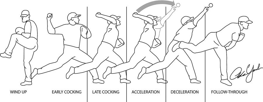
Wind-Up
Definition: Begins the throwing sequence and establishes the body's position, balance, and timing.
Important details: The lead leg is lifted while the thrower maintains balance over the supporting leg; the pelvis and trunk are positioned for subsequent rotation. Muscles of the lower limb (quadriceps, hamstrings, hip rotators) establish and control this base, while trunk musculature maintains pelvis/torso position. The throwing arm is positioned in preparation for cocking; muscles like the deltoid, rotator cuff, biceps, and scapular musculature show early activity.
Stride / Early Cocking
Definition: The body begins moving toward the target as the lead leg moves forward and the pelvis begins to rotate.
Important details: The hip, knee, and ankle of the lower limbs coordinate to create a stable base and produce movement toward the target. The pelvis is especially important as the mechanical connection between the lower limbs and trunk; as it rotates, the trunk can subsequently rotate relative to it. The throwing shoulder begins moving into the cocking position (supraspinatus, infraspinatus, teres minor, deltoid, trapezius, biceps).
Late Cocking
Definition: Begins after the stride foot contacts the ground and continues until the shoulder reaches its maximum external rotation.
Important details: The lead foot's ground contact allows movement transfer from the lower extremities through the pelvis and trunk; the throwing arm is positioned behind the body. The humerus undergoes substantial external rotation while elevated, placing large rotational demands on the glenohumeral joint. The rotator cuff (supraspinatus, infraspinatus, teres minor, subscapularis) dynamically stabilizes the shoulder during this large range of motion. At the elbow, the joint remains flexed (biceps brachii contributing) while positioned for acceleration. The phase ends at maximum shoulder external rotation.
Acceleration
Definition: The extremely rapid portion of the throw beginning at maximum shoulder external rotation and continuing until ball release.
Important details: Movement begins proximally — rotation of the pelvis and trunk transfers to the shoulder complex, which rapidly moves toward internal rotation while the elbow undergoes rapid extension; the forearm and wrist then contribute to the final movement leading to release (proximal-to-distal sequence). Major muscles — pectoralis major, latissimus dorsi, subscapularis, serratus anterior (propulsive contributors); triceps brachii (elbow extension). The subscapularis is a major internal rotator of the shoulder. Research found evidence for sequential activity from scapular protractors → shoulder horizontal flexors → elbow extensors, but did not establish the same clean sequential pattern all the way to the wrist flexor or forearm pronator — so this final portion should not be oversimplified.
Deceleration
Definition: The phase immediately after release when the throwing arm, moving at very high velocity, must be slowed down.
Important details: Places substantial demands on the shoulder and scapular musculature. The posterior rotator cuff (infraspinatus, teres minor) and posterior deltoid contribute to controlling the rapidly moving humerus, performing important eccentric actions (generating tension while lengthening to resist movement). Scapular muscles (trapezius, rhomboids, serratus anterior) help stabilize the scapula and control the shoulder girdle; the biceps also shows marked eccentric activity. The muscles here are not simply "opposite muscles" turning on after acceleration — they actively control the movement and dissipate the mechanical energy generated earlier.
Follow-Through
Definition: The final portion of the throwing motion, where the arm and trunk continue moving after release as the body dissipates remaining momentum.
Important details: Not an attempt to generate additional ball velocity — the ball has already been released. Continued movement allows the body to decelerate and dissipate momentum gradually rather than stopping instantaneously. The shoulder, scapular muscles, trunk musculature, and lower limbs all contribute to this continued control.
Example: A pitcher's arm continuing to swing across their body well after the ball leaves their hand is the follow-through safely dissipating the throw's momentum.
Problems With Incorrect Throwing Technique
Poor Lower-Body Contribution
Definition: A thrower relying predominantly on the throwing arm while failing to use the legs, hips, and trunk effectively.
Important details: Reduces the efficiency of energy transfer through the kinetic chain and increases the work required from the upper extremity.
Poor Timing or Sequencing
Definition: When the pelvis, trunk, shoulder, elbow, and distal segments fail to coordinate their movements rather than accelerating independently.
Important details: When the timing of one segment is altered, subsequent segments may have to compensate, increasing unnecessary joint loading.
Poor Trunk or Pelvic Movement
Definition: Reduced or poorly timed contribution from the pelvis and trunk, which normally connect the lower extremities to the upper extremity.
Important details: More demand may be placed on the shoulder and elbow to produce the required throwing velocity — this is why throwing should not be taught as an isolated arm movement.
Poor Shoulder Mechanics
Definition: Excessive loading on the shoulder, which already experiences particularly high demands during late cocking, acceleration, and deceleration.
Important details: During deceleration, the posterior shoulder muscles must work eccentrically to slow the rapidly moving arm; an inefficient kinetic chain increases these mechanical requirements further.
Elbow Overload
Definition: Increased stress on the elbow when throwing mechanics are inefficient.
Important details: Overhand throwing places substantial forces and torques on the elbow, particularly during cocking and acceleration; a breakdown elsewhere in the kinetic chain can increase demands on the distal upper extremity.
Scapular Dyskinesis
Definition: Poor scapular control or altered scapular motion.
Important details: Because the scapula provides an important foundation for shoulder movement, altered scapular motion can interfere with normal shoulder mechanics and is associated with altered shoulder mechanics.
Poor Deceleration and Follow-Through
Definition: Inadequate control during the phase after ball release, when the arm is still moving rapidly and must be slowed through coordinated eccentric muscle activity.
Important details: Inadequate control here can increase the demands placed on the shoulder and elbow.