Week 6: Locomotor System (L19)
finished
🐮 Locomotor System Functions
The locomotor system, also known as the musculoskeletal system, plays a critical role in:
Supporting the body for normal locomotion and posture.
respiration, mastication, urination, and defecation.
Diseases of the Locomotor System
Generalized diseases that involve in this system, such as metabolic disorders, nutritional deficiencies (osteomalacia, rachitis, muscular dystrophy), toxicosis (fluorine, Se, drugs), purulent-septic infections, parasitosis or primary disease confined to the limbs → fever, pyraemia. And metastatic spread of pus-forming bacteria to organs.
Hoof-related diseases are complex and influenced by factors like trimming, housing, and feeding.
Lameness affects 25-35% of dairy cows annually, potentially leading to recumbency.
Foot lameness accounts for 88% of all lameness in cattle.
Hind leg lesions make up 86% of foot lameness cases
85% is lateral claw of hind leg accounting for hind leg lesions
Environmental factors also contribute to locomotor issues:
Poor stall design can cause swollen hocks, especially in heavy cows or those with walking difficulties.
Neck rails obstructing a cow's high step.
Obstacles and uneven slats
Slippery concrete and uneven floors
🩺 Clinical Examination
A thorough clinical examination involves:
Inspection (posture, Position, Movement), Palpation, Percussion, Passive movement - moving of the animal`s limb, Auscultation, Probing and puncture - depends on situation, comparison - size + feel of affected vs. unaffected, skin sensitivity tests, diagnostic anesthesia, radiography, surgical intervention and lab testing (blood and urine testing).
Symptoms to watch out for: Stiffness, weakness in limbs, lameness (limping), Paralysis, abnormal posture/position, not wanting to put weight on limbs, skin problems - sores/swelling.
Inspection: Assessing posture, position, and movement.
Individual assessment of musculoskeletal components: muscles, bones, joints, and feet.
A. Posture
Regular
Irregular
Examples:
Stiffness - Muscle disorders, inflammation (myositis), degeneration (WMD, paralytical myoglobinuria), parasites (Trichinella).
Bone disorders - rickets, osteomalacia, osteomyelitis
Congenital causes - spastic paresis

Kneeling posture: Indicates pain in distal limbs, rickets, osteomalacia, infectious diseases (MFD, footrot), tendon disorders (neuromyodystrophy), or contracture of tendons.
Crossing of the legs: Can be a sign of phalanx fracture or hereditary chondrodysplasia.
Adduction: Limbs placed close together, elbows pointing inwards.
Abduction: Limbs held wide apart, elbows pointing outwards.
Other observations include:
Changes in the size of chest or flank.
"Dog sitting" posture may indicate acute gastric distension in horses or rupture of the hamstring tendon.
pain indications: if front limbs are shifted forward, then it means there is pain in the front of the foot (laminitis, infection). If hind legs are pushed backward → pain in back of foot or heel.
crossing front legs: crossing, mean there is a fracture in a toe on inner claw.
head position: lowered head → neck problem, raised head → pressure in brain.
B. Position
Normal: Ability to stand up without difficulty.
Abnormal: Recumbency (inability to stand).
Recumbency can be:
Sternal
Lateral
Differential diagnosis of recumbency in dairy cows:
Classical milk fever (parturient paresis).
Hypomagnesemia (lactation, grass tetany).
Severe toxemia (acute diffuse peritonitis, coliform mastitis).
Maternal obstetrical paralysis (MOP).
Fat cow syndrome.
Physical injuries.
Ruptured gastrocnemius.
Dislocation of hip.
Osteoporosis.
Abnormal positions:
Hind legs extending forward - rupture of muscle
hind legs flexed - paralysis of nerve
fore legs stretched out - muscle problems
deviation of limb - not in its normal position, indicating a fracture or dislocation at that point.
Torticollis - abnormal twisting of neck can be caused by issues with cervical vertebrae, or muscles, tendons, or nerves in that area.
Process of standing up:
Horse: 1. fore legs 2. hind legs
Cattle: 1. hind legs 2. fore legs
in pregnancy, heavy fetus, short chain - changes leg order
C. Movement
Limb movements, or gait, should be:
In the same direction.
Of equal length.
assess rate, range, force, direction
To detect gait abnormalities in cattle?
needs to be led on hard surface (or soft if needed). 2 main types:
Supportive lameness - problem while its foot is on the ground
Suspensory lameness - when cow has a problem with swinging its leg forward off the ground.
mixed Lameness - issue in both phases.
Lameness is also classified based on severity:
Grade 1: slight
Grade 2: moderate
Grade 3: pronounced
Grade 4: severe
Grade 5: extremely severe lameness
Abnormal movements include:
Lameness
Stiffness
Shortened stride
Exaggerated flexion
Stumbling
Staggering
Swaying of the hindquarters
Duration can be constant or intermittent of these movements.
Gait or postures can indicate pain, with etiology stemming from:
Infectious diseases (listeriosis, scrapie, etc.).
Inherited diseases.
Non-infectious diseases affecting muscles, joints, bones, or feet.
Ten signs of lameness:
Uneven weight bearing
Not tracking up
Short strides
Arched back
Head nod
Lowered head carriage
Abnormal foot placement
Leg swing (wide or narrow)
Unsymetrical body posture
Reduced walking speed or reluctance to move
Locomotor scoring system:
Score | Description |
|---|---|
1 | Normal: Stands and walks normally with a level back, making long confident strides. |
2 | Mildly Lame: Stands with a flat back but arches when walking; gait is slightly abnormal. |
3 | Moderately Lame: Stands and walks with an arched back and short strides. |
4 | Lame: Arched back when standing and walking, favoring one or more limbs. |
5 | Severely Lame: Pronounced arching of the back, reluctant to move, with almost complete weight transfer off the affected limb. |
Head movements in lame horses:
Forelimb lameness: Head lowers when the sound leg is placed on the ground and raises when the lame leg bears weight.
Hindlimb lameness: Head lowers when the lame leg contacts the ground and rises when the sound leg is on the ground.
Bilateral lameness: Nodding of the head is absent when equal weight is placed on equally painful limbs.
🦴 Components of the Musculoskeletal System
We continue with examination of muscle (after examining movement)
1) The Muscles 💪
Skeletal muscles are responsible for posture and movement. Smooth muscles facilitate processes like blood flow and digestion.
easier to spot changes in muscles when we compare the same muscles on both sides of body.
Methods of clinical examination:
Inspection:
Size (hypotrophy, hypertrophy, atrophy)
if normal size, then we call it “normotrophy”
shape, symmetry
Palpation: Myotonus (muscle tonous, if we touch it, there is tonous as a normal finding, normal tonous of the muscle), pain, trauma.
we can detect hypertonous, hypotonous, increased or decreased.
consistency - normal is firm-elastic
in some cases of myosities, then we can find differences in the temperature, increased in the affected muscle tissue.
Muscle conditions:
Atrophy: Reduction in muscle size due to limited use. You may see atrophy in cases like:
radial nerve paralysis - animal can`t move the limb forward. The elbow drops because the muscle that straightens the elbow (triceps brachii) is not working, and the wrist bends (flexed). The muscles that extend the wrist start to shrink within a few weeks.
Painful conditions - if bone/joints hurt, the animal may avoid moving → atrophy.
Ankylosis - movement is physically restricted → muscles waste away.
Myopathy: non-inflammatory degenerative diseases of voluntary muscles → Muscle weakness affecting posture, recumbency and cardiac functions.
Acute cases: Swollen and hard muscles, fish flesh appearance at autopsy (hyaline degeneration).
White Muscle Disease/enzootic muscular dystrophy (WMD):
Etiology: Vitamin E or selenium deficiency, increased polyunsaturated FA.
Occurrence: Cattle, pigs, sheep.
tend to be tired, don`t want to get up, walks with stiffness. Can have trouble with breathing + circulation.
Equine Paralytic Myoglobinuria ("Tying-Up" Syndrome):
Etiology: Resting for several days, fed on full ration, stress.
causes hard board-like muscles in gluteal + lumbar areas when touched due to tissue damage.
In saddle horses → muscle fibers break down → pain, stiffness, sweat, reluctance to continue working.
Myositis: Inflammatory changes in muscles.
Causes: Trauma, infection (blackleg), parasites (sarcosporidiosis + trichinosis).
Muscle Hypertrophy: Hereditary condition.
in breeds like friesian, ayrshire, piedmont, hereford → selecting for better beef traits has led to genetic change → muscular hypertrophy, meaning that muscles grow larger than normal. It can range from mild to severe (hindquarters mainly).
condition cause difficulties during calving (dystocia).
Developmental Defects:
calves/lamb: Tendon contracture (limbs stuck in a bent/straight position), arthrogryposis (in calves, similar issues but not inherited, can also cause brain abnormality + hydranencephaly)
cattle: periodic spasticity, spastic paresis - inherited functional disease.
pig: myofibrillar hypoplasia (splayleg)
2) The Bones 🦴
Bones provide structure, protect organs, house bone marrow, and maintain calcium reserves.
Methods of clinical examination:
Inspection & Palpation: Shape, contour, consistency, sensitivity.
Radiological examination - structure of bone
Bone conditions:
Developmental Defects: Ankylosing lesions, achondroplastic dwarfism, hydrocephalus, spina bifida, osteogenesis imperfecta.
Bone Degeneration: Osteodystrophia fibrosa, rickets (limb joints + junctions → forward + outward curvature), osteomalacia (inability to stand on hindlegs), hypertrophic osteopathy (dog, bilateral symmetrical enlargement)
Inflammatory Diseases: Osteomyelitis (infectious or non-infectious, clinical signs depends - lameness, pain, temp.)
3) The Joints 🔗
Joints determine the degree and direction of motion.
Methods of clinical examination:
Inspection.
Palpation.
Radiological examination.
Puncture.
In practice, we will only do inspection and palpation, can also do flexion of the joint.
Joint conditions:
Congenital Defects: Contracture, ankylosis, dislocation, acetabular dysplasia.
Inflammatory Diseases:
Arthritis (purulent or non-purulent, clinical signs - lameness, heat, pain, resentment to passive movement, swelling).
bursitis (acute or chronic inflammation of a bursa in response to irritation or injury).
acute: swelling, edema, pain on site of mucous sac, within - serous/purulent exudate and also blood in rare cases
affects horse + cattle mostly.
causes: lying on hard floor, poorly adjusted harness, traumatic injuries, etc.
signs: painful swelling in region of inflammation, high temperature in purulent bursitis, limping in extreme cases.
Degenerative Diseases: Artropathy (patchy thinning of joint cartilage)
Normal synovial fluid is clear, colorless (yellowish opalescence), odorless, and coagulates in 48 hours. This fluid can be analysed for cell count + differential, crystals, culture + sensitivity, cytology.
4) The Feet 🦶
Methods of clinical examination:
Inspection.
Palpation.
Percussion.
Probing.
Pressure applying (hoof tongs).
Healthy hooves have the hoof wall and entire sole weight-bearing.
Defects arise from:
Trauma.
Inflammation.
Developmental defects.
Common diseases:
Laminitis (eq + cattle)
Infectious footrot (Spaerophorus necrophorus).
Digital and interdigital dermatitis.
Rusterholz's ulcer (cattle)
Descriptions of Foot Conditions
Condition | Description |
|---|---|
Footrot - eczema like infection beginning in cleft bw. claws → heel. 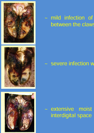 | a) Mild infection of the skin between the claws with a putrid yellow discharge; b) severe infection affects the heels causing cracks. c) extensive moist infection of heels, extends into inter digital space. |
Strawberry Footrot - discrete area of infection → skin at coronary band. 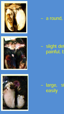 | a) A discrete, round lesion causing slight pain b) slight deterioration of hoof tissue at coronary band, pain, bleeds easily. c) large, strawberry-like, extremely painful lesion, bleeds easily. |
Laminitis - yellow (serum leak) discoloration of sole, red due to bleeding. 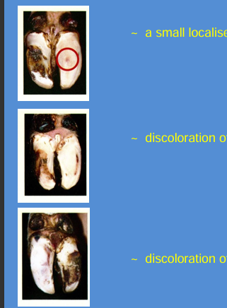 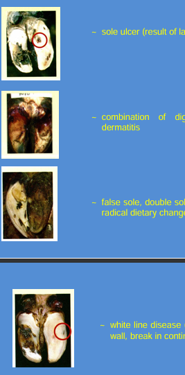 | a) small localized discoloration b) discoloration of 1/3 of sole c) discoloration of entire sole e) sole ulcer - result of laminitis or bruising d) combo of digital dermatitis + interdigital e) false sole, double sole after acute laminitis, or radical dietary changes. f) white line disease (connection bw. sole and wall, break in continuity) |
Tyloma interdigitale | Interdigital growths between the claws due to long-standing lesions in the cleft. |
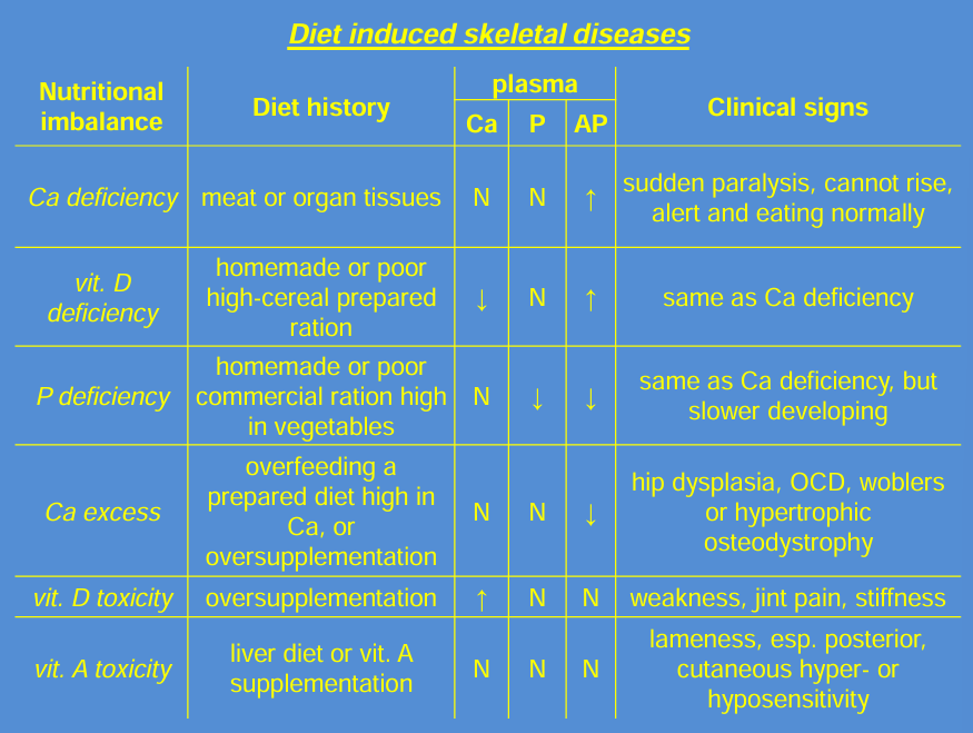
Diet-Induced Skeletal Issues | Sudden paralysis or inability to rise due to calcium or vitamin D deficiency; developmental issues due to calcium excess; weakness, joint pain, and lameness due to vitamin A toxicity. |
Pathological changes of dog paw:
cat`s paw
spoon paw
rabbit`s paw
open paw
practical VIEW:
Inspection: Assessing posture, position, and movement.
Individual assessment of musculoskeletal components: muscles, bones, joints, and feet.
A. Posture
Regular: Normal stance.
Irregular: Indicative of underlying issues.
B. Position
evaluate the animal`s ability to stand without difficulty
normal position: can stand up easily
abnormal position: recumbency indicating inability to stand, with assessments on whether it is sternal or lateral.
C. Movement
Evaluation of gait: direction and length
animal should move in same direction with equal stride lengths
Examination of the Musculoskeletal System
1) The Muscles
Inspection:
Assess size (hypotrophy, hypertrophy, atrophy). normal=normotrophy.
shape, and symmetry.
Palpation:
Check for myotonus, pain, trauma.
Assess consistency (normal is firm-elastic)
detect temperature differences (in cases of myositis)
2) The Bones
Inspection: shape, contour
Palpation: consistency, sensitivity and presence of crepitation
3) The Joints
Inspection & palpation
flexion of the joint to assess function
4) The Feet
Inspection
palpation
percussion
probing
pressure application (using hoof tongs)