Tissues
Tissue
epithelial tissue- covers/lines another organ. it also serves as protection, secretion, absorption, and excretion
connective tissue- supports and binds
muscle tissue- movement
nerve tissue- conducts impulses
Epithelial tissues
covers all body surfaces
major tissue of glands
anchored to basement membrane
avascular
reproduces quickly
4 basic shapes
1) squamous
2) cuboidal
3) columnar
4) transitional
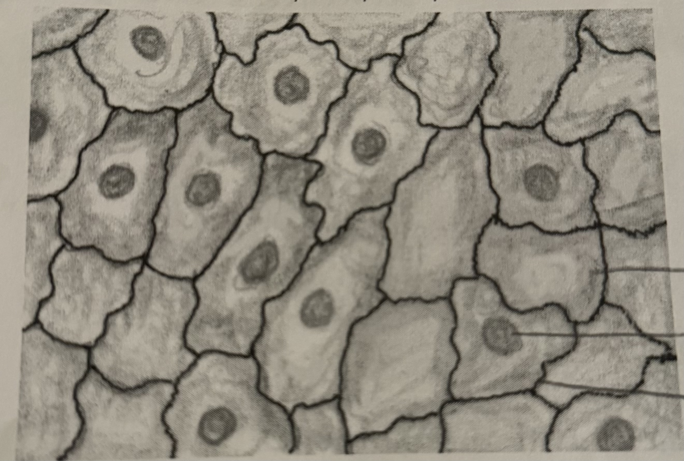
Simple Squamous Epithelium
location: air sack of the lungs, inner linings of blood vessels
endothelium- inner lining of blood vessels
function: filtration, diffusion, osmosis, exchange
This is what it looks like:
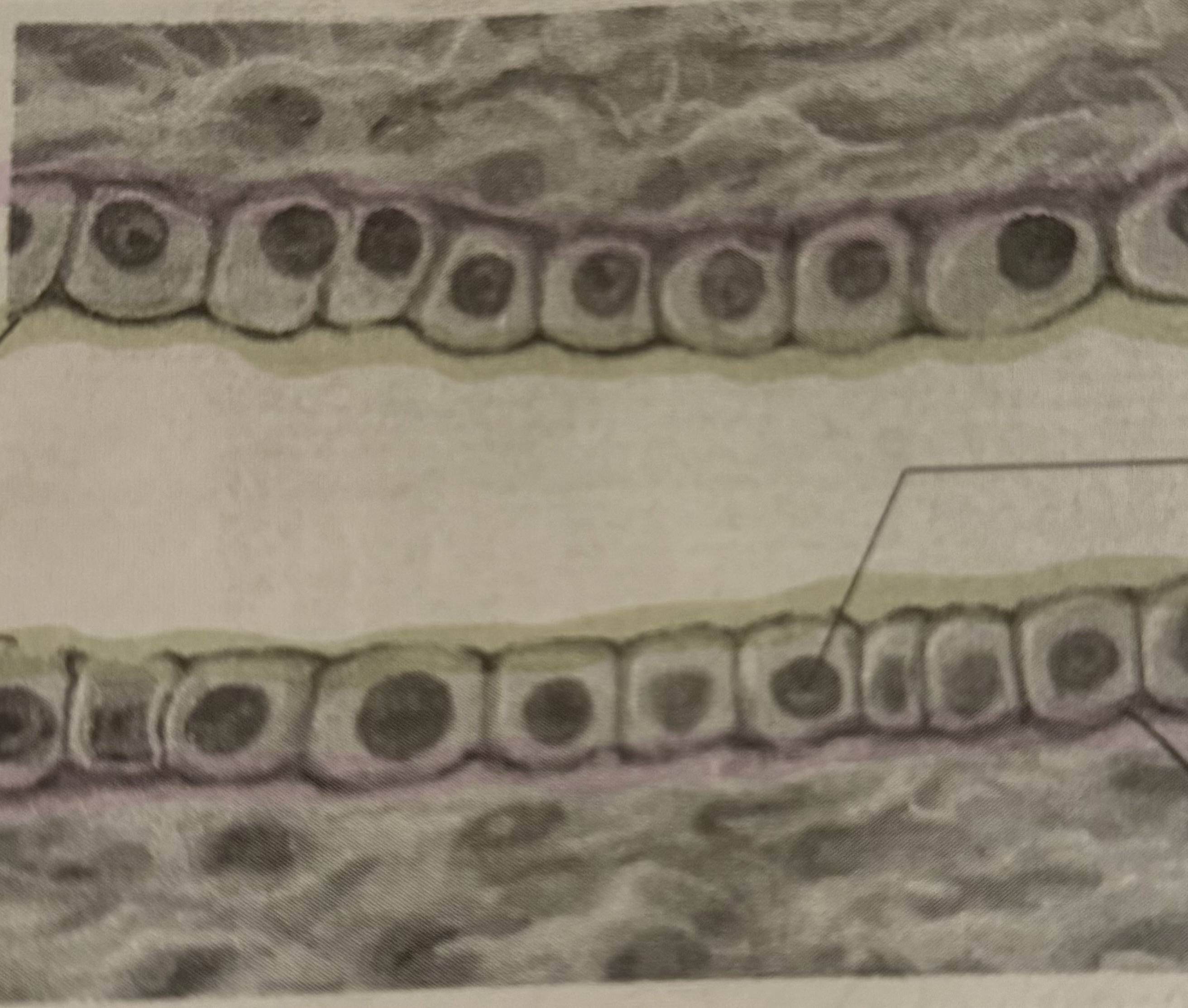
Simple Cuboidal Epithelium
location: lining of the kidney, ducts of glands
function: secretion, absorption, filtration
this is what it looks like:
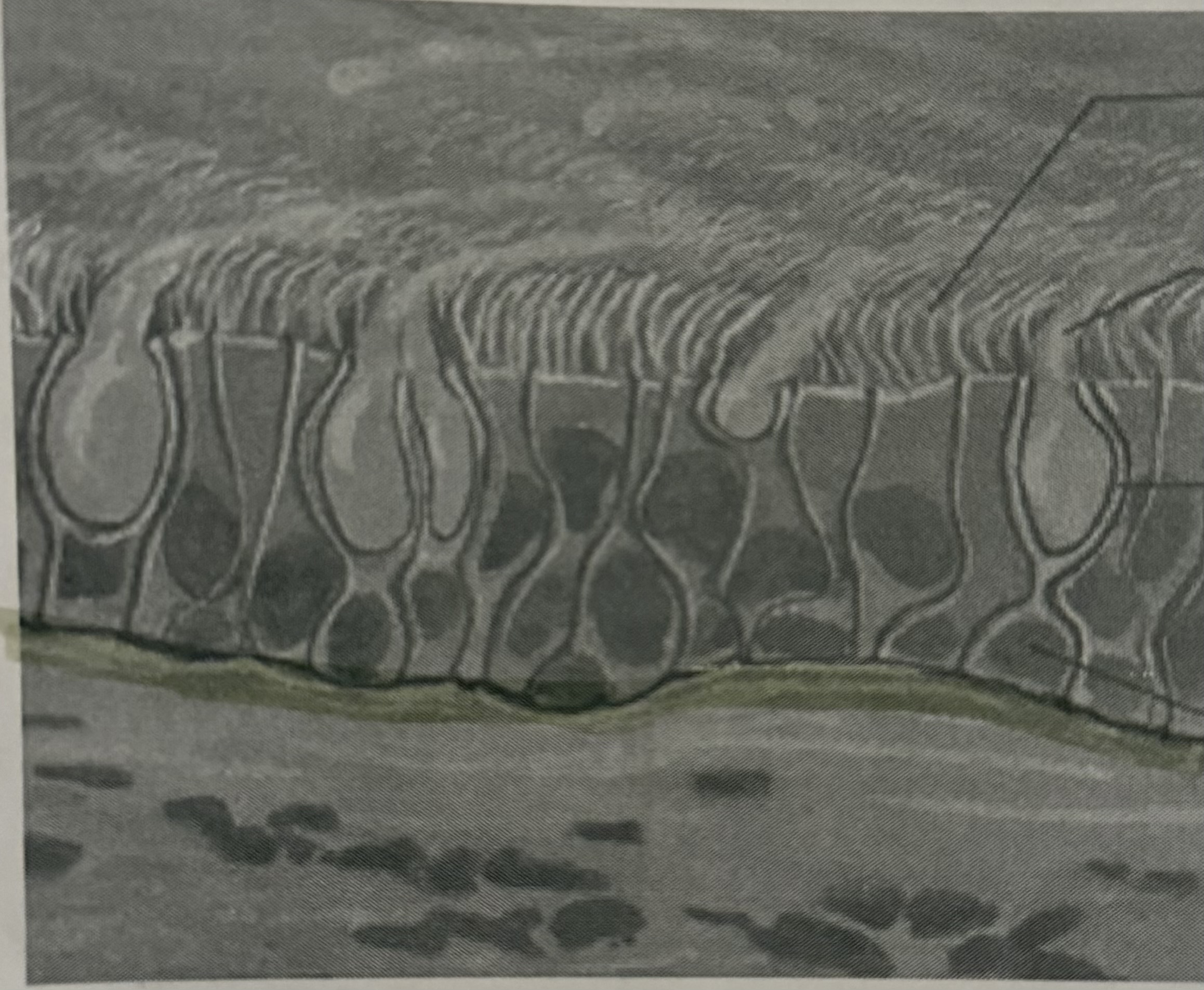
Simple Columnar Epithelium
location: lining of the digestive track and uterus
function: protection, secrete digestive fluid, absorb nutrients
microvilli- the valleys involved in absorption
goblet cells- secrete mucus
this is what it looks like:
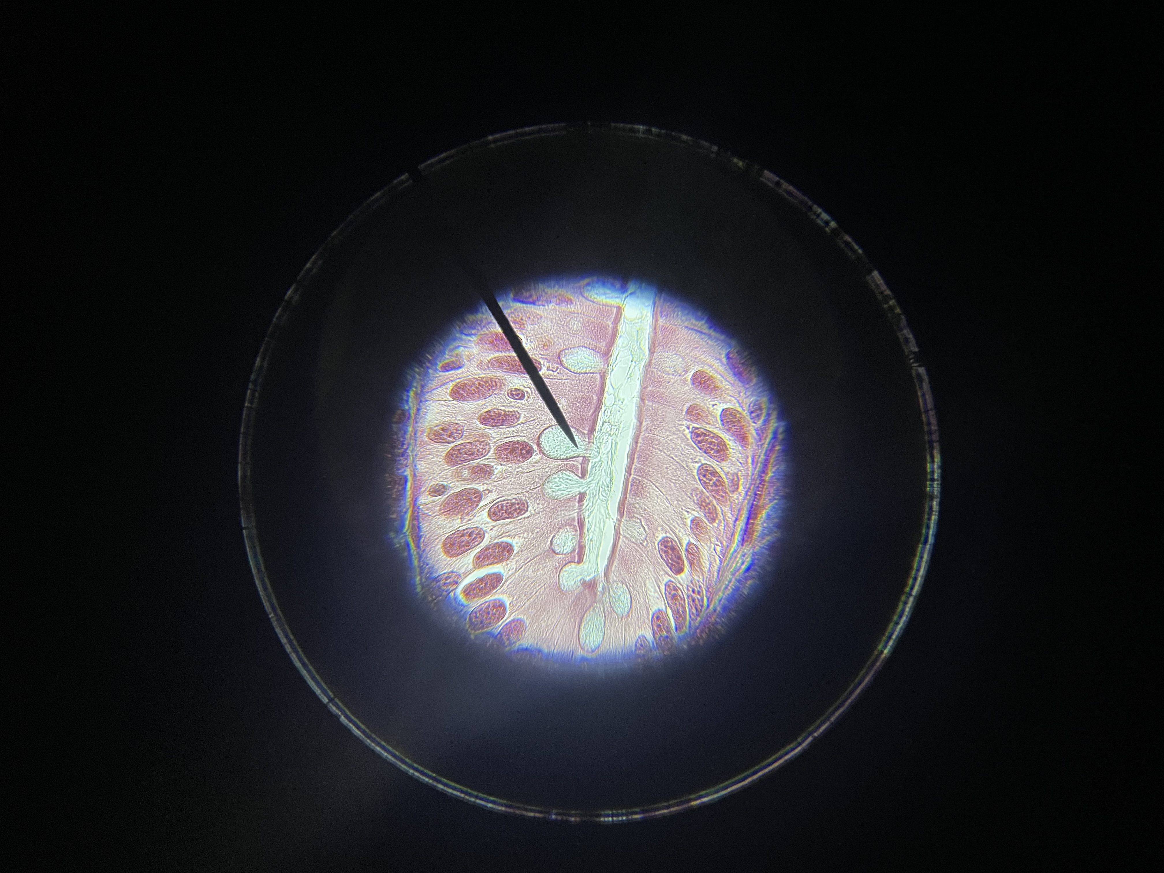
Pseudostratified Columnar Epithelium
location: respiratory tract, tubes of reproductive system
function: protection, movement of mucus and sex cells
contains goblet cells and cilia
This is what it looks like:
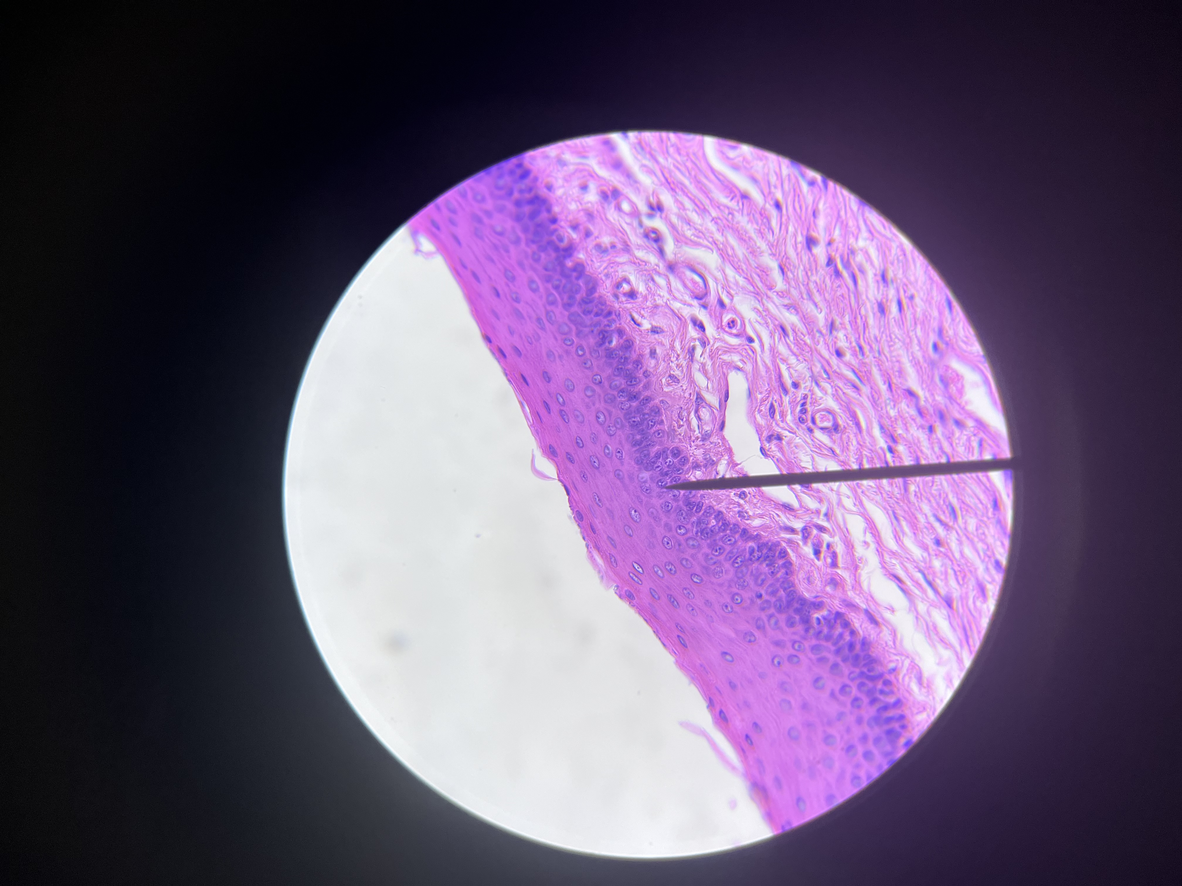
Stratified Squamous Epithelium
location: outer layer of skin, lining of mouth
aged skin cells fill with a protein called keratin and become hardened and die
function: protection
this is what it looks like:
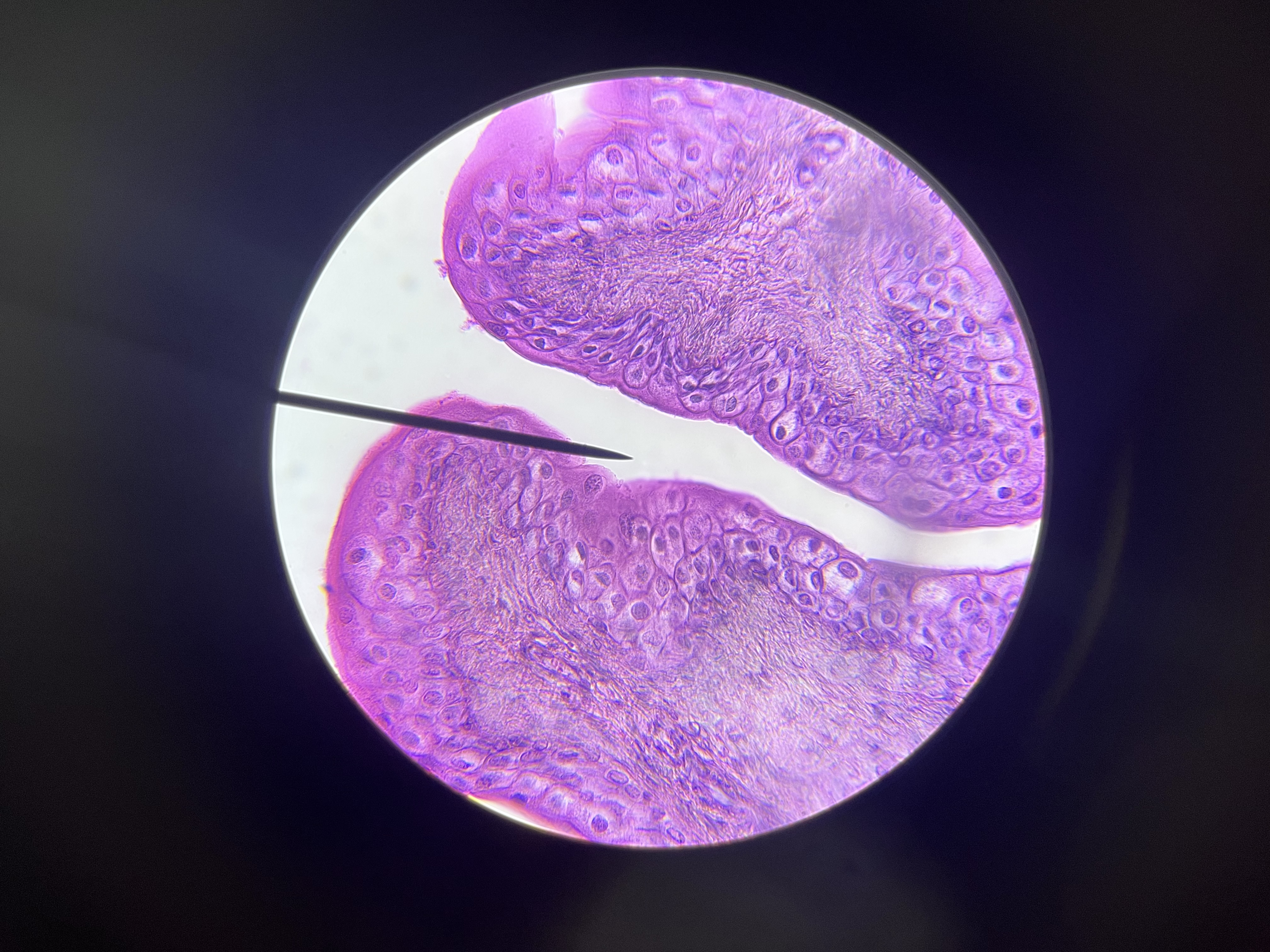
Transitional Epithelium
location: urinary bladder and tract
function: stretch during times of increased tension (distensibility)
This is what it looks like:
Glands
Glandular epithelium
gland- one or more glandular epithelium cells
exocrine glands- secretes into ducts
endocrine glands- secret into tissue fluid or blood
location: within columnar cuboidal epithelium
Multicellular exocrine glands
merocrine glands
release fluid without loss of cytoplasm
can be subdivided into serous cells and mucous cells
serous cells- secrete watery fluid containing enzymes
mucous cells- secrete mucus
apocrine glands
lose small portion of cells during secretion
holocrine glands
glands that release entire cells filled with secretory products
connective tissues
cells that lie in a matrix
matrix: consists of fibers and a ground substance tyhat is a fluid, semi-fluid or solid
2 major cell types
resident cells and wandering cells
Resident cells
produce in stable numbers
fibroblast
function: produce fibers by secreting proteins into matrix
macrophage
function: phagocytosis
phagocytosis- clear away foreign particles from tissue
Wandering cells
appear temporarily in tissues
usually when fighting injury or infection
mostly white blood cells
collagenous fibers
grouped in long, parallel buyndles, strong but flecible
function: to hold structures together
collagen- protein that makes up thick threadlike fibers
elastic fibers
branching, not very strong, flexible, appear yellow
elastin- protein with embedded microfibrils
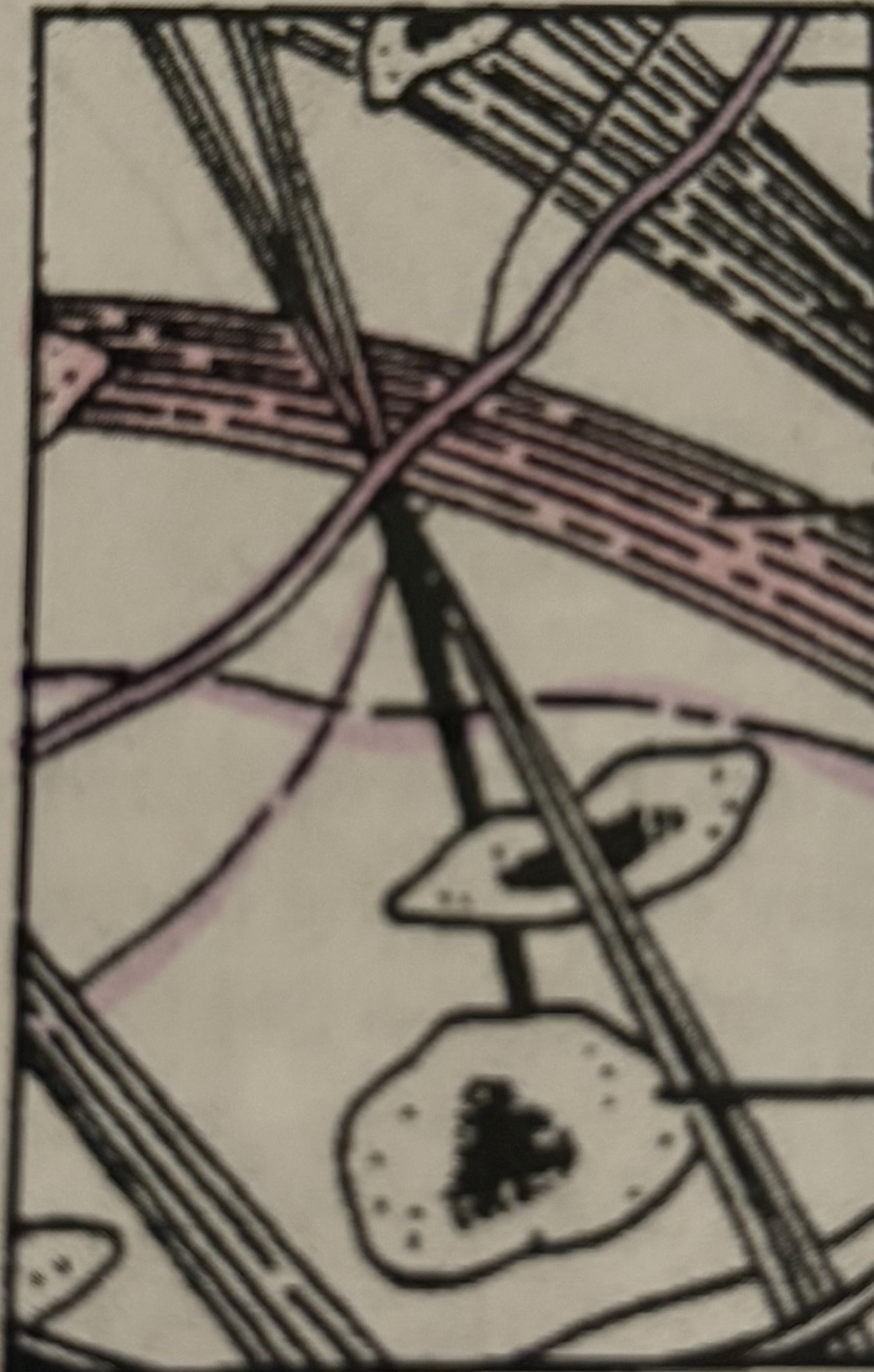
loose connective tissue
location- layers of epithelium and between muscles
function- binds skin to underlying organs, fills spaces, holds tissue fluids
this is what it looks like:
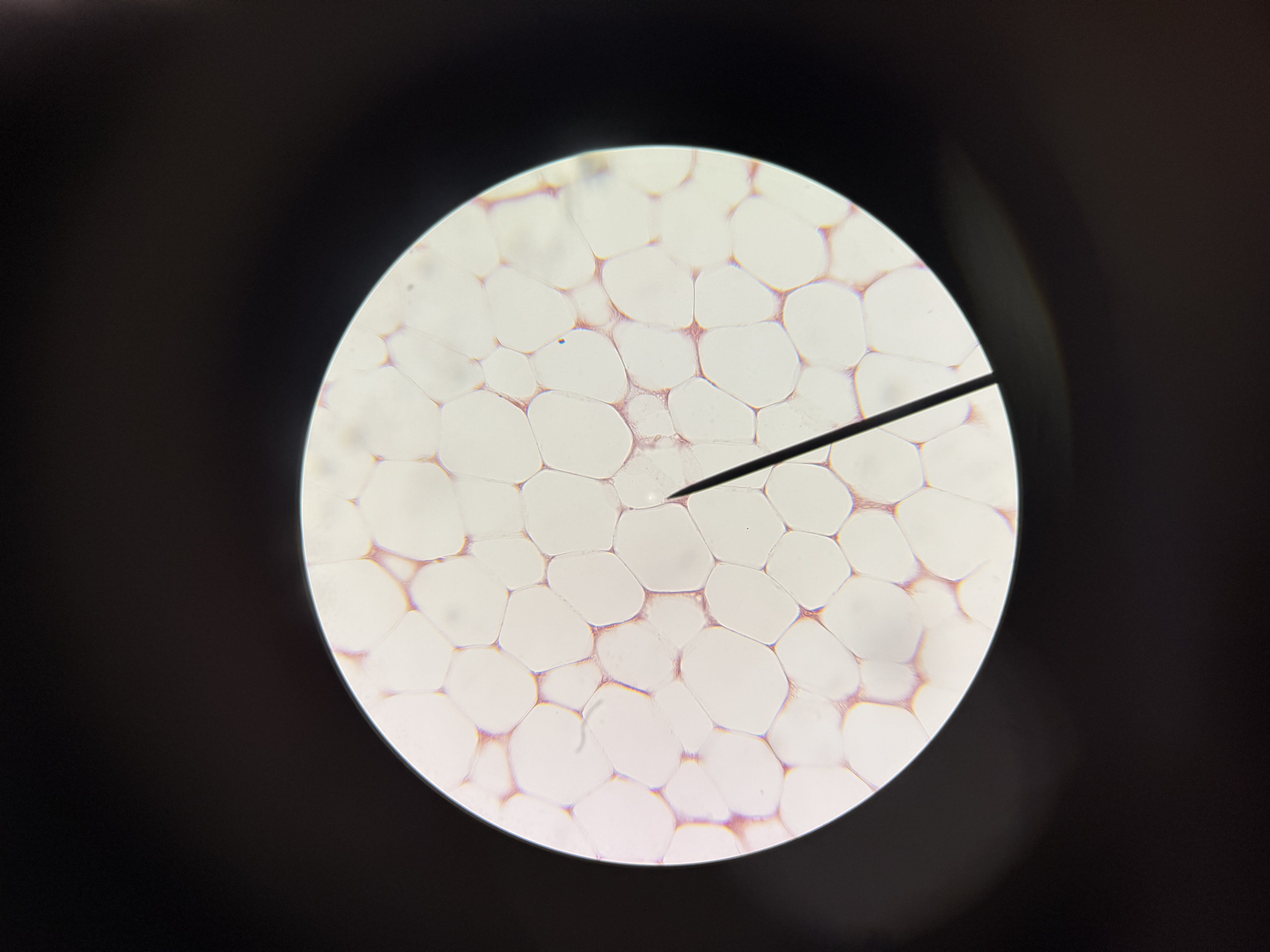
adipose tissue
location: beneath skin, around kidneys, behind eye balls, surface of heart
function: protective cushion, insulation and fat storage
this is what it looks like:
fibrous connective tissue (aka dense connective tissue)
location: tendons, ligaments, white layer of eye, skin
function: binds body parts, protection
this is what it looks like:
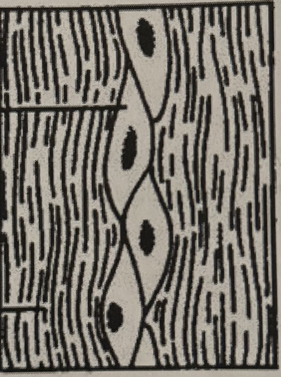
cartilage
cartilage- rigid connective tissue
structure: matrix of fibers
chondrocytes- cartilage cells
lacunae- small chambers containing chondrocytes (little lakes)
perichondrium- fibrous tissue surrounding cartilage and containing blood vessels
cartilage is avascular
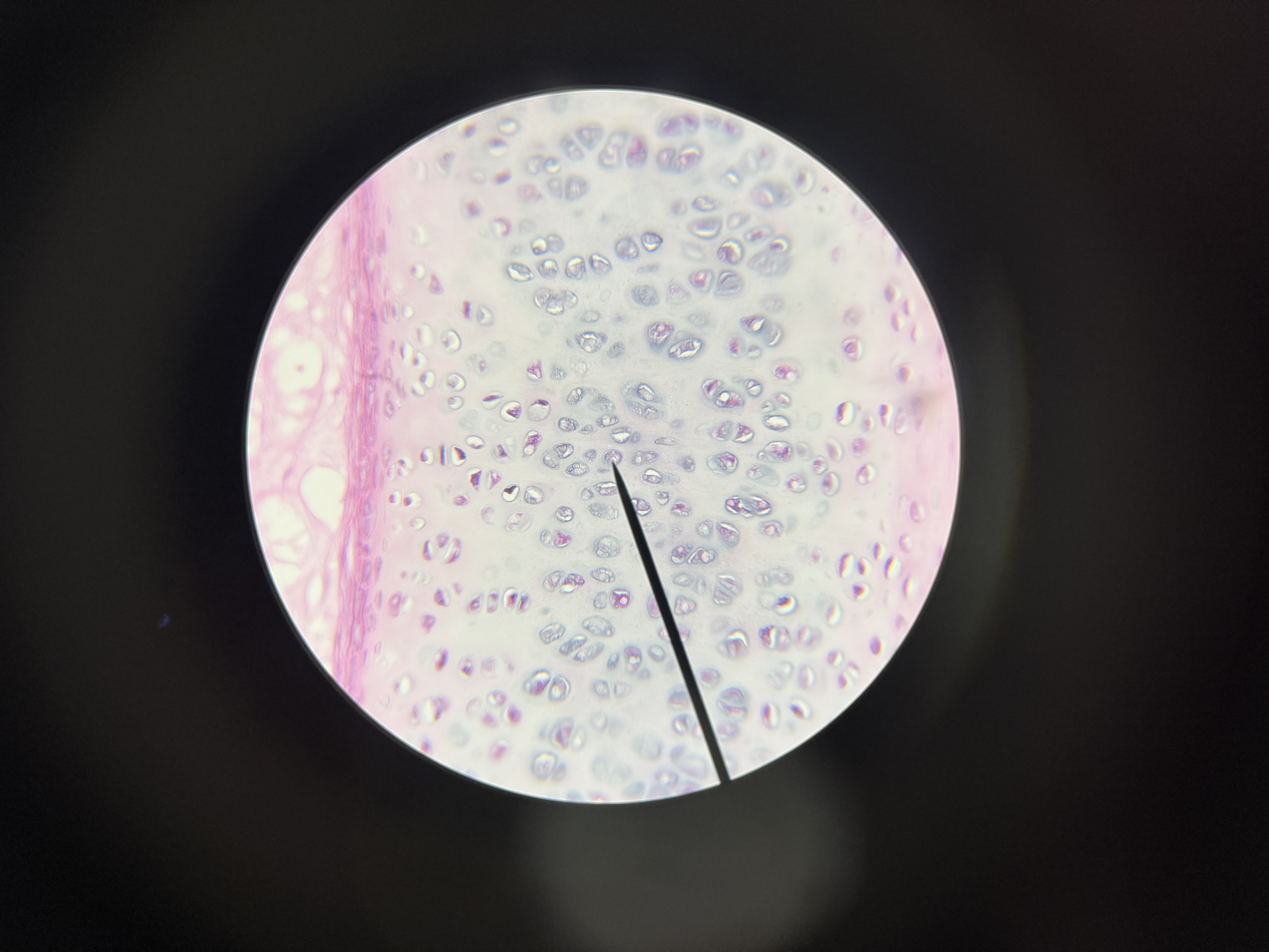
hyaline cartilage
location: end of bones (joints), soft part of the nose, suporting rings of respiratory tract, trachea
function: support, protection, provides framework of developing bones
this is what it looks like:
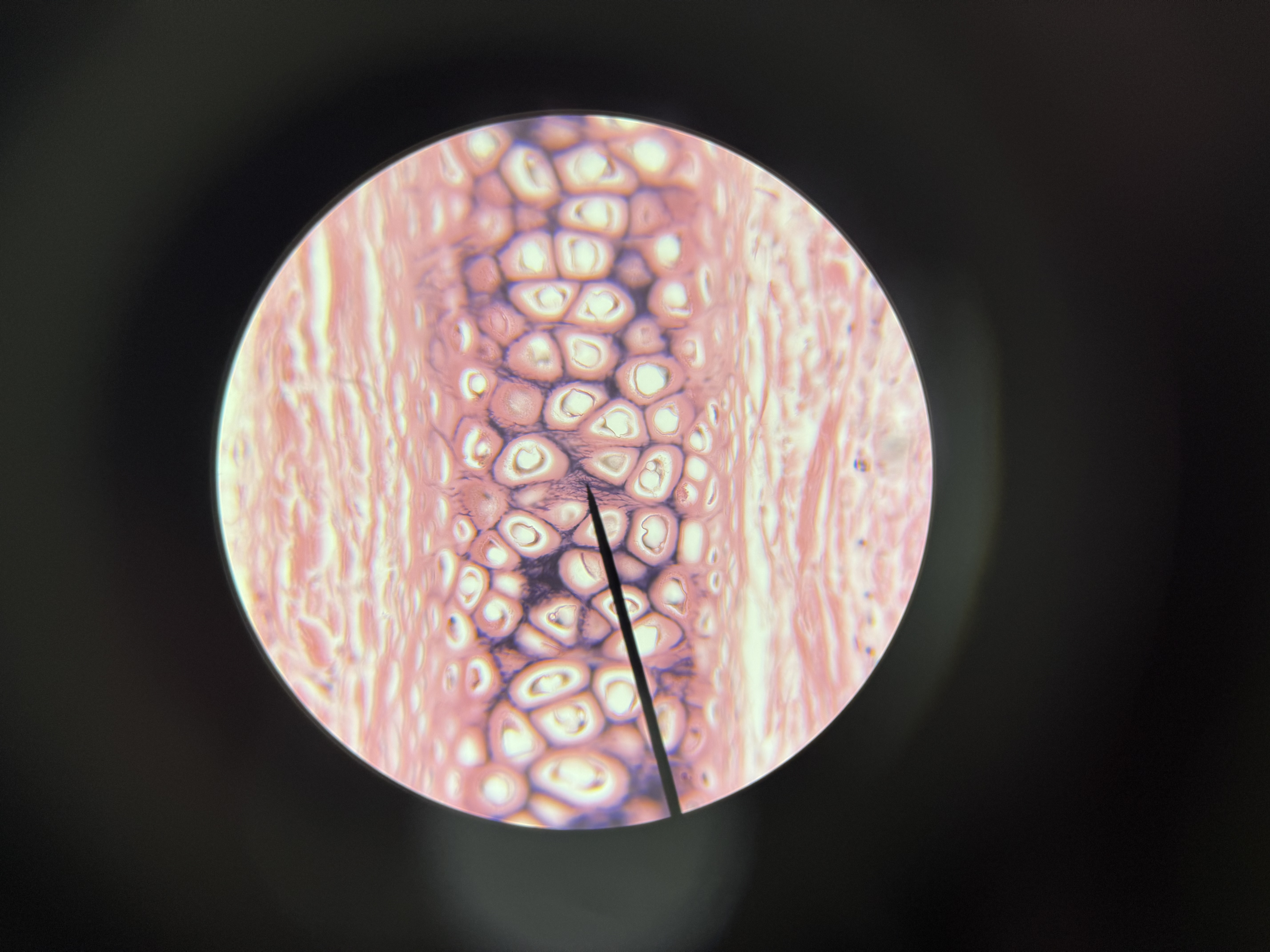
elastic cartilage
location: external framework of ear, part of larynx
function: support, protective framework
this is what it looks like:
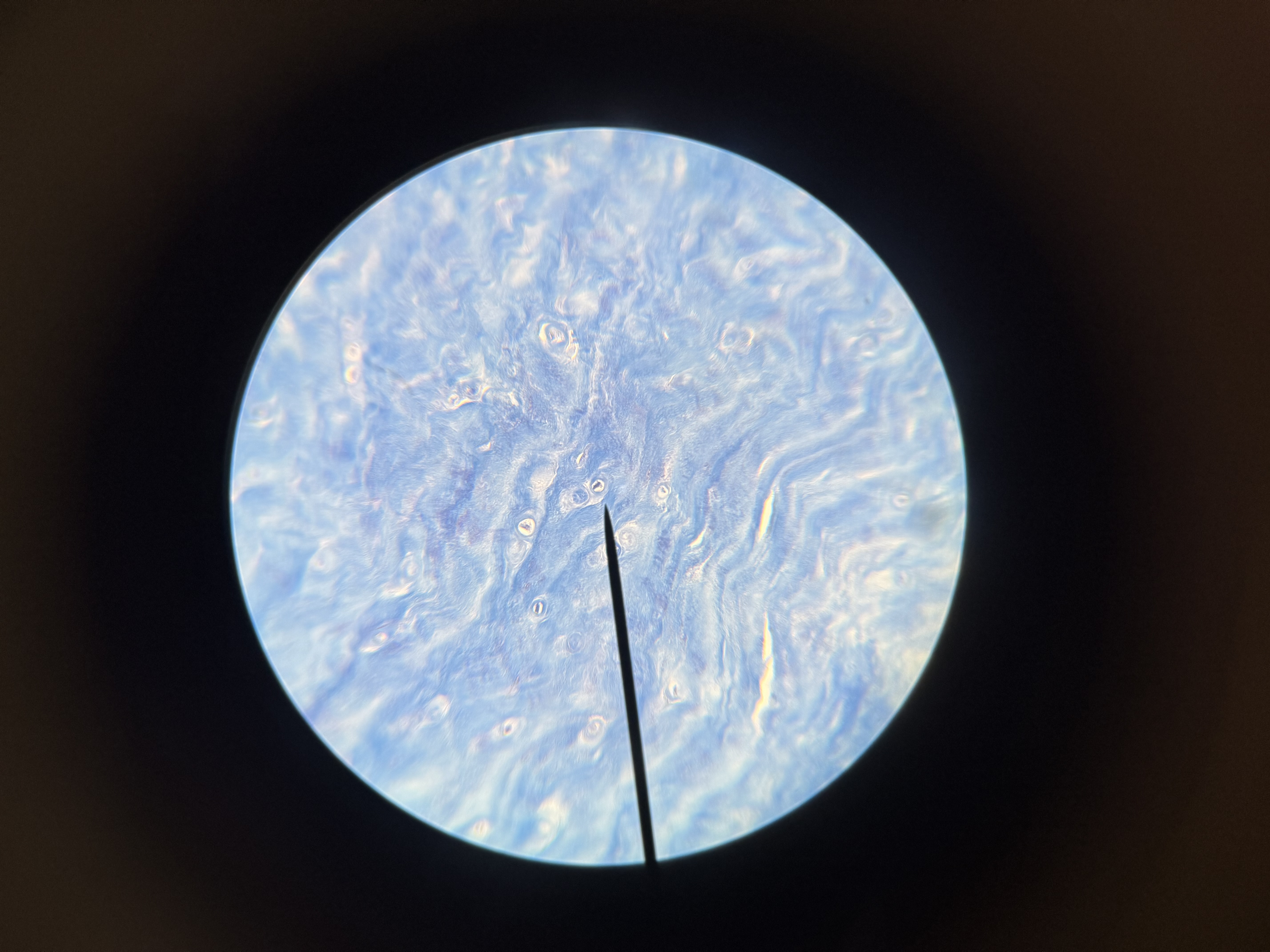
fibrocartilage
location: between vertebrae, bone in the knees, pelvic girdle
function: support, shock absorber
this is what it looks like:
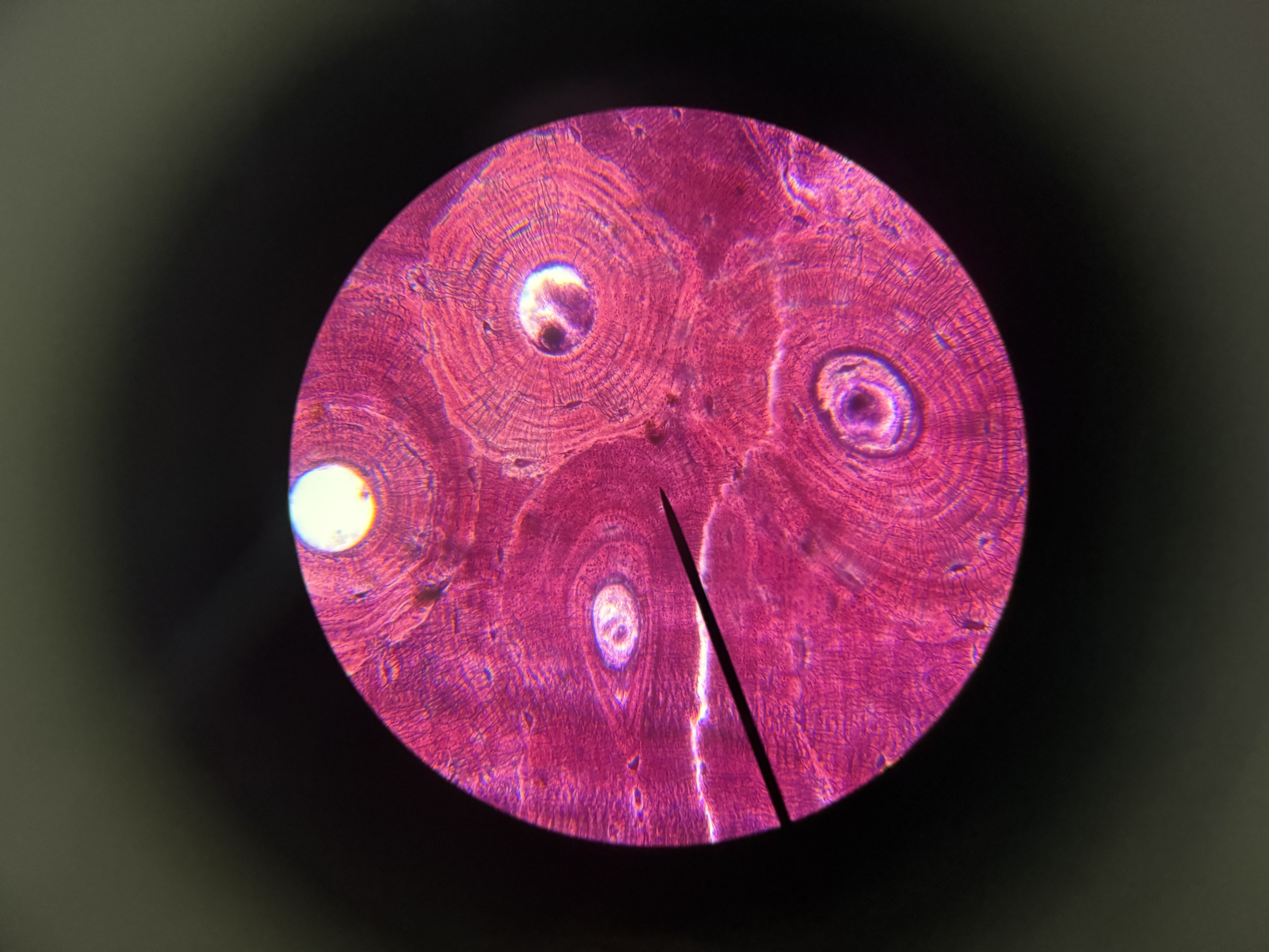
bone
osteocytes (bone cells located in lamellae
osteonic canals (aka haversian canals) - channels containing blood vessels
osteon- each cylinder shape dunit containing lamellae and a haversian canal
cannaliculi- tubes in the matrix where the osteocytes pass through to get blood supply
location: skeleton
function: support for body structures, protection, attachment for muscles
this is what it looks like:
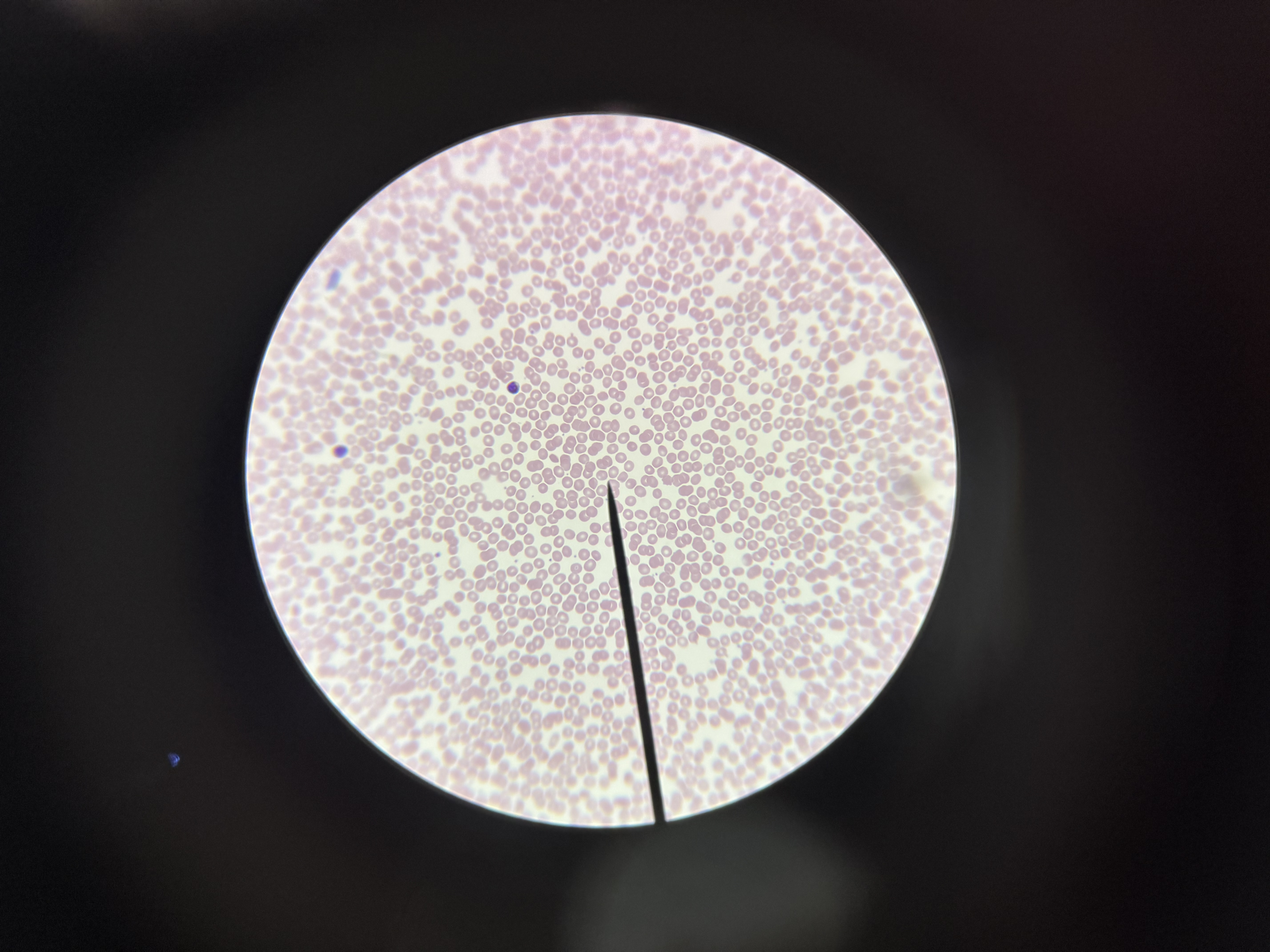
blood
liquid matrix called plasma
contains no fibers
location: blood vessels throughout the body
function: transport materials, fight infection, prevent bleeding
this is what it looks like:
muscle tissue
3 main types of muscle
skeletal muscle, smooth muscle, and cardiac muscle
skeletal muscle
voluntary
have striations (stripes)
location: attatched to bones
function: movements of skeleton and face
smooth muscle tissue
involuntary
no striations
location: walls of hollow internal organs
function: movements of materials through internal organs
cardiac muscle tissue
involuntary
has striations
location: heart
function: pump blood
nerve tissue
neurons
nerve cells that conduct electricity
location: entire body
function: transmit impulses, regulate body functions, store information, thoughts, emotions
sends chemical electrical impulses
neuroglial cells
do not make electricity
location: with nerves
function: bind nerve tissue together, phagocytosis, and connect neurons to blood vessels