Hindlimb Muscle Origin and Insertion
Osteology
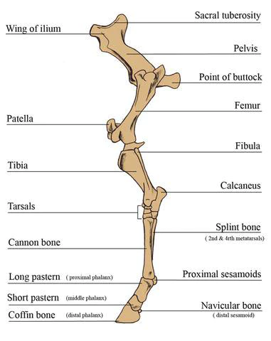
Pelvic girdle
Ilium
Pubis
ischium
Thigh
Femur
Patella
Leg
Tibia
Fibula
Pes
Tarsus
Metatarsus
Digit
Ilium
Wide part of the bone is the wing
Gluteal surface dorsolaterally
Sacropelvic surface
Iliac surface
Lateral quadrilateral part which contains the wing and body
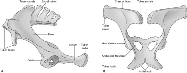
Constricted portion is the body/shaft
Ventral surface with psoas tubercle
Lateral angle
Tuber coxae
Medial angle
Tuber sacrale
Ischium
Pelvic surface is convex in the stallion and concave in the mare
Medial border meets the opposite bone to form the ischial symphysis
Lateral border
forms the lesser ischiatic notch
Cranial border
forms the cranial margin of the obturator foramen
Craniolateral angle joins the other two bones at the acetabulum.
Caudolateral angle
Forms the ischiatic tuberosity
Pubis
Smallest of the three bones
Cranial border bears iliopubic eminence
Ventral pubic tubercle near the symphysis
Caudal border
Cranial margin of obturator foramen
Bony Pelvis
Dorsal wall formed by the sacrum and first three caudal vertebrae
Ventral wall formed by the pubis and ischiatic bones
Lateral walls formed by the ilia
Femur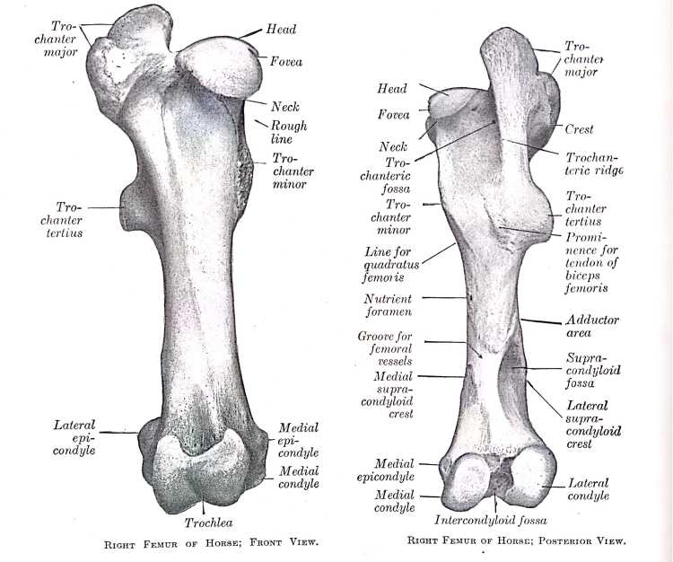
Long bone
Medial border
Lesser trochanter
Medial supracondylar tuberosity
Lateral border
Third trochanter
Supracondylar fossa
Lateral supracondyloid tuberosity
Proximal extremity
Head on the medial side
Cut medially by fovea capitis femoris
Neck
Greater trochanter on the lateral side
Distal extremity
Trochlea on the cranial side
Medial and lateral ridges to form patella surface
Medial ridge is wider and extends higher
Medial and lateral condyles
Intercondylar fossa separates the two
Proximal to the condyles is a deep supracondylar fossa
Medial and lateral epicondyles
Horse | Ox, Sheep | Dog | |
|---|---|---|---|
Greater trochanter | Divided | Undivided | Undivided |
Trochanteric ridge | Straight, vertical and long | Oblique and connects between the major and lesser trochanter | Oblique and connects between the major and lesser trochanter |
Third trochanter | Present | Absent | Absent |
Supracondyloid fossa | Deep | Shallow | Absent |
Ridges of the trochlea | Medial ridge is wider and higher than the lateral | Medial ridge is slightly larger | Nearly equal |
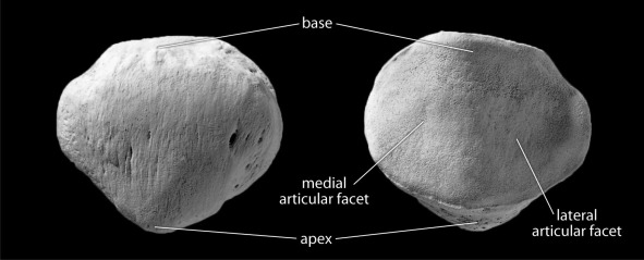
Patella
Large sesamoid bone
Articulates with the trochlea with the femur
Cranial free surface
Articular surface
Medial and lateral borders
Base
Apex
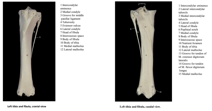
Tibia
Long bone
Caudal surface
2 parts divided by the popliteal line
Cranial border prominent forming the crest of the tibia
Lateral border forms interosseous space with fibula
Proximal extremity
Lateral and medial condyles
Intercondylar eminence
Separated caudally by deep popliteal notch
Lateral condyle bears a facet for articulation with fibula
Tibial tuberosity
Separated from lateral condyle by the extensor groove
Distal extremity
Articular surface consisting of two grooves separated by a ridge
Grooves bound by malleoli
Medial malleolus- more prominent
Sulcus malleolaris for flexor digitorum longus muscle tendon
Lateral malleolus- broader
Vertical groove for lateral digital extensor tendon
Fibula
Terminates one half or two thirds down the lateral border of the tibia
Head presents narrow area along the proximal border for articulation with lateral condyle of tibia
Distal extremity fuses with tibia, constituting the lateral malleolus
Horse | Ox, Sheep | Dog | |
|---|---|---|---|
Tibial tuberosity | Grooved | Not grooved | Not grooved |
Popliteal notch | Deep | Shallow | Shallow |
Caudal surface | Divided by popliteal line | Not divided, many muscular lines | Not divided |
Tibial crest | Prominent | Blunt | Sharp |
Distal extremity | Articular grooves and ridge are oblique | Articular grooves and ridge are sagittal | Articular grooves and ridge are sagittal |
Fibula | Reduced bone | Reduced bone | Elongated bone |
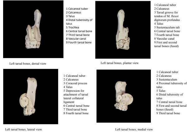
Tarsal bones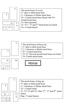
Proximal row
Talus and calcaneus
Distal row
1-4
Talus
Proximal and dorsal surfaces form trochlea for articulation with tibia
Caudal surface has facets for articulation with calcaneus
Medial surface has a large tuberosity
Calcaneus
Largest bones of the hock
Proximal end forms the calceneal tuber
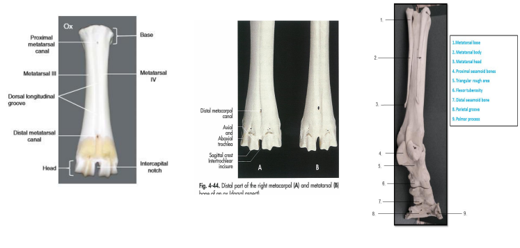
Metatarsal Bones
Horses
3 metatarsal bones
II, III, and IV
II and IV are reduced to splint bones
III is the cannon bone
Caudal surface forms a groove for suspensory ligament
Dogs
5 metatarsal bones
I-V
I is reduced to a dew claw
Pigs
4 metatarsal bones
I is absent
II and V are reduced
III and IV are full developed
Ruminants
3 metatarsal bones
III and IV form the large metatarsal bone
V is reduced
Vertical grove connects two canals at the ends of the shaft on the dorsal surface
Distal extremity is divided by sagittal groove
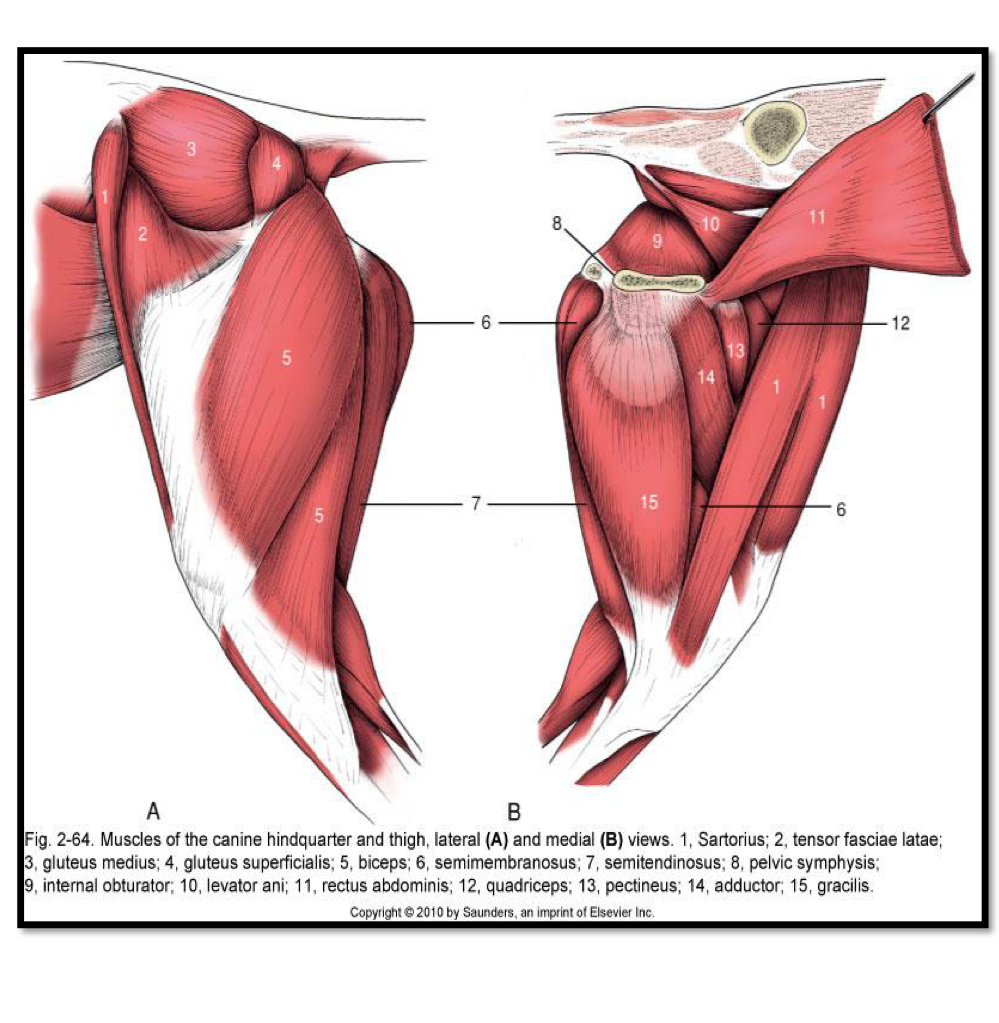
Muscles (Blood supply and Innervation)
Muscle | Origin/Insertion | Innervation | Blood Supply | Action |
|---|---|---|---|---|
Psoas major- Psoas minor and iliacus | O: Caudal thoracic and lumbar vertebrae I: Os coxae and femur | Lumbar | Flex or stabilize the vertebral column. | |
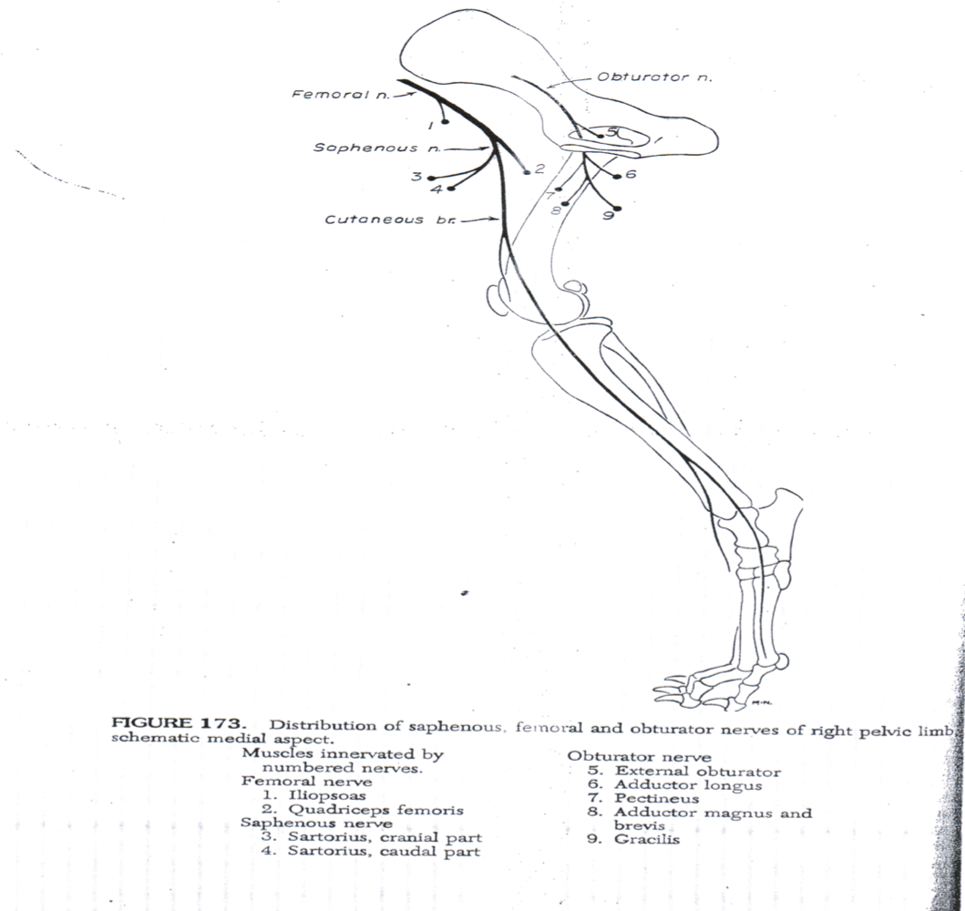
Iliopsoas (Psoas major and Iliacus) | Femoral | Chief flexor at hip joint | ||
Tensor fasciae latae | O: Coxal tuber I: Fascia latae | Cranial gluteal | Femoral | Flex the hip joint Extend the stifle |
Superficial gluteal muscle | O: Coxal tuber I: Trochanter tertius | Caudal gluteal | Inferior gluteal | Abduct the limb |
Gluteus medius | O: Sacroiliac and sacrotuberal ligaments I: Greater trochanter of the femur | Cranial gluteal | Superior gluteal | Extend the hip joint and abduct the limb |
Gluteus profundus | O: Ischiatic spine (shaft of ilium- dog) I: Greater trochanter of femur | Cranial gluteal | Gluteal | Abduct the hip joint and extend the hip joint |
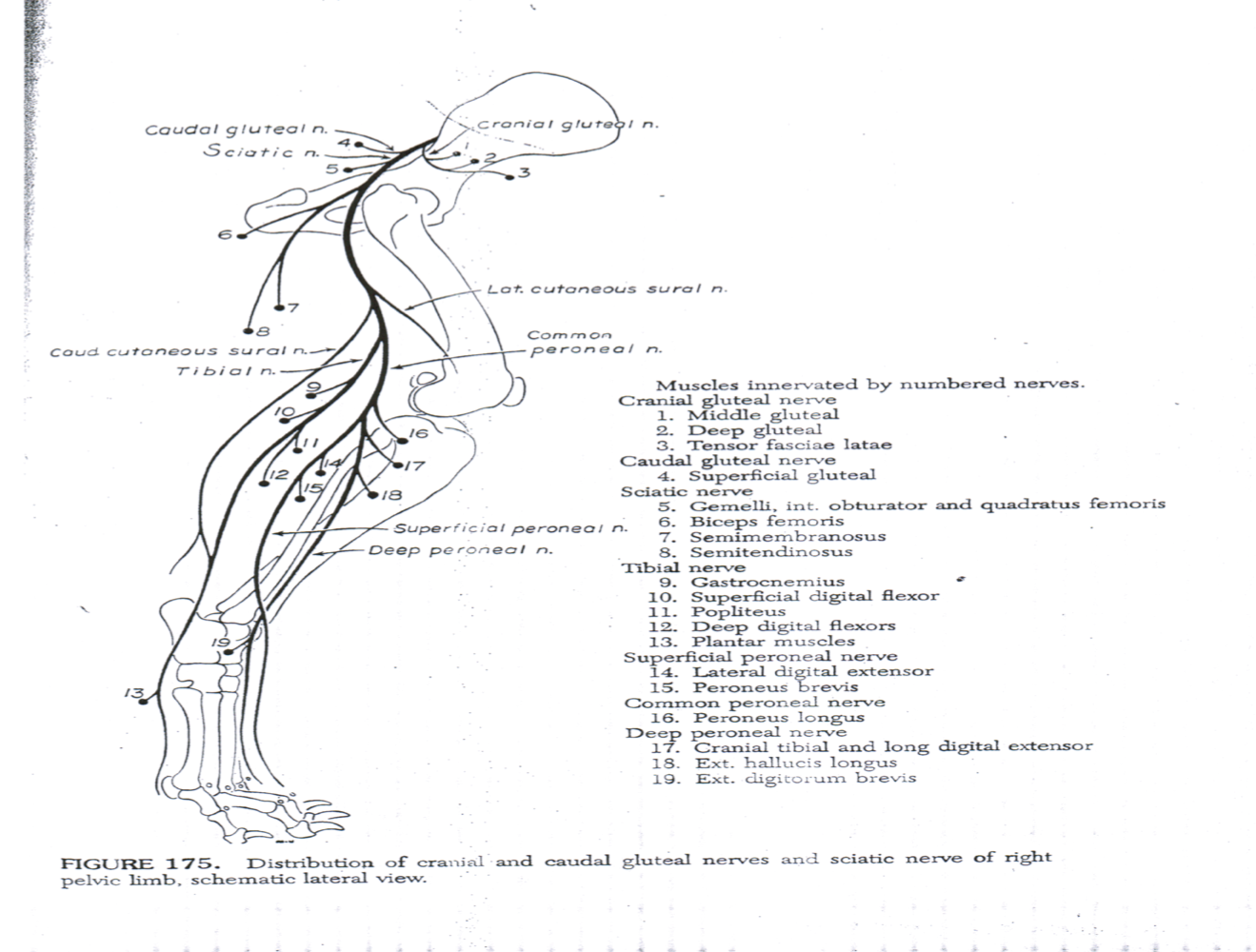
Biceps femoris | O: Ischiatic tuberosity I: Patella and tibia and calcaneus | Caudal gluteal Ischiatic | Distal caudal femoral | Extend the limb |
Semitendinosus | O: Ischiatic tuber I: Calcaneus | Caudal gluteal Ischiatic | Distal caudal femoral | Extend the hip and hock joints, flex the stifle |
Semimembranosus | O: Ischiatic tuber and sacrotuberal ligament I: Medial epicondyle of femur | Caudal gluteal Ischiatic | Middle caudal femoral Distal caudal femoral | Extend the hip joint and adduct the limb |
Sartorius | O: Iliac fascia and tendon of psoas minor I: Tibial tuberosity and medial patella ligament | Saphenous | Saphenous | Flex the hip joint and adduct the limb |
Gracilis | O: Pelvic symphysis I: Medial surface of tibia | Obturator | Obturator | Adduction of the hip joint |
Pectineus | O: prepubic tendon I: medial border of femur | Obturator | Femoral | Adduct the limb and flex the hip joint |
Adductor | O: Pubis and ischium I: Caudal surface of femur | Obturator | Distal caudal femoral | Adduct the limb and extend the hip joint |
Quadratus femoris | O: Ventral surface of the ischium I: Caudal surface of the femur | Ischiatic | Lateral circumflex femoral | Extend the hip joint and adduct the thigh |
Obturator externus | O: Ventral surface of the pubis I: Trochanteric fossa | Obturator | Adduct the thigh | |
Obturator internus | O: Pubis and ischium I: Trochanteric fossa | Obturator | Rotate the limb laterally | |
Gemelli | O: Lateral border of ischium I: Trochanteric fossa and crest | Ischiatic | Rotate the femur laterally | |
Rectus femoris | O: Body of the ilium I: patella | Femoral | Femoral iliacofemoral medial circulmflex femoral | Extend the stifle joint Flex the hip joint |
Vastus lateralis | O: lateral surface of femur I: patella, rectus femoris tendon | Femoral | Iliacofemoral caudal femoral popliteal | Extend the stifle joint |
Vastus medialis | O: medial surface of femur I: patella, rectus femoris tendon | Femoral | Medial circumflex femoral Femoral | Extend the stifle joint |
Vastus intermedius | O: Cranial surface of femur I: patella | Femoral | Iliacofemoral Popliteal | Extend the stifle joint |
Articularis coxae | O: Ilium I: Cranial surface of femur |
|
The femoral triangle is formed
Cranially: Sartorius
Caudally: Pectineus
Laterally: Iliopsoas and vastus medialis
Medially: Femoral fascia and gracilis
It contains
Femoral artery
Femoral vein
Saphenous nerve
Deep inguinal lymph nodes
Clinical significance:
Pulsation (femoral and saphenous artery (cattle))
Vein puncture: Lateral saphenous vein
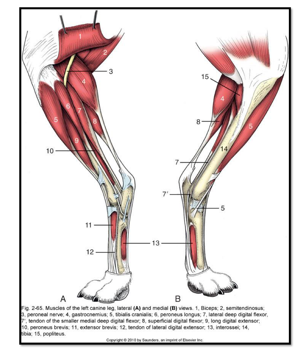
Muscle | Origin/Insertion | Innervation | Blood Supply | Function |
|---|---|---|---|---|
Tibialis cranialis | O: lateral condyle (tibia) I: Proximal end of metatarsal bone | Fibular nerve | Cranial tibial | Flex the hock |
Long digital extensor muscle | O: Extensor fossa (femur) I: Extensor process of third phalanx | Fibular | Cranial tibial | Extend the digit and flex the hock |
Peroneus longus | O: Lateral condyle of tibia I: Middle phalanx of IV digit | Fibular nerve | Cranial tibial | Flex the hock Rotate paw medially |
Lateral digital extensor | O: Fibula and lateral surface of tibia I: tendon of long digital extensor | Fibular nerve | Cranial tibial | Extend the digit Flex the hock Assist long digital extensor |
Peroneus tertius (horse and cattle) | O: extensor fossa of femur I: metatarsal bone, third tarsal, calcaneus and fourth tarsal. | Fibular nerve | Cranial tibial | Flex the hock (stay apparatus) |
Peroneus brevis (dog) | O: distal third of lateral fibula I: metatarsal V | Flexes tarsus | ||
Extensor digitorum brevis | O: dorsal surface of talus I: insertion tendon of long digital extensor | Fibular | Dorsal metatarsal artery | Assist extensor digitorum longus |
Gastrocnemius | O: lateral and medial supracondyloid tuberosities (femur) I: calcaneus | Tibial | Popliteal Caudal femoral | Extend the hock Flex the stifle |
Soleus (horse and cattle) | O: head of fibula I: tendon of gastrocnemius | Tibial | Cranial tibial Popliteal | Extend the hock Flex the stifle |
Superficial digital flexor | O: supracondyloid fossa (femur) I: calcaneus | Tibial | Caudal femoral Popliteal | Flex the digits Extend the hock |
Deep digital flexor muscle | Tibial | Caudal tibial | Flex the digit and extend the hock | |
Flexor digiti I longus (of the deep digital flexor) | O: lateral condyle (tibia) I: cartilage of distal phalynx | Flex the digit and extend the hock | ||
Popliteus | O: Lateral epicondyle (femur) I: caudal surface of tibia | Tibial | Caudal Popliteal | Flex the stifle joint Rotate the leg medially |
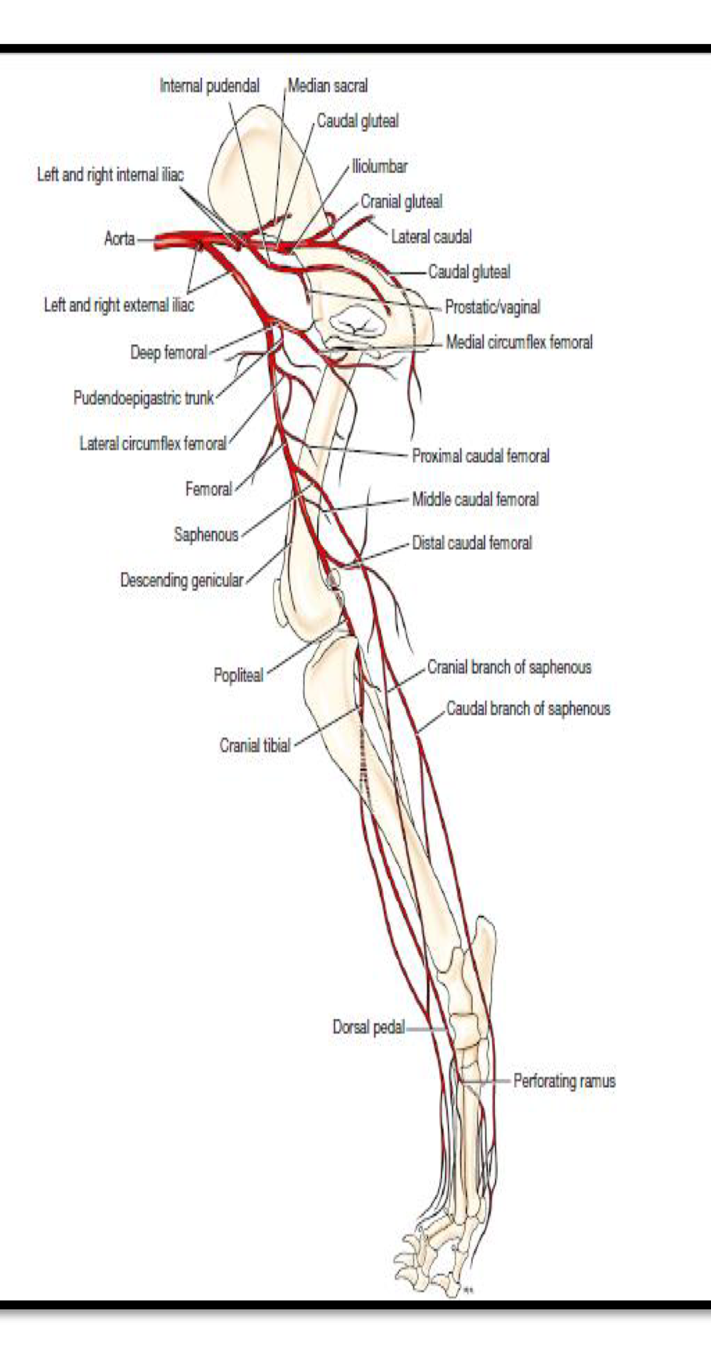
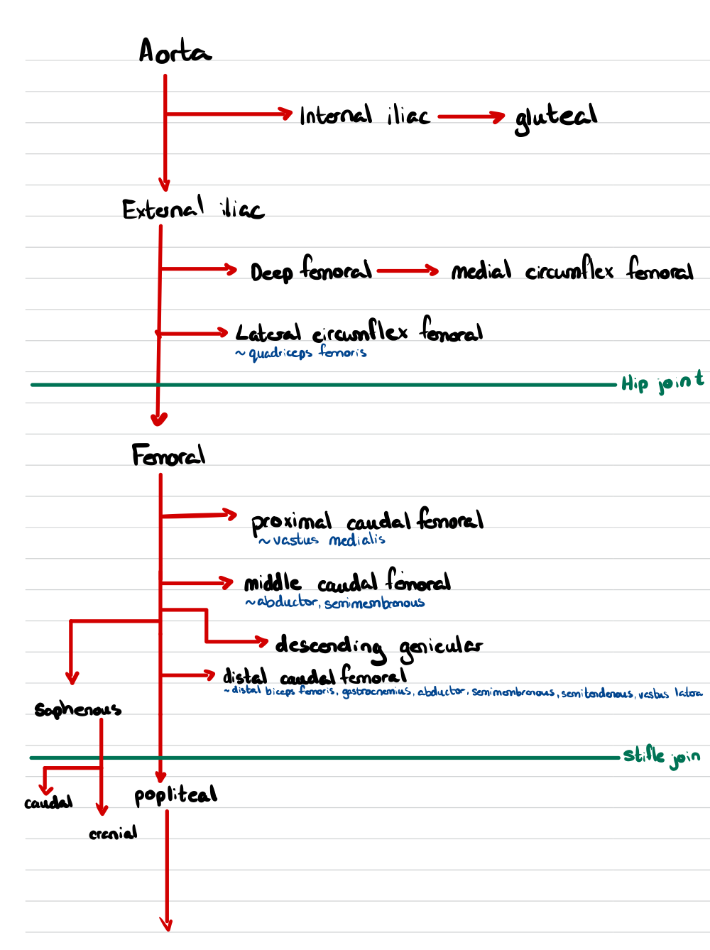
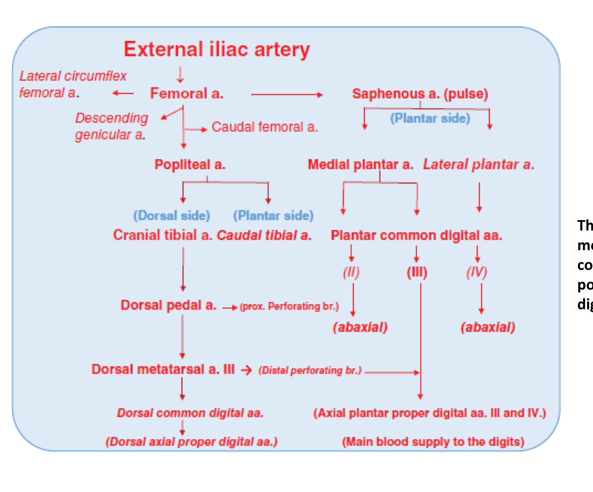
Arterial Pathways
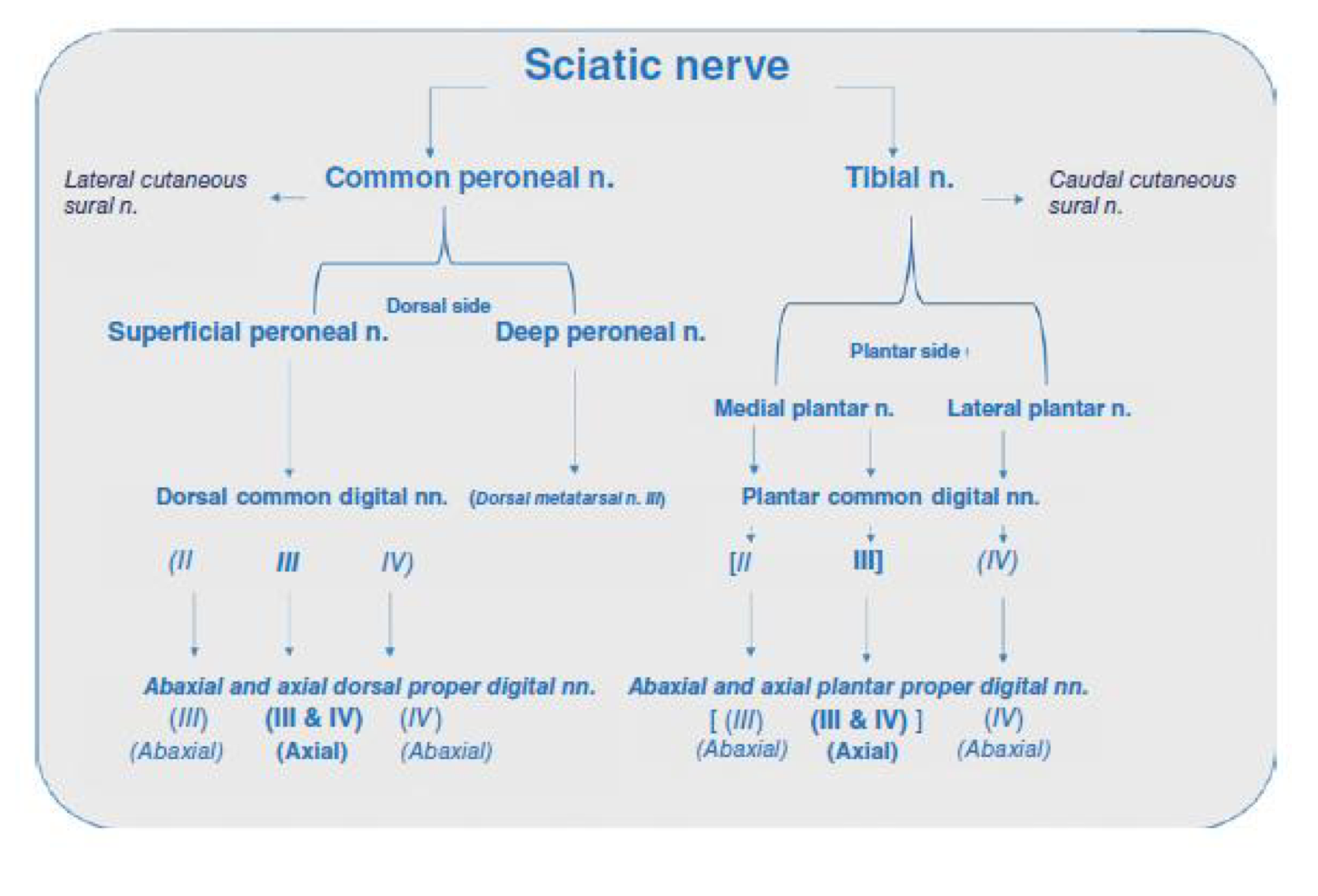
Nerve Pathway