Untitled Flashcard Set
Senses
All sensory organs contain sensory receptors (dendrites) that react to a certain external or internal stimulus.
There are 5 major types of sensory receptors:
Mechanoreceptors: touch, pressure, vibrations, sound
Thermoreceptors: Temperature variations
Nocireceptors: Pain stimulus
Chemoreceptors: Chemical stimuli, taste, smell, blood chemistry
Photoreceptors: Detect light
Special senses: Senses that have a specific organ associated with them
⭐Each sensory organ has one or more of these receptors
🤺Senses: Touch
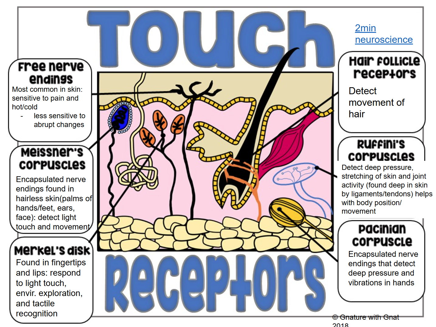
Cutaneous sensations: impulses produced in the skin (touch, heat, cold, pressure, & pain)
Not equally distributed throughout the body
Meissner’s Corpuscles: Encapsulated nerve endings found in hairless skin (palms, feet, face) that detect light, touch, and movement
Merkel’s Disk: Found in fingertips and lips that respond to light touch, environment, and tactile recognition
Hair follicle receptors: Detect movement of hair
Ruffini’s Corpuscles: Detects deep pressure, stretching of skin and joint activity
Pacinian Corpuscle: Encapsulated nerve endings that detect deep pressure and vibrations in hands
👅Senses: Taste
Taste buds: Specific receptors for the tounge that contain chemoreceptors
Most taste buds (10,000+) are located on the tounge, but some are also on the soft palate and inner cheeks
Chemoreceptors bind to certain molecules and then initiate an impulse through the nerves
The dorsal tounge is filled with bumps called papillae
Sharp filiform papillae
Rounded fungiform papillae
Circumvallate papillae
5 basic taste sensations:
Sweet
Sour
Bitter
Salty
Umami
The spicy sensation we feel when eating certain foods isn’t actually a taste, ut a burning sensation caused by capsaicin, a compound that treggers heat receptors in the mouth, tricking the brain into thinking it’s hot
Children have more taste buds than adults, so they taste more strongly
this is why they are picky eaters
over time we lose taste buds, which is why older people cover food in salt
The sense of taste is closely related to the sense of smell because they both rely on chemoreceptors

👃Senses: Smell
Olfactory receptors: in the upper nasal cavities help to distinguish thousands of different molecules
“Sniffing” helps to bring these molecules into contact with the olfactory receptors, intensifying the smell
Olfactory receptors are closely ties to the limbic system (emotional-visceral brain)
So smell impressions are long lasting and strongly tied to memories / emotions
Also, survival function to determine if something is good or bad based on smell (ex: expiered meat or smoke)
Accomidation: When olfactory receptors contact a substance continually, they become insensitive to the smell

👂Senses: Hearing
The structures of the ear work together to collect and transfer sound vibrations to the auditory nerve
Ear is also important for maintaining balance
The ear is divided into 3 regions:
Outer ear
Middle ear
Inner ear
Outer Ear
Sound waves are collected by the auricle in the outer ear
Auricle: outer ear where sound waves first enter
Sound is then passed through the external auditory canal
The sound waves then vibrate the tympanic membrane (eardrum)
Lightly stretched membrane that vibrates when vibrating air passes

Middle Ear
Sound waves then travel through the middle ear to a set of 3 tiny, jointed bones: malleus, incus, stapes
aka the hammer, the anvil, and the stirrup
They amplify sound waves to the inner ear through a membrane called the oval window
Oval window: a gateway to the inner ear and cochlea

MIS jointed bones: Malleus, Incus, Stapes
MIS jointed bones function: Amplify the vibrations and sends them to the inner ear
Inner Ear
Cochlea: A bony snail-shaped structure containing membranes filled with fluid and tiny hairs in the inner ear
Sterecocilia: Tiny hair cells that vibrate when sound waves travels depolarizing the ends of the nerve cells, which begins an action potential, transferring the impulse to the brain through the auditory nerve
The brain registers these impulses as sounds
Cochlear implants: Implants are surgically placed into the ear to improve hearing, direct connection to the cochlea
Hearing Issues
Deafness: Hearing loss of ANY degree
Conduction Deafness: Results when something interferes with the conduction of sound vibrations to the fluid in your ear
examples of conduction deafness: earwax buildup, ruptured eardrum, or incorrect bone fusion in the skul
Sensorineural Deafness: Degeneration or damage to the receptors, cochlear nerve, or neurons
examples of sensorineural deafness: extended time listening to excessively loud soung (earbuds on max)
Balance
In addition to hearing, the ear also helps us with balance
Three semicircular canals are located above the cochlea and are situated at right angles to each other
Semicircular canals: cancals are filled with fluid and lined with hair cells.
Otoliths: Tiny crystals located above the hair cells on the semicircular canals. When the head moves, these otoliths slide, stimulating the hair cells and sending an impulse to your brain
Because the the 3 cancals are at right angles, all types of motion can be recognized.
Types of motion recognized by semicircular canals: pitch, roll, yaw
👁Senses: Vision

Sclera: White part of the eye that protects and shapes the eye
Choroid: Blood rich nutrients, dark pigment to prevent light from scattering in the eye
Retina: Absorbs light through photoreceptors and turns light energy into impulses to transmit to brain
How you see
Similar to sound, you see light through a series of steps that involves various parts of the eye and brain
Parts include:
pupil
iris
retina
cornea
optic nerve
rods and cones

Step 1: The Cornea
Cornea is made of thick, transparent tissue
Important for protecting the pupil and iris
It is responsible for the refraction of the image
This lens makes adjustments to its shape depending on how close or far an image is
Distant object: eye muscle relaxes = lens is thinner
Close objects: eye muscle contracts = lens is thicker
Step 2: Light enters the through the pupil
Pupil: The black opening in the center of the iris that regulates the amount of light entering the eye
Low light: pupil wide open
High light: pupil nearly closed
Step 3: The iris controls the amount the amount of light that enters the pupil
The iris is located behind the cornea. It is the colored part of the eye that works with the pupilt o regulate light entering the eye

Step 4: Light that enters the eye is refracted on the rear curved surface of the retina on the back of the eye
Since the retina has concave shape, the image is upside down and smaller than the original image
The retina is connected to the optic nerve which sends the upside down image to your brain
The brain then flips the image rightside up

Step 5: Rods and Cones are connected to the nervous system which connects to the brain
Rods and cones are light sensitive nerves that help your brain decode what you are seeing
Rods: sensitive to light and darkness
Cones: responsible for seeing color
Humans have 3 types of cones that can only detect 3 types of light, red, green, and blue.
The mantis shrimp has 12-16 types of cones that can detect colors we can’t even imagine exists
There are no photoreceptors where the optic nerve meets the eye, this is called a blind spot. Because this blind spot affects different parts of each eye, it is not regularly noticed. The brain “fills in” the missing images
The lens is a semi-solid disc that directs light waves towards the retina.
Controlled by ciliary muscles and suspensory ligaments
These muscles and ligaments help bend or flatten the lens based on the distance of the image being viewed.
The lens becomes less elastic with age, causing some adults to need corrective lenses to have difficulty focusing at certain distances
The lens is responsible for focusing the light on the retina, to be intercepted by the photoreceptors and sent to the brain
In front of the lens is a fluid called Aqueous humor, which nourishes the cornea and behind the lens is the vitreous humor, which is a thick, jelly-like fluid that refracts light and fills the space between the lens and retina.

In some individuals, the eye is too long or too short, causing near-sightedness or far-sightedness
Corrective lenses help refract the light so they accurately converge on the retina
Nearsighted: A person has difficulty seeing object far away, but has no trouble seeing things up close because the lens is too convexly shaped
Occurs because the convex cornea is too long is too long, causing the image to fall in front of the retina
Their lens is basically too convex
To fix this, nearsighted people need a corrective concave lense to adjust the image before it enters the eye
Myopia: Nearsightedness
Farsighted: Person has difficulty seeing objects close up, but has no trouble seeing things far away
Occurs because the convex cornea is too short causing the rays of light to converge at a point beyong the retina
Basically the lens is not convex enough, creating blur
Correcting lenses would be more convex in shape to converge the light better before it enters the eye
Hyperopia: Farsightedness