Muscle Anatomy
Part 1: The "Big Picture" Hierarchy
Muscle anatomy is like a set of Russian nesting dolls. Each layer is wrapped in a specific "mysium" (connective tissue).
Muscle (Whole Organ): Wrapped in Epimysium.
Fascicle (A bundle of cells): Wrapped in Perimysium.
Muscle Fiber (The individual cell): Wrapped in Endomysium. Its cell membrane is the Sarcolemma.
Myofibril: Rod-like structures inside the cell.
Myofilaments: The actual proteins (Actin and Myosin).
Part 2: The Three Muscle Types
Feature | Skeletal | Smooth | Cardiac |
Location | Attached to bones | Walls of hollow organs | The Heart |
Control | Voluntary | Involuntary | Involuntary |
Appearance | Striated (Striped) | No striations | Striated & Branched |
Nuclei | Multi-nucleated | Uni-nucleated | Uni-nucleated |
Special | Fast & powerful | Slow & rhythmic | Intercalated discs |
Image | 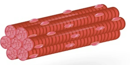 |  |  |
Part 3: The Sarcomere (The Contractile Unit)
This is the most technical part of your quiz. The sarcomere is the segment from one Z-line to the next.
Myosin (Thick Filament): Shaped like a golf club with a head. It sits in the center (A-band).
Actin (Thin Filament): Attached to the Z-lines.
H-Zone: The center area where there is only myosin (no actin overlap).
I-Band: The area near the Z-lines where there is only actin (no myosin).
The Sliding Filament Theory
When a muscle contracts:
Myosin heads grab onto Actin.
They pull the actin toward the center (M-line).
The Result: The H-zone and I-band disappear/shrink, the Z-lines move closer together, but the A-band (length of myosin) stays exactly the same length.
1) Sarcomere
2) H-zone
3) Actin
4) Myosin
5) Z-lines
6) A-band
7) I-band
8) M-line

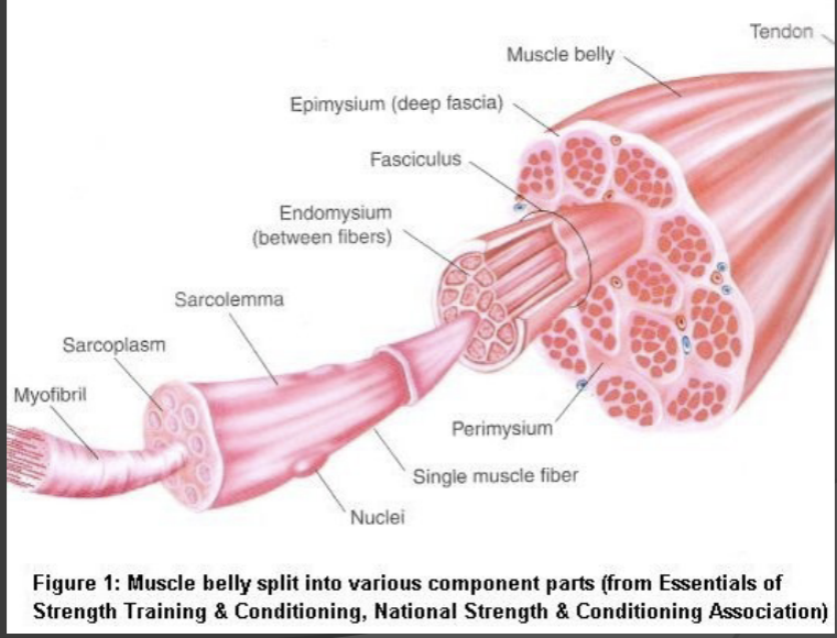


The diagrams associated with muscle anatomy illustrate the hierarchical structure of muscles and the types of muscle tissues:
Muscle Hierarchy Diagram
- Muscle (Whole Organ): The entire muscle is encased in a protective layer called the epimysium.
- Fascicle (A bundle of cells): Each fascicle, which consists of multiple muscle fibers, is surrounded by perimysium.
- Muscle Fiber (The individual cell): Each muscle fiber is wrapped in endomysium, and the cell membrane itself is termed the sarcolemma.
- Myofibril: These rod-like structures are found inside each muscle fiber.
- Myofilaments: These are the actual proteins responsible for muscle contraction, mainly actin and myosin.Muscle Types Diagram
- This diagram compares the three main types of muscle tissues (Skeletal, Smooth, and Cardiac) based on various features such as location, control, appearance, number of nuclei, and special characteristics:
- Skeletal Muscle:
- Location: Attached to bones
- Control: Voluntary
- Appearance: Striated
- Nuclei: Multi-nucleated
- Special: Fast & powerful
- Smooth Muscle:
- Location: Walls of hollow organs
- Control: Involuntary
- Appearance: No striations
- Nuclei: Uni-nucleated
- Special: Slow & rhythmic
- Cardiac Muscle:
- Location: The Heart
- Control: Involuntary
- Appearance: Striated & Branched
- Nuclei: Uni-nucleated
- Special: Intercalated discs.Sarcomere Diagram
- This is a technical diagram that highlights the contractile unit of muscle fibers:
- The Z-line demarcates the ends of a sarcomere.
- Myosin (Thick Filament): Forms the center band (A-band) and resembles a club.
- Actin (Thin Filament): Attaches to the Z-lines on either side.
- H-Zone: The middle area with only myosin.
- I-Band: The outer area with only actin.
- The Sliding Filament Theory explains muscle contraction: Myosin heads pull on actin filaments, causing them to slide towards the center, making the H-zone and I-band disappear while the A-band remains the same length.