Chapter 4 - Tissues
4.1
Nervous Tissue: Internal Communication
- Brain
- Spinal Cord
- Nerves
Muscle Tissue: Contracts to cause Movement
- Muscles attached to bones (skeletal)
- Muscles of the heart (cardiac)
- Muscles of walls of hollow organs (smooth)
Epithelial Tissue: Forms boundaries between different environments, protects, secretes, absorbs, filters
- The lining of digestive tract organs and other hollow organs
- Glands (e g., pancreas)
- Skin surface (epidermis)
Connective Tissue: Supports, protects, and binds other tissues together.
- Bones
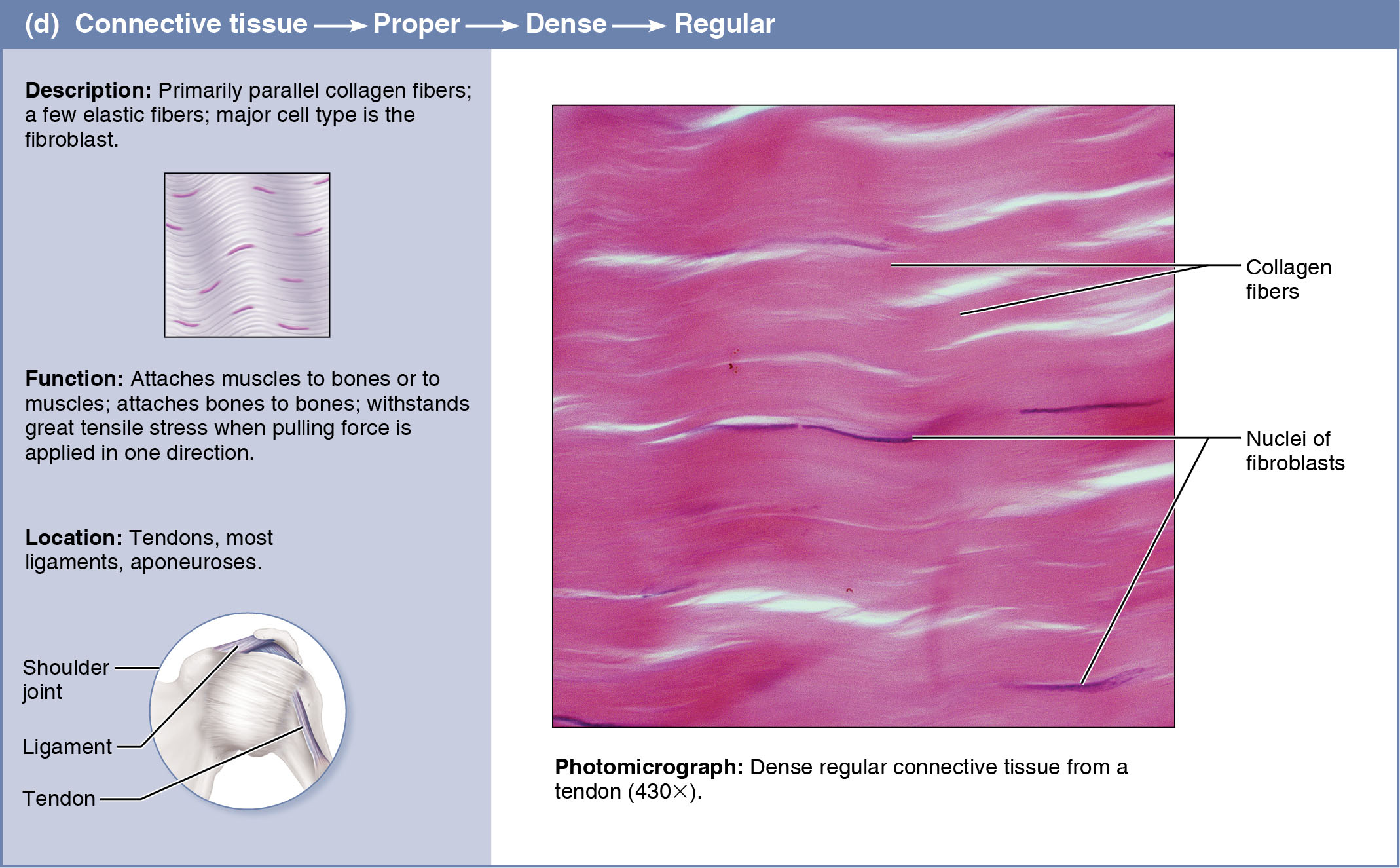
- Tendons
- Fat and other soft padding tissue
Microscopy allows us to study tissue structure. It must be fixed and cut into sections thin enough to transmit light (light microscopy) or electrons (electron microscopy). Finally, the specimen must be stained to enhance the contrast between structures.
In light microscopy, the stains are dyes, many of which consist of negatively charged molecules (acidic stains) or positively charged molecules (basic stains) that bind within the tissue to macromolecules of the opposite charge.
For Transmission Electron Microscopy (TEM), tissue sections are stained with heavy metal salts. Scanning Electron Microscopy (SEM), provides three-dimensional pictures of an unsanctioned tissue surface. TEM = 2D, SEM = 3D.
4.2
2 Forms of Epithelial Tissue:
- Covering and lining epithelium, which forms the outer layer of the skin; lines the open cavities of urogenital, digestive, and respiratory systems; and covers the walls and organs of the closed ventral body cavity.
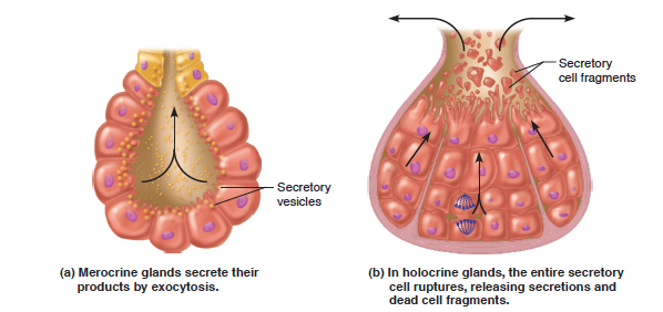
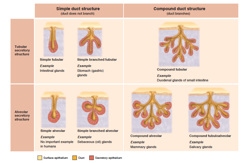
- Glandular epithelium, which fashions the glands of the body.
Functions of Epithelium:
- Protection
- Absorption
- Filtration
- Excretion
- Secretion
- Sensory Reception
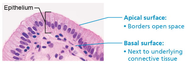
Apical Surface:
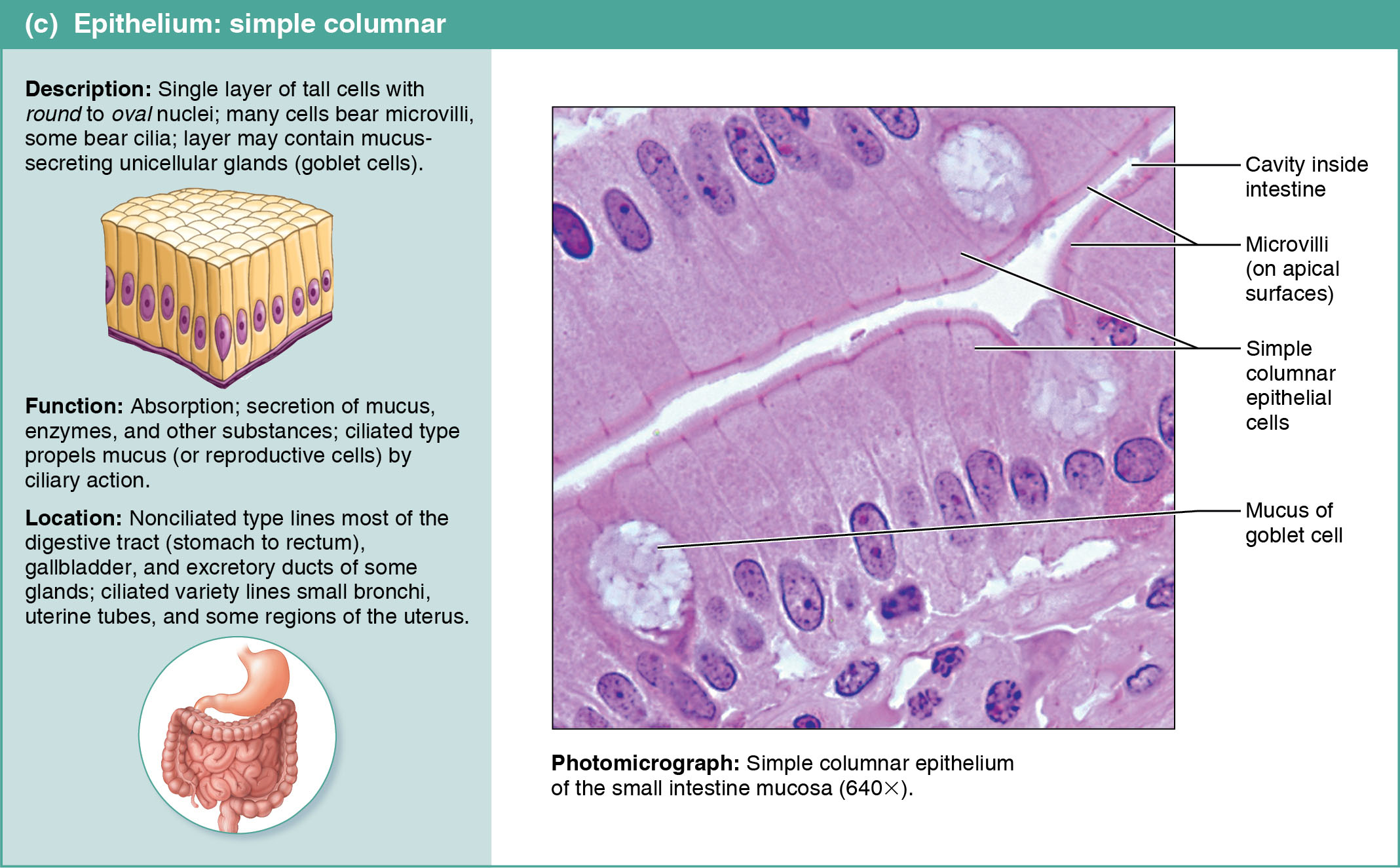
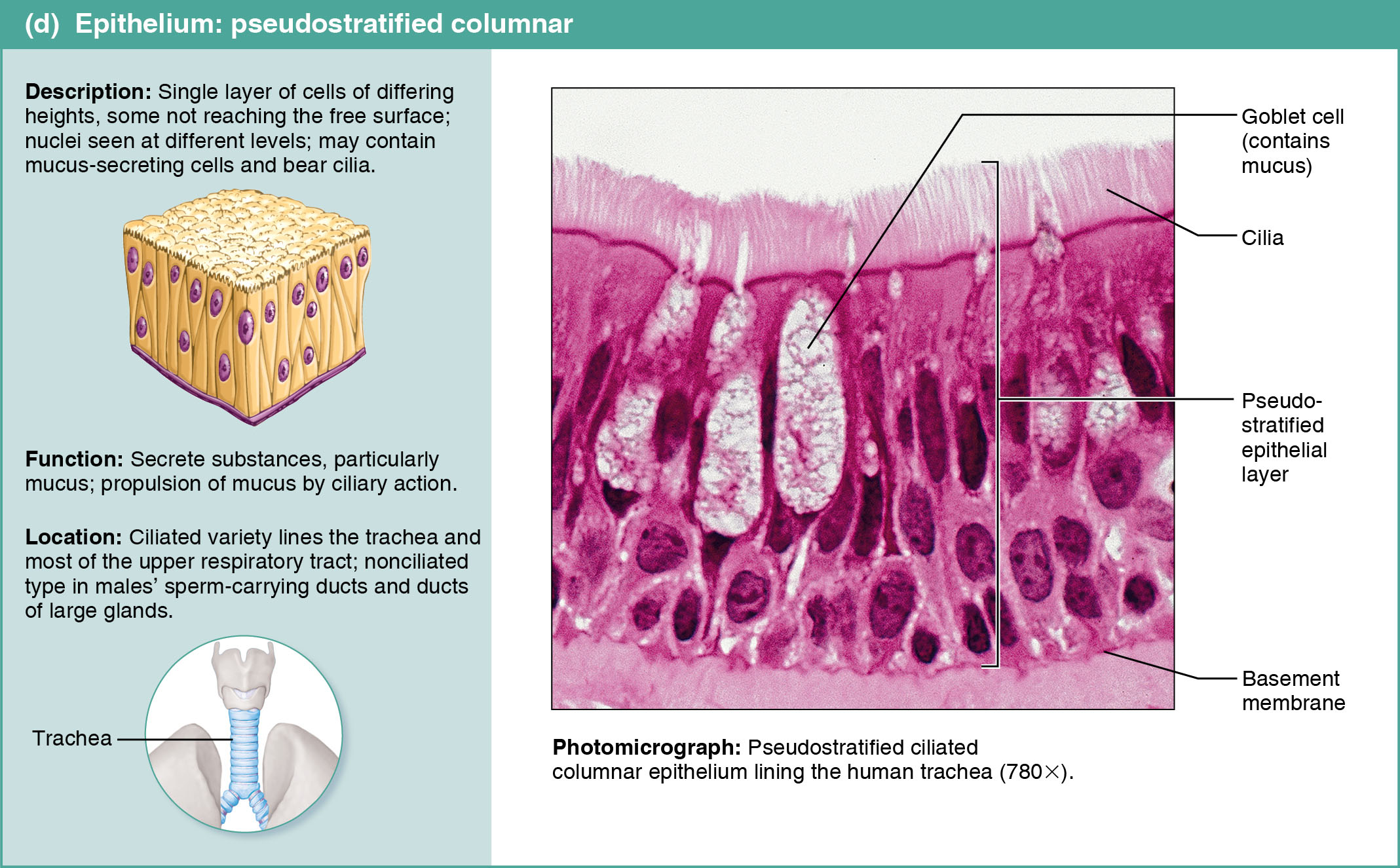
Contains Microvilli, fingerlike extensions of the plasma membrane that increase exposed surface area. Microvilli are often so dense that the cell has a fuzzy appearance called a brush border. Cilia propels substances along their free surface.
Basal Surface:
Adjacent to the Basal Surface is a thin sheet called the Basal Lamina. Basal Lamina is a noncellular sheet that consists largely of glycoproteins. Basal Lamina acts as a selective filter that determines which molecules diffusing from the connective tissue are allowed the enter the epithelium.
The sides of adjacent cells are tied together by tight junctions and desmosomes. All epithelial sheets are supported by connective tissue.
The epithelium is avascular (contains no blood vessels), and innervated (supplied by nerve fibers).
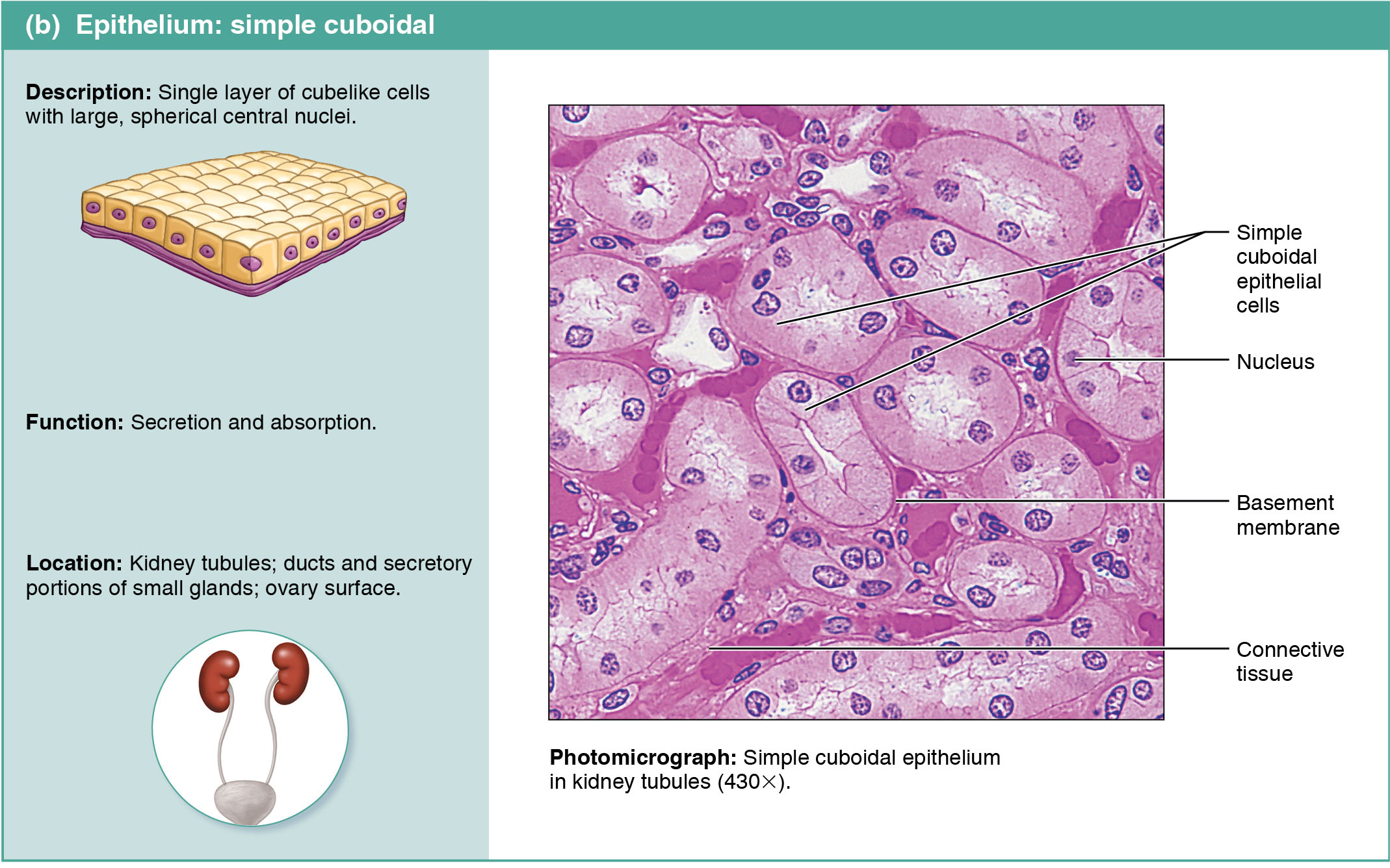
Simple Epithelia - a single cell layer.
Stratified Epithelia - two or more cell layers.
Squamous Cells - flattened and scale-like
Cuboidal Cells - boxlike, as tall as they are wide
Columnar Cells - tall and column-shaped
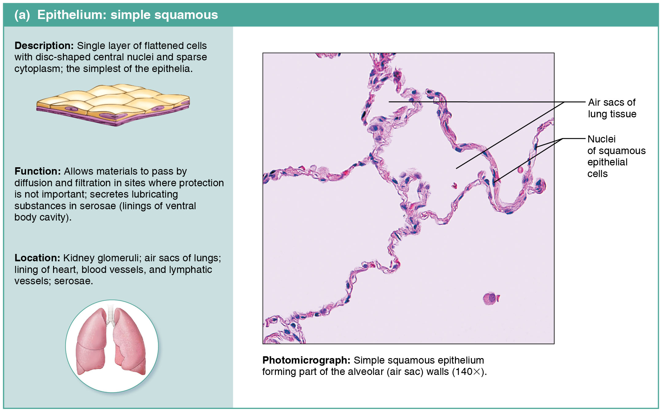
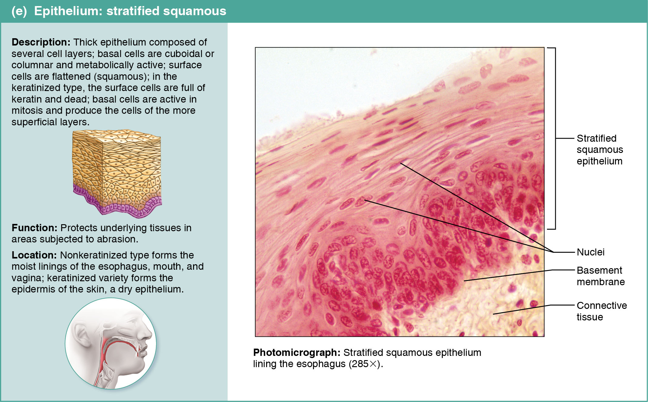
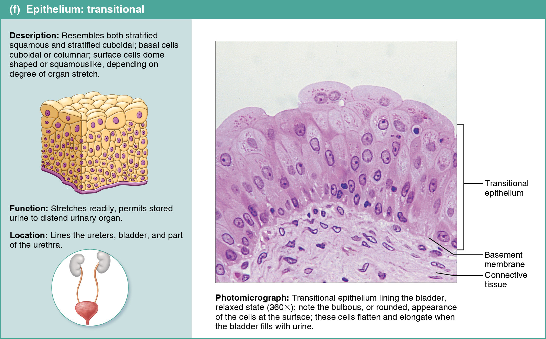
- Endothelium - provides a slick, friction-reducting lining in lymphatic vessels and in all hollow organs of the cardiovascular system.
- Mesothelium - the epithelium found in serous membranes. lining the ventral body cavity and covering its organs.
4.3
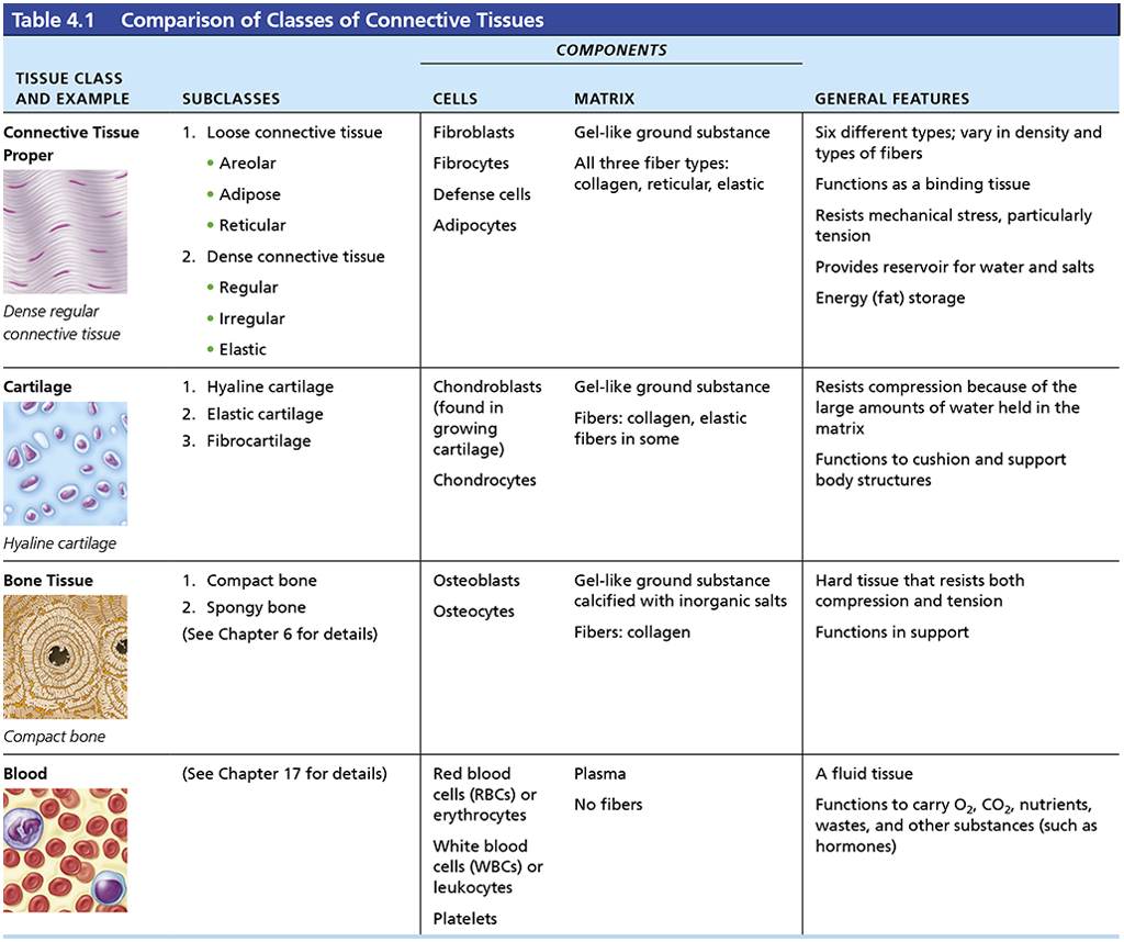
Functions of Connective Tissue:
- Binding & Supporting
- Protecting
- Insulating
- Storing reserve fuel
- Transporting substances within the body
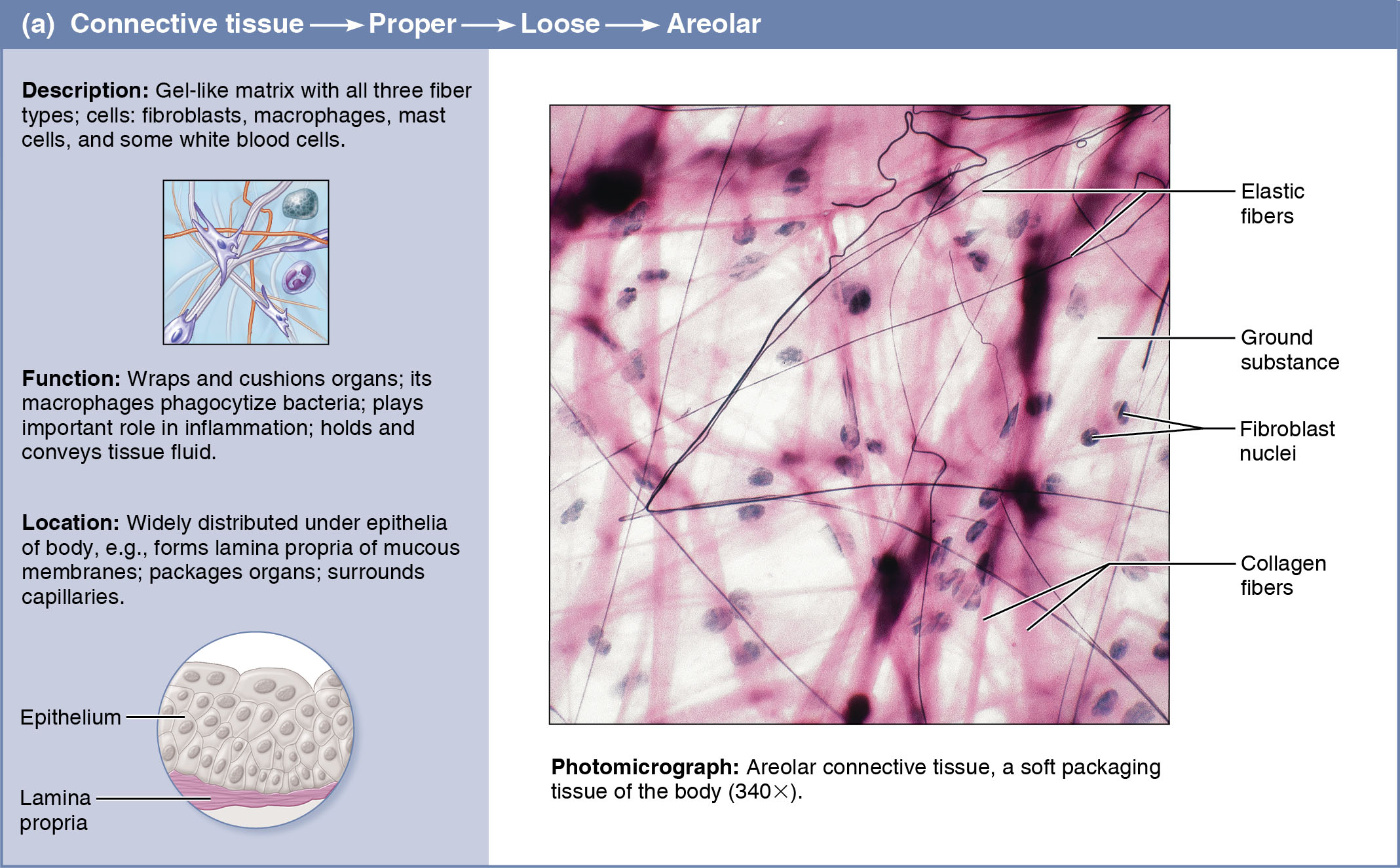
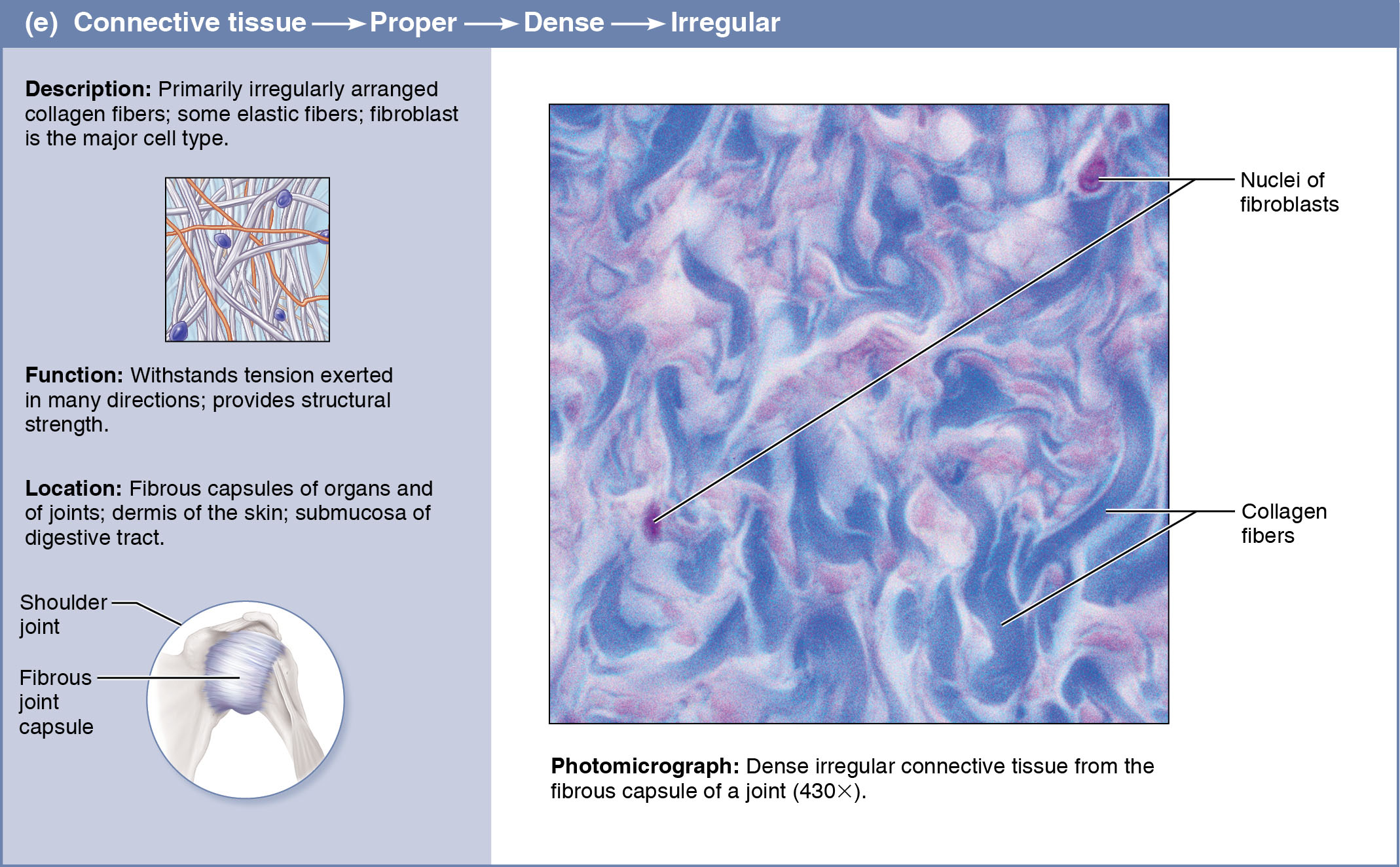
Connective Tissues consist largely of extracellular matrix. All connective tissues arise from mesenchyme. Cartilage is avascular. Connective Tissues have 3 main components: ground substance, fibers, and cells.
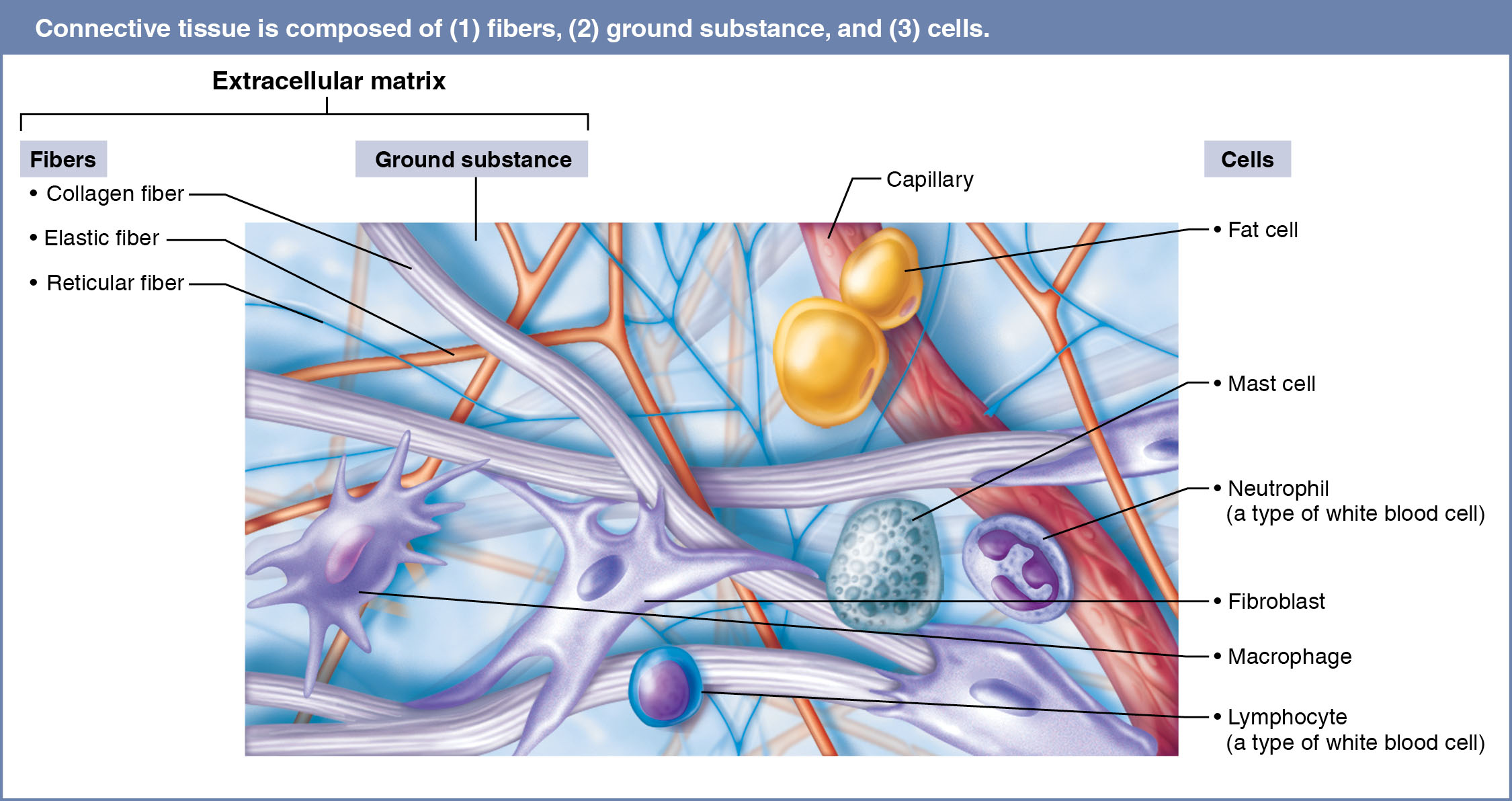 Ground Substance - unstructured material that fills the space between the cells and contains the fibers (3 components)
- Interstitial Fluid - The ground substance consists of large amounts of fluid and functions as a molecular sieve through which nutrients and other dissolved substances can diffuse between the blood capillaries and the cells.
- Cell Adhesion Proteins - These proteins serve mainly as a connective tissue glue that allows connective tissue cells to attach to the extracellular matrix.
- Proteoglycans - The proteoglycans consist of a protein core to which large polysaccharides called glycosaminoglycans are attached.
Collagen Fibers -
These fibers are constructed primarily of the fibrous protein collagen. Collagen molecules are secreted into the extracellular space
Elastic Fibers -
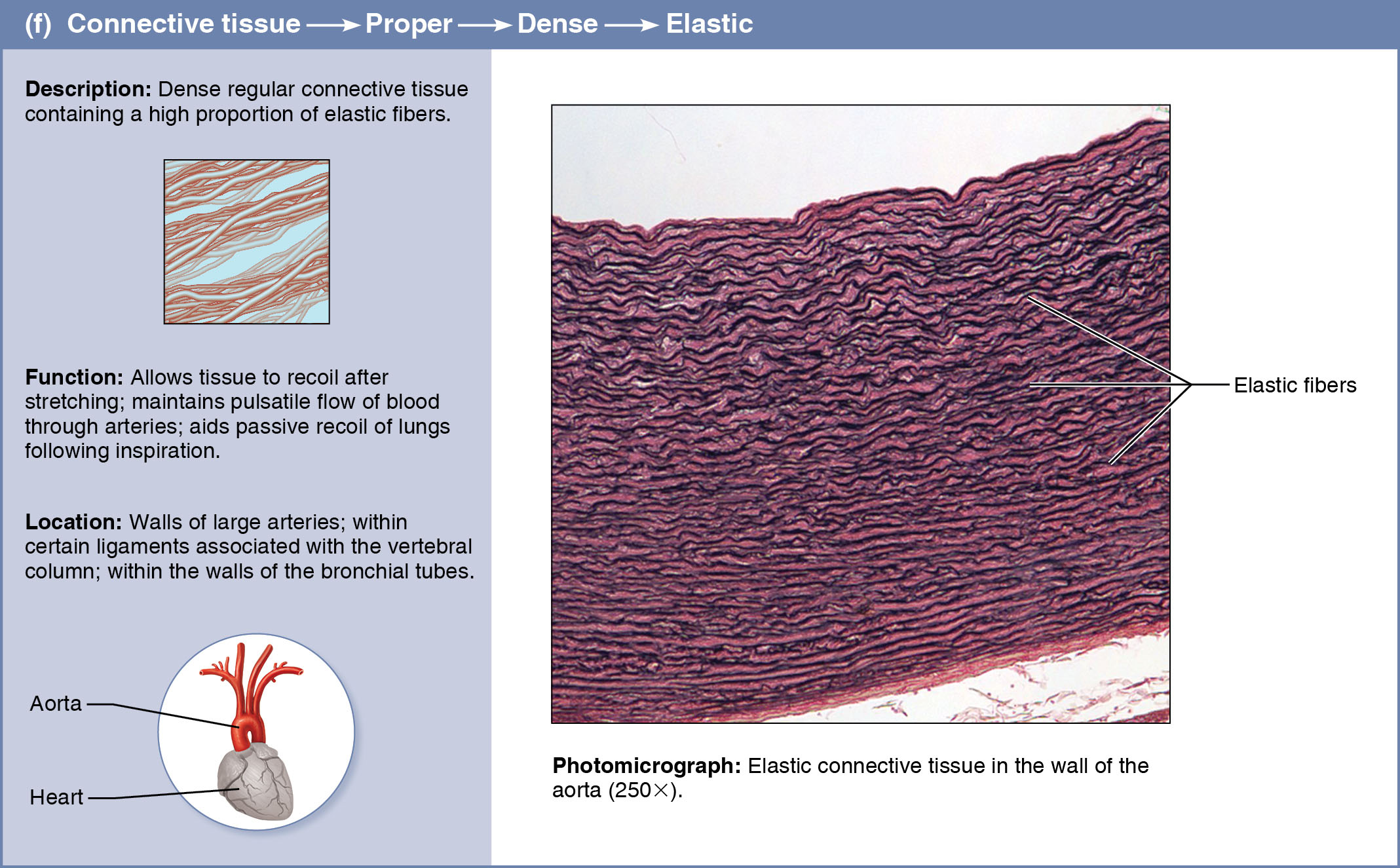
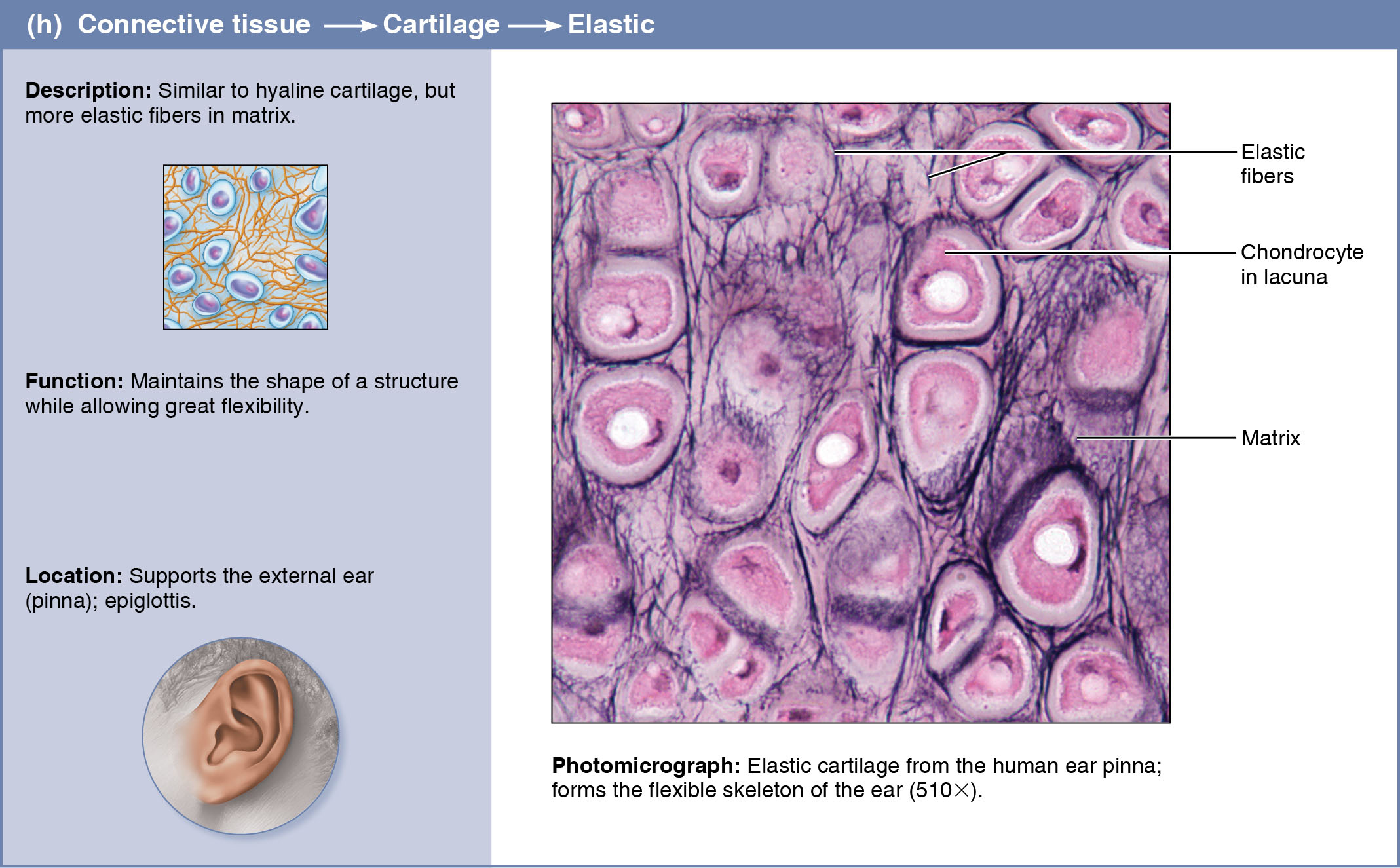
Long, thin, elastic fibers form branching networks in the extracellular matrix. These fibers contain a rubberlike protein, elastin, that allows them to stretch and recoil like rubber bands.
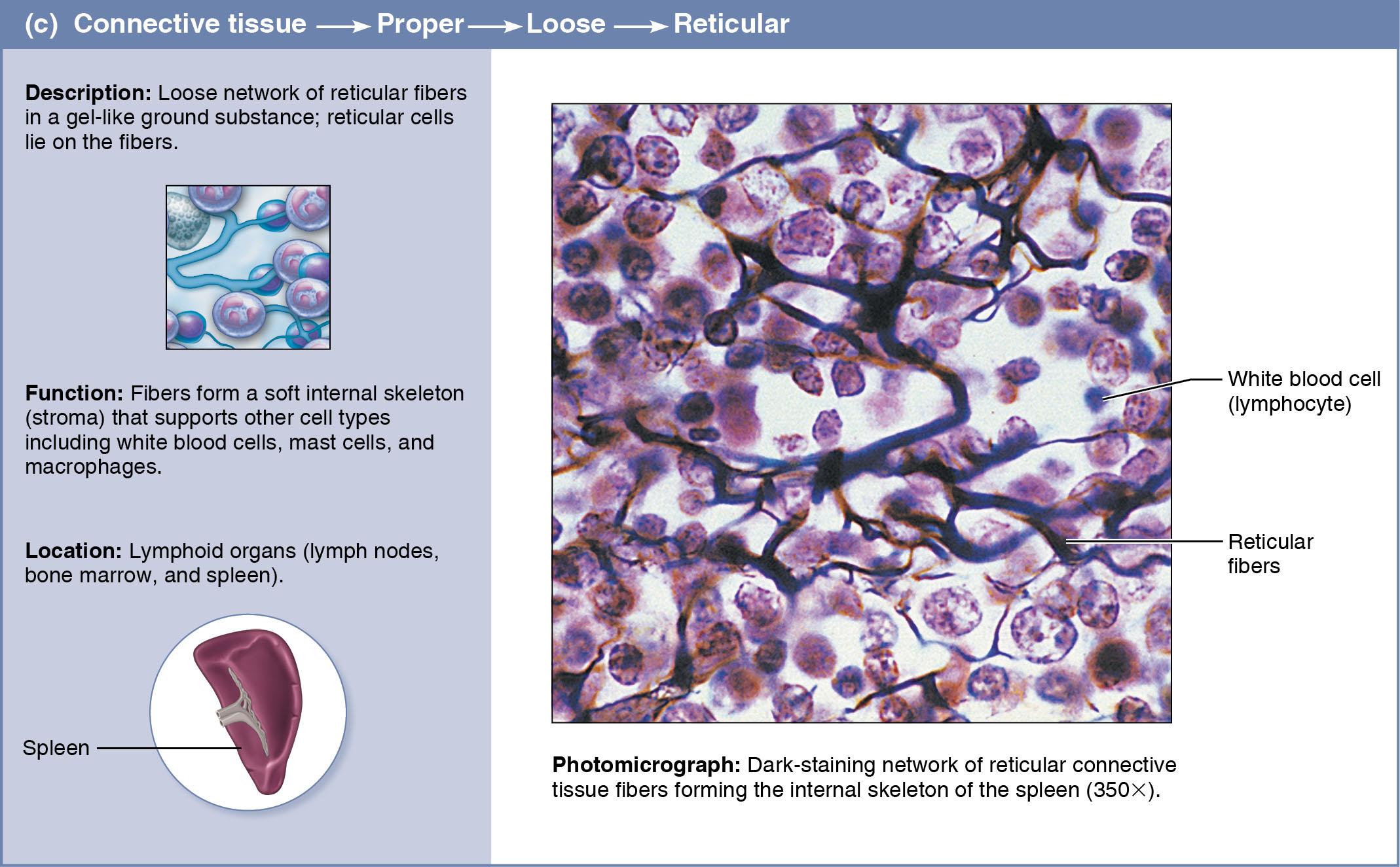
Reticular Fibers -
These short, fine fibers are made of a different type of collagen than the more common, thicker collagen fibers. They connect to the coarser collagen fibers, but they branch extensively, forming delicate networks
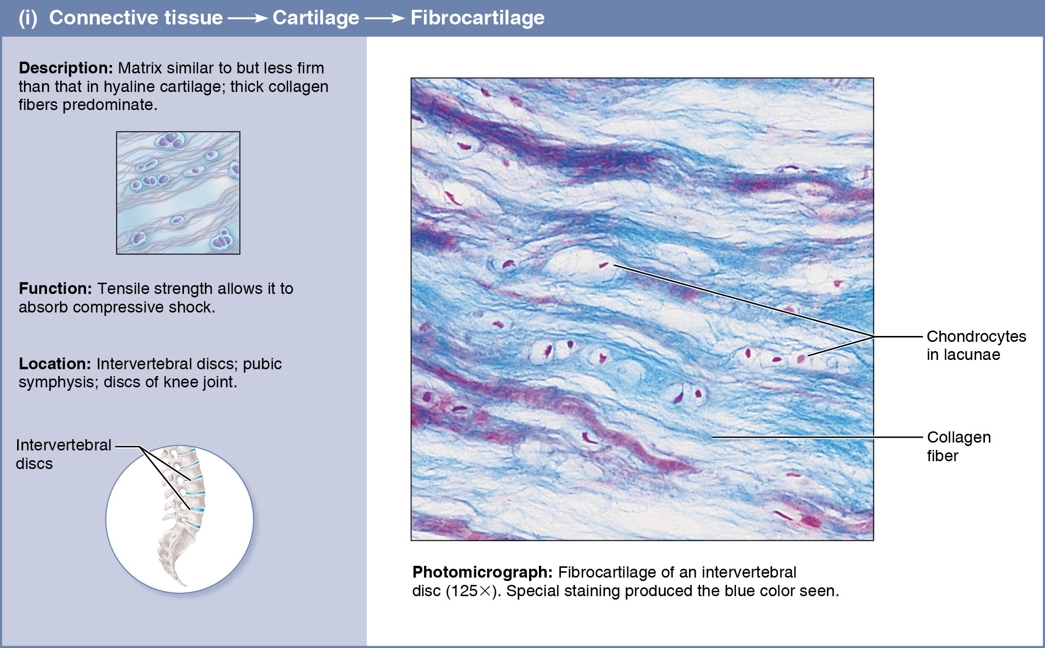
Fibroblasts in connective tissue properly become fibrocytes.
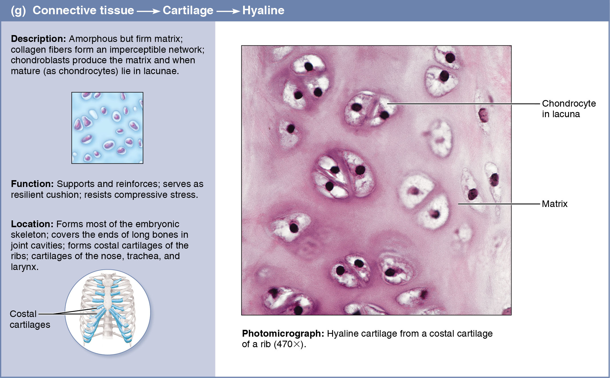
Chondroblasts in cartilage become chondrocytes.
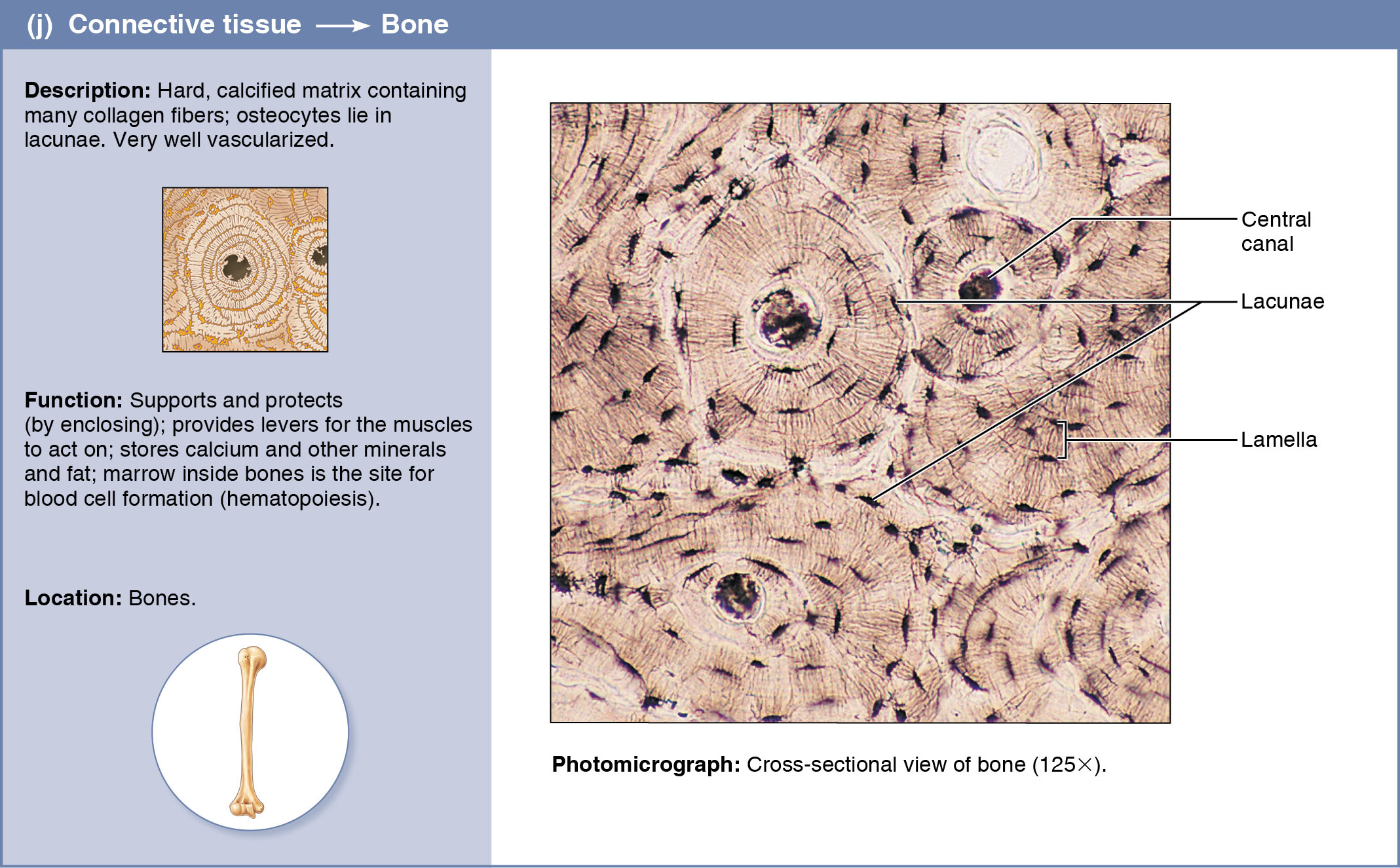
Osteoblasts in bone become osteocytes.
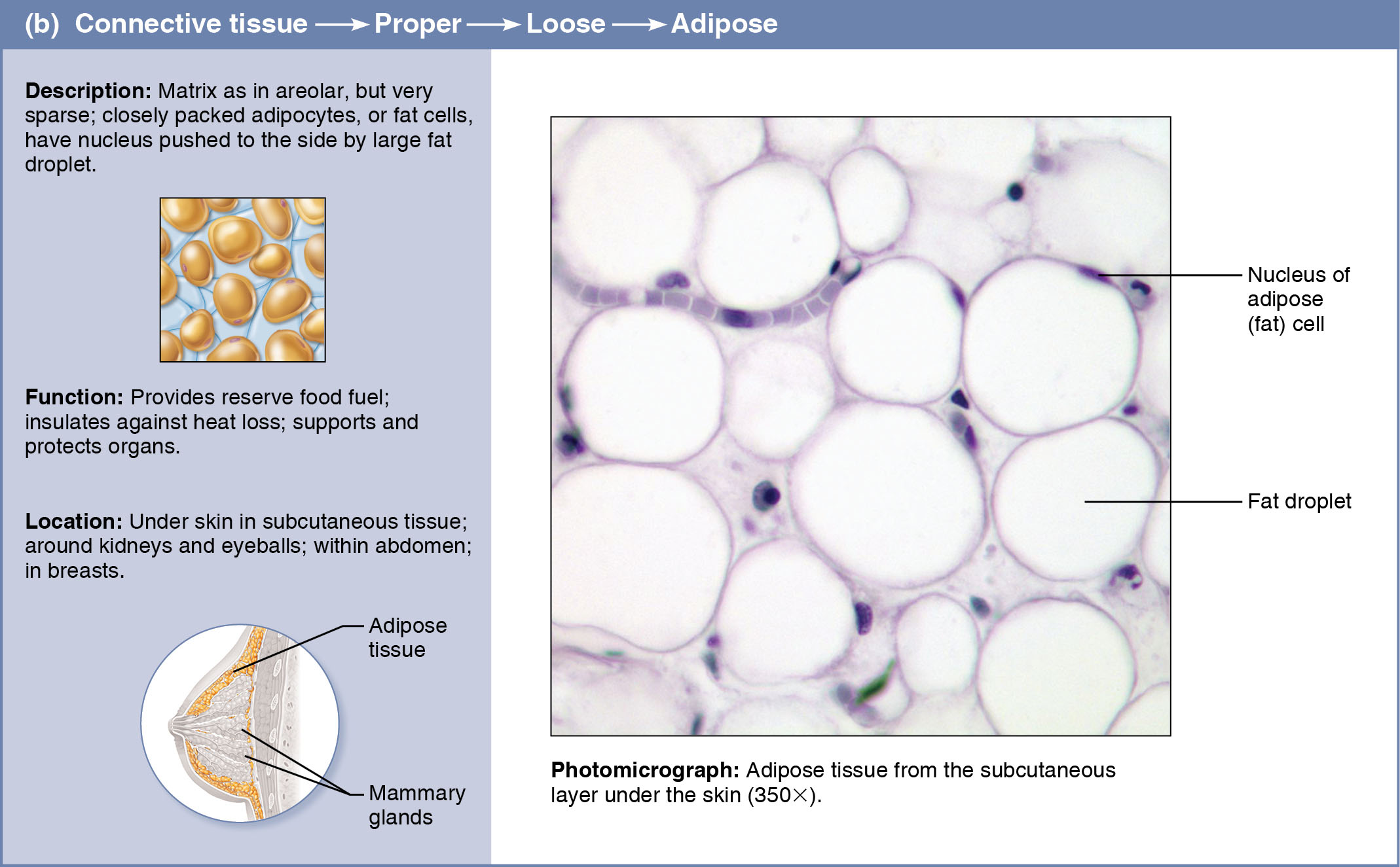
Adipocytes, commonly called adipose or fat cells, store energy as fat.
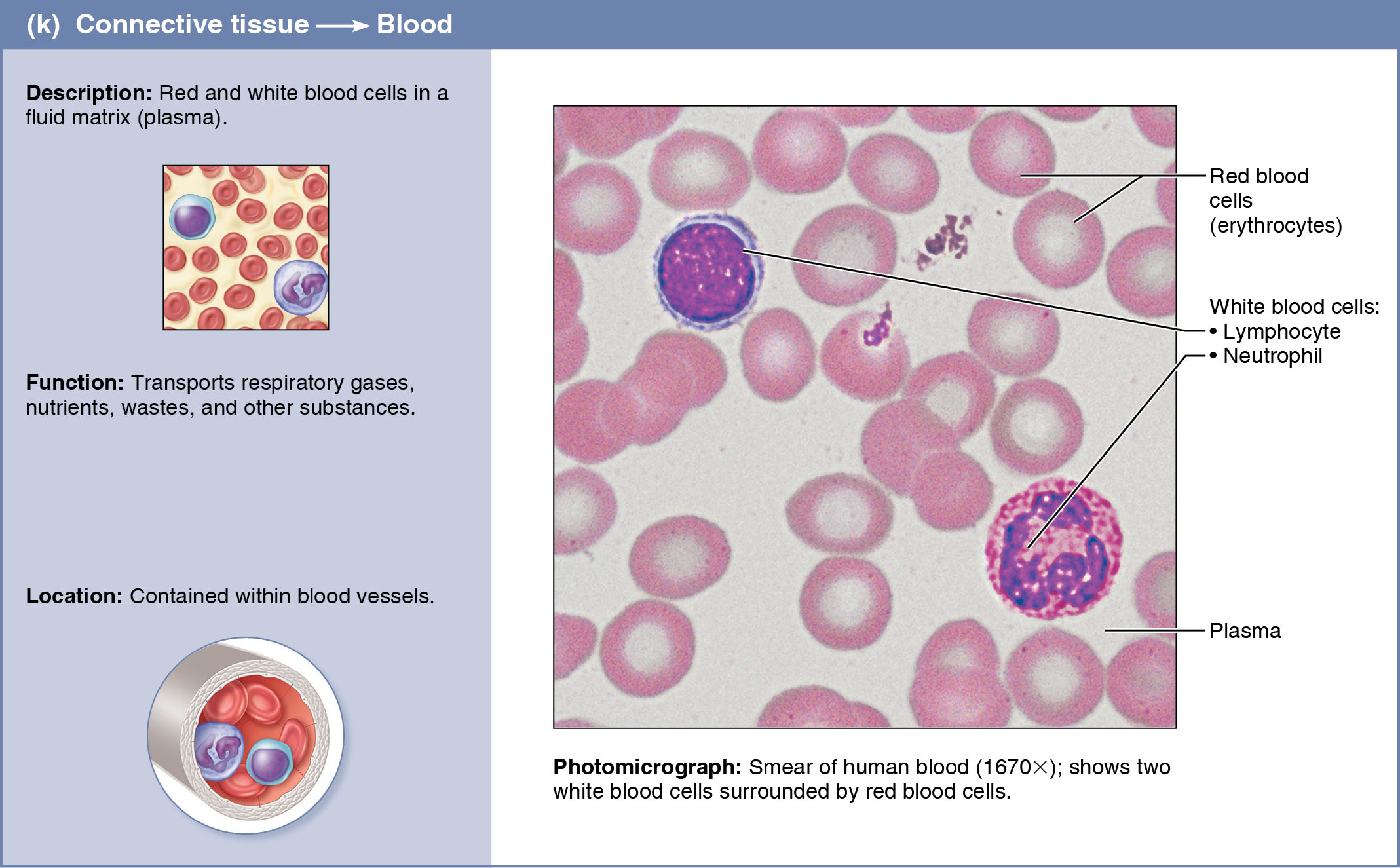
White blood cells (WBCs or leukocytes, including neutrophils, eosinophils, and lymphocytes) and other cell types are concerned with tissue response to injury.
Mast cells, typically cluster along blood vessels. These oval cells detect foreign microorganisms (e.g., bacteria, fungi) and initiate local inflammatory responses against them. Mast cell cytoplasm contains secretory granules (mast = stuffed full of granules) with chemicals that mediate inflammation, especially in severe allergies. These chemicals include:
Heparin is an anticoagulant chemical that prevents blood clotting when free in the bloodstream (but in human mast cells it appears to regulate the activity of other mast cell chemicals)
Histamine, a substance that makes capillaries leaky
Proteases (protein-degrading enzymes)
Other enzymes
Macrophages are large, irregularly shaped cells that avidly devour various foreign materials, ranging from foreign molecules to entire bacteria to dust particles. These also dispose of dead tissue cells, and they are central actors in the immune system. Macrophages are throughout loose connective tissue, bone marrow, and lymphoid tissue.