4a: Renal, nephron structure, renal blood flow, glomerular filtration
Renal Physiology
Learning Outcomes
Produce a diagram showing the structures and major functions of the nephron.
Explain the relationship between renal blood flow and glomerular filtration rate.
Discuss the role of the sympathetic nervous system in the regulation of renal blood flow.
Describe the role of the renin-angiotensin system as it relates to renal blood flow.
List several factors that affect glomerular filtration rate.
Functions of the Kidneys
The kidneys have numerous functions beyond urine production:
- Key role in regulating blood pressure.
- Produce erythropoietin vital for red blood cell production.
- Maintaining calcium and phosphate balance via nephrons and Vitamin D activation.
- Maintaining acid/base balance.
- Maintaining electrolyte balance such as potassium and sodium.
- Removal of waste products including urea, creatinine, drugs, and toxins.
Kidney Anatomy Overview
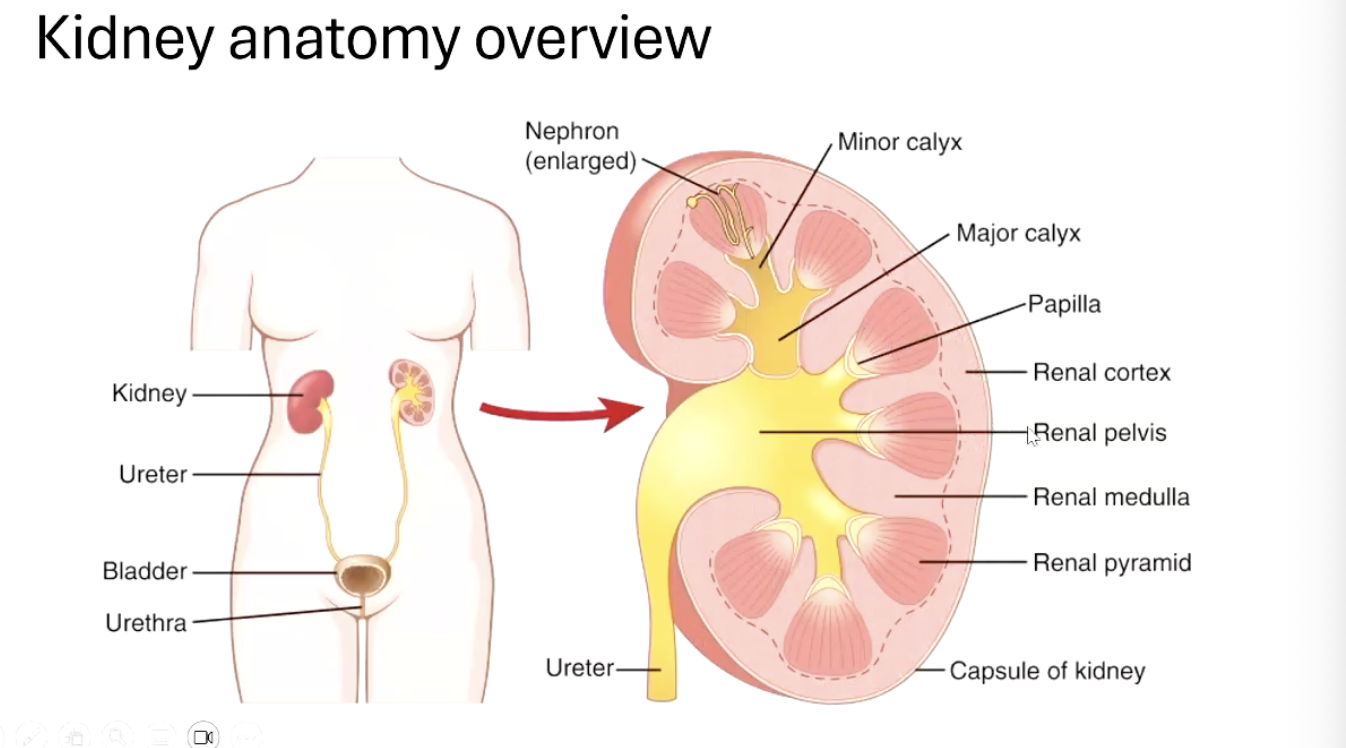
Components of the urinary system (urine formation and excretion):
Ureter
muscular tube connecting kidney to bladder
uses peristalsis to push urine to bladder
prevents backflow with one-way flow dynamics
peristalsis: involuntary constriction and relaxation of the muscles of a canal, creating wave-like movements that push content forward
kidney
filters blood
remove waste
regulates electrolytes
maintains acid-base balance
controls blood pressure (via renin
produces hormones
bladder
urethra
lined with mucosa (protective, mucus-secreting liner of hollow organs. shields bladder and urethra from urine while allowing stretch and smooth flow)
surrounded by sphincters (specialised ring-shaped muscle)
internal urethral sphincter (smooth, involuntary)
external urethral sphincter (skeletal muscle, voluntary)
nephron (enlarged)
the functional unit of the kidney (~1 million per kidney)
filtration (glomerulus)
reabsorption (proximal tubule, loop of henle, distal tubule)
secretion (distal tubule)
concentration (collecting duct)
minor calyx
a small cup like structure that collects urine from one papilla
passes it into a major calyx
major calyx
formed by merging several minor calyx
channels urine toward the renal pelvis
papilla
tip of each renal pyramid
where urine exits the medulla
drains into the minor calyx
the first point where fluid is officially “urine”
renal cortex
outer region of kidney, (but within the capsule)
contains most of the nephrons
where blood first becomes “filtrate”
primary site of filtration and initial urine formaion
renal pelvis
funnel shaped reservoir inside the kidney
collects all urine from the major calyces
narrows to become the ureter
renal medulla
contains loops of henle/nephron loops and collecting ducts
preserves water
creates osmotic gradient to concentrate urine
renal pyramid
cone shaped section OF the medulla
houses loops of henle and collecting ducts
transports urine from nephrons to papilla “urine funnels”
capsule of kidney
tough fibrous outer layer
protects kidney from trauma
maintains shape
acts as barrier against infection spreading into the kidney
Structure of the Nephron
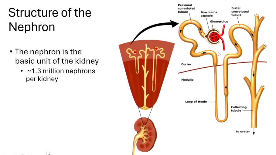
The nephron is the basic functional unit of the kidney.
Approximately 1.3 million nephrons per kidney.
glomerulus: a tuft of capillaries inside bowman’s capsule
proximal convoluted tubule (PCT)
Alternative Diagram of Nephron
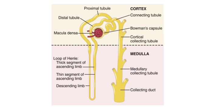
Key Structures filtrate - small solutes filtered out of the glomerulus
Cortex:
glomerulus (inside bowman’s capsule)
performs filtration
blood pressure forces water + small solutes out of the blood
large proteins and cells stay in the bloodsteream
bowman’s capsule
surrounds the glomerulus
collects the filtrate produced by the glomerulus
funnels it into the proximal tubule
proximal convoluted tubule (PCT)
highly folded segment of the tubule
“workhouse” of the nephron
reabsorbs most of filtered water and Na
reabsorbs all glucose and amino acids
secretes some wastes (H, drugs)
distal convoluted tubule (DCT)
more selctive, hormune-regulated segment
fine-tunes electrolyte balance
reabsorbs Na
regulates Ca
secretes H and K
macula densa
specialised grouop of DCT cells that sense NaCl in the filtrate and adjusts GFR and renin release to maintain stable kidney function
connecting tubule
cortical collecting tubule
determines final urine concentration
rebasorbs water
reabsorbs Na
carries urine through the medulla to papilla to calyces
Medulla:
loop of henle: overall, creates the medullary osmotic gradient (concentrates the medulla) which allows the kidney to produce concentrated urine.
descending limb first: permeable to water, not permeable to solutes. water leaves, filtrate becomes more concentrated
thick segment of ascending limb
thin segment of ascending limb
ascending limbs: permeable to solutes (Na, K, Cl). not permeable to water. solute leave. filtrate become more dilute
medullary collecting tubule
collecting duct
Renal Blood Flow (RBF)
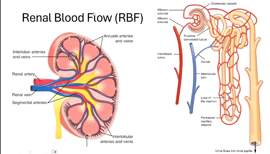
Key components:
interlobar arteries and veins
between lobes
renal artery
blood comes in from here
renal vein
blood goes out form here
segment arteries
arteries that are brnched off
afferent and efferent arteriole
afferent before glomerulus
efferent after glomerulus
- Interlobar arteries and veins
- Renal artery and vein
- Segmental and arcuate arteries and veins
- Interlobular arteries and veins
- Peritubular capillary network
- Urine flows into renal papilla
The Renin-Angiotensin System (RAS)\
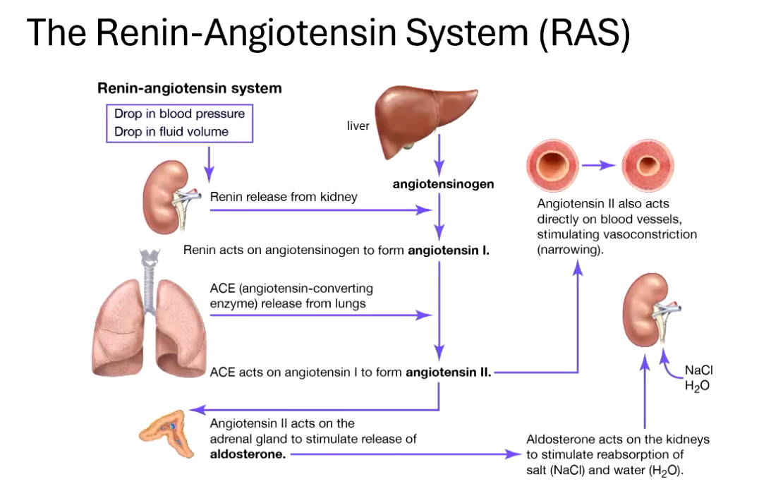
a hormonal system that regulates:
blood pressure
fluid balance
electrolyte homeostasis
Processes:
1. Drop in blood pressure
2. Drop in fluid volume
3. Renin release from kidney
4. Renin acts on angiotensinogen (produced by the liver) to form angiotensin I.
5. ACE (angiotensin-converting enzyme) released from lungs converts angiotensin I to angiotensin II.
6. Angiotensin II effects:
- Vasoconstriction of blood vessels.
- Acts on the adrenal gland to stimulate release of aldosterone.
- Aldosterone acts on kidneys to stimulate reabsorption of sodium chloride (NaCl) and water (H₂O).
Urine Formation - Overview
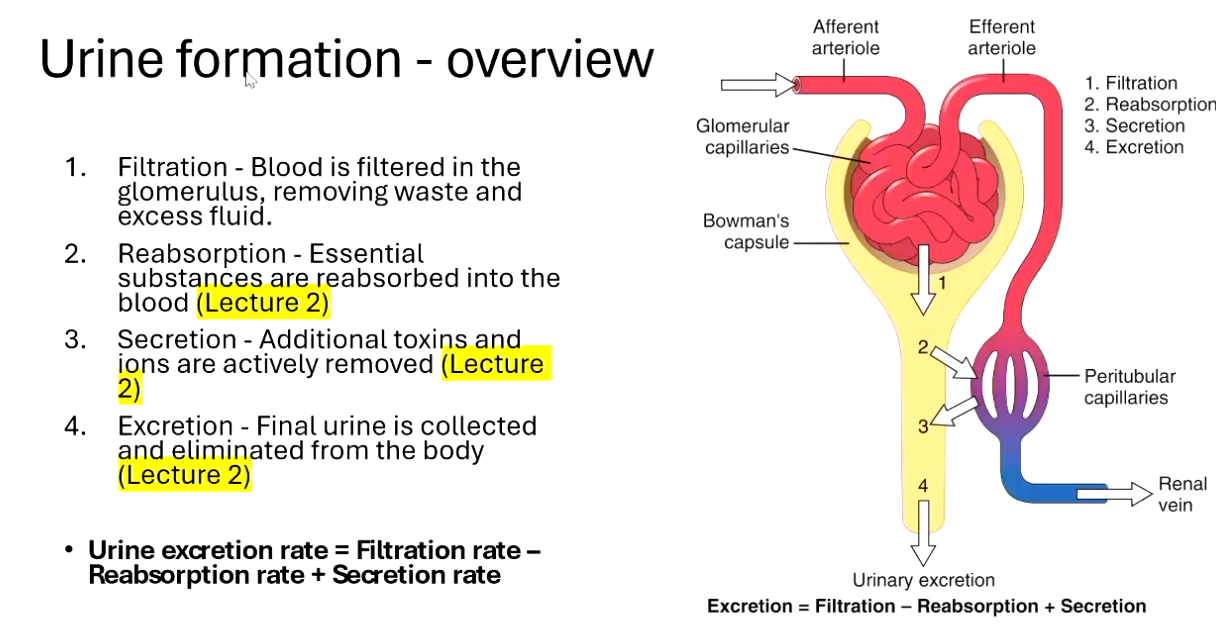
Filtration:
- Blood is filtered in the glomerulus (coming in afferent, exiting efferent), removing waste and excess fluid.Reabsorption:
- Essential substances are reabsorbed into the blood (to be covered in Lecture 2).Secretion:
- Additional toxins and ions are actively removed (to be covered in Lecture 2).Excretion:
- Final urine is collected and eliminated from the body (to be covered in Lecture 2).
urine excretion rate = filtration rate - reabsorption rate + secretion rate
Glomerular Filtration – Cell Membrane
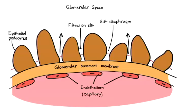
Working from the inside out, the capillary walls consist of three layers:
- Endotheliumlarger pores
solutes, plasma can pass through but not blood cells
- Basement membrane
- Epithelium
Glomerular Filtration – Permeability
Permeability metrics:
- Glomerular capillary is 50 times more permeable than skeletal muscle capillary. (very very permeable)
- Selective permeability based on:
- Size
- Charge (likes neutral molecules more than anionic and cationionic)
- For neutral molecules:
- Less than 4.0 nm is freely filterable.
- More than 8.0 nm is almost non-filterable.
Glomerular Filtration Rate (GFR) Control
Note: There is a Bowman's colloidal osmotic pressure, but it usually is 0 mm Hg; thus, it is often omitted in equations.
In certain disease states, the presence of proteins in the Bowman's capsule can raise this pressure above 0 mm Hg.
rate at which the kidneys filter blood
net filtration pressure = glomerular hydrostatic pressure - bowman’s capsule pressure (~18 mm Hg) - glomerular colloid osmotic pressure (usually 32 mm Hg)
glomerular hydrostatic pressure:
pressure of the blood as it forces its fluid into the bowman’s capsule
~60 mm Hg
bowmans colloidal osmotic pressure, usually 0mm Hg unless disease present
Factors That Affect GFR
Changes in filtration coefficient (Kf):
- Filtration coefficient (Kf):
- Defined as the product of a biological membrane's permeability to water and the surface area of the membrane.Changes in glomerular capillary hydrostatic pressure (GCHP)
Changes in Bowman's capsule hydrostatic pressure (BCHP)
Changes in glomerular colloidal osmotic pressure (GCOP)
- Notably, changes in Bowman's colloidal osmotic pressure
Factors That Affect GFR – Filtration Coefficient (Kf)
affected by Surface Area of the Glomerular Membrane:
- Increase in surface area (↑ surface area) leads to (↑ Kf) and subsequently (↑ GFR).
- Relaxation of mesangial cells increases surface area.
- Decrease in surface area (↓ surface area) leads to (↓ Kf) and subsequently (↓ GFR), connected to:
- Contraction of mesangial cells.
- Decreased number of glomerular capillaries.
- Uncontrolled diabetes impacting Kf.
affected by Permeability of Glomerular Membrane:
- Increase in thickness (↑ Thickness) leads to (↓ Kf) and subsequently (↓ GFR) (impacted by uncontrolled diabetes and hypertension).
Factors That Affect GFR – Glomerular Capillary Hydrostatic Pressure
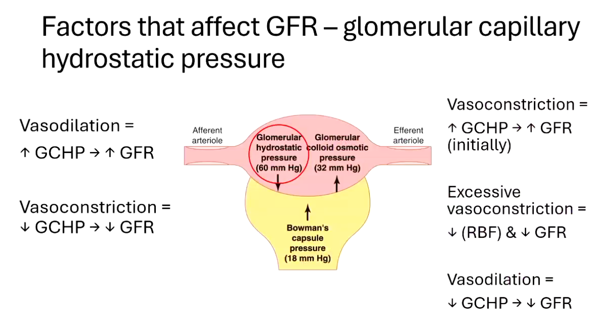
Effects of Vasodilation vs. Vasoconstriction:
- Vasodilation leads to (↑ GCHP) which elevates (↑ GFR).
- Vasoconstriction leads to (↓ GCHP) reducing (↓ GFR).
- Initial vasoconstriction raises GCHP, potentially leading to increased GFR, but excessive vasoconstriction decreases renal blood flow (RBF) and decreases GFR.
- Hormonal and neural control:
- Integrated with autoregulation mechanisms, including myogenic regulation.
blood pressure
tubuloglomerular feedback
hormonal and neural control
blood pressure provides the driving force
tubuloglomerular feedback stabilises GFR locally
hormonal/neural systems adjust afferent and efferent arterioles to raise or lower GFR depending on the body’s needs
Factors That Affect GFR – Bowman's Capsule Hydrostatic Pressure
Urinary Obstruction:
- Increased Bowman's capsule hydrostatic pressure (BCHP) leads to decreased GFR.
- Common examples include:
- Kidney stones.
- Prostate enlargement.
Factors That Affect GFR – Glomerular Colloidal Osmotic Pressure
Effects of Changes in GCOP:
- An increase in GCOP (↑ GCOP) results in a decrease in GFR (↓ GFR).
- Example: Dehydration increases GCOP.
- A decrease in GCOP (↓ GCOP) results in an increase in GFR (↑ GFR).
- Example: Hypoproteinemia.
pressure | location | what causes it | pushes fluid _ of blood | effect on GFR |
|---|---|---|---|---|
glomerular hydrostatic | glomerular capillaries | blood pressure | out | rises |
capsule pressure | bowmans capsule | fluid already in capsule | in | decreases |
glomerular colloid osmotic | glomerular capillaries | plasma proteins | in | decreases |
capsule colloid osmotic | bowmans capsule | proteins in filtrate | out (but normally 0) | increase (theoretical) |
glomerular hydrostatic pressure pushes filtrate out
capsule pressure and glomerular oncotic pressure pushes back in
bowmans oncotic pressure is normally 0 because proteins don’t filter.