Lymphatic System Anatomy
Overview of the Lymphatic Network and Drainage Systems
The lymphatic system is a complex network of vessels, nodes, and organs responsible for fluid homeostasis and immune defense.
Body regions are divided based on which major duct they drain into:
Right Lymphatic Duct Drainage: This duct drains lymph from the upper right quadrant of the body, including the right side of the head and neck, the right upper limb, and the right thoracic region.
Thoracic Duct Drainage: The thoracic duct is the primary collector for the rest of the body. It drains the entire lower half of the body (both legs and pelvic region), the abdomen, the left side of the thorax, the left upper limb, and the left side of the head and neck.
Major Lymphatic Vessels and Vascular Junctions
Right Lymphatic Duct: Small duct that empties into the right subclavian vein.
Thoracic Duct: The largest lymphatic vessel in the body. It ascends through the thorax and empties into the left subclavian vein.
Cisterna Chyli: A dilated sac at the lower end of the thoracic duct that serves as a collection point for lymph from the lower limbs and intestines.
Internal Jugular Veins (Right and Left): Major veins in the neck that meet with the subclavian veins to form the brachiocephalic veins where lymph is returned to the blood.
Subclavian Veins (Right and Left): Located under the clavicle, these provide the entry point for the lymphatic ducts to return fluid to the venous circulation.
Brachiocephalic Vein: Formed by the union of the internal jugular and subclavian veins.
Mammary Vessels: Lymphatic vessels associated with the breast tissue that drain into local nodes.
Femoral Vessel: Significant lymphatic pathway in the upper thigh.
Tibial Vessel: Lymphatic pathway associated with the lower leg.
Anatomy and Histology of the Lymph Node
Capsule: A dense connective tissue outer layer that protects and sustains the internal structure of the lymph node.
Afferent Lymphatic Vessels: Multiple vessels that enter the lymph node at various points on its convex surface, bringing lymph into the node for filtration.
Efferent Lymphatic Vessel: Typically a single vessel that carries filtered lymph away from the node; it exits the node at the hilum.
Hilum: A recessed area or depression on the concave side of the lymph node where the efferent vessel exits and blood vessels enter or leave.
Cortex: The outer portion of the lymph node located beneath the capsule.
Cortical Sinus: A space just beneath the capsule through which lymph flows.
Cortical Nodules (Follicles): Dense clusters of lymphocytes within the cortex.
Germinal Centers: Pale-staining central regions within the cortical nodules where B-cell proliferation and maturation occur.
Medulla: The innermost portion of the lymph node.
Medullary Cord: Tropical columns of tissue in the medulla containing B-cells and plasma cells.
Medullary Sinus: Large, irregular channels in the medulla that collect lymph from the cortical sinuses and direct it toward the efferent vessel.
Regional Lymph Nodes
Submandibular Nodes: Located beneath the mandible (jawbone), draining the floor of the mouth and nearby structures.
Cervical Nodes: Located along the sides of the neck.
Axillary Nodes: Located in the armpit (axilla), draining the upper limb and the mammary region.
Inguinal Nodes: Located in the groin area, draining the lower limb and external genitalia.
Popliteal Nodes: Located behind the knee, draining the lower leg and foot.
Lymphatic Organs and Specialized Lymphoid Tissues
Thymus: A primary lymphoid organ located in the upper mediastinum (thorax), responsible for the maturation of T-lymphocytes.
Spleen: The largest lymphatic organ, situated in the left upper quadrant of the abdomen, involved in blood filtration and immune response.
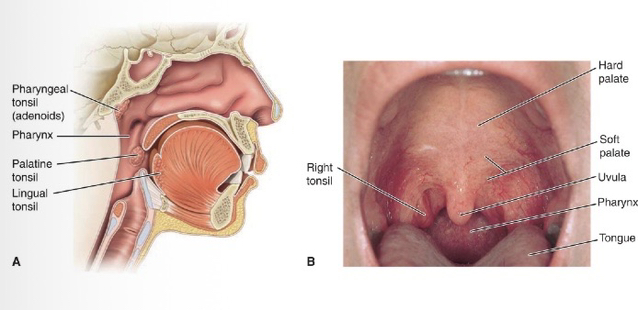
Tonsils: Lymphoid tissue located in the pharynx that provides an initial immune defense against inhaled or swallowed pathogens.
Pharyngeal Tonsil: Located in the posterior wall of the nasopharynx (also known as the adenoid when swollen).
Palatine Tonsil: Located on either side of the posterior oral cavity.
Peyer’s Patches: Specialized clusters of lymphoid nodules found in the lining of the small intestine (specifically the ileum) to monitor intestinal bacteria.
Appendix: A small, finger-like projection attached to the cecum of the large intestine, containing significant masses of lymphoid tissue.